-
Ivana Karanovic, Stefan Eberhard, Giulia Perina
Zookeys
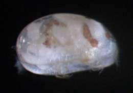
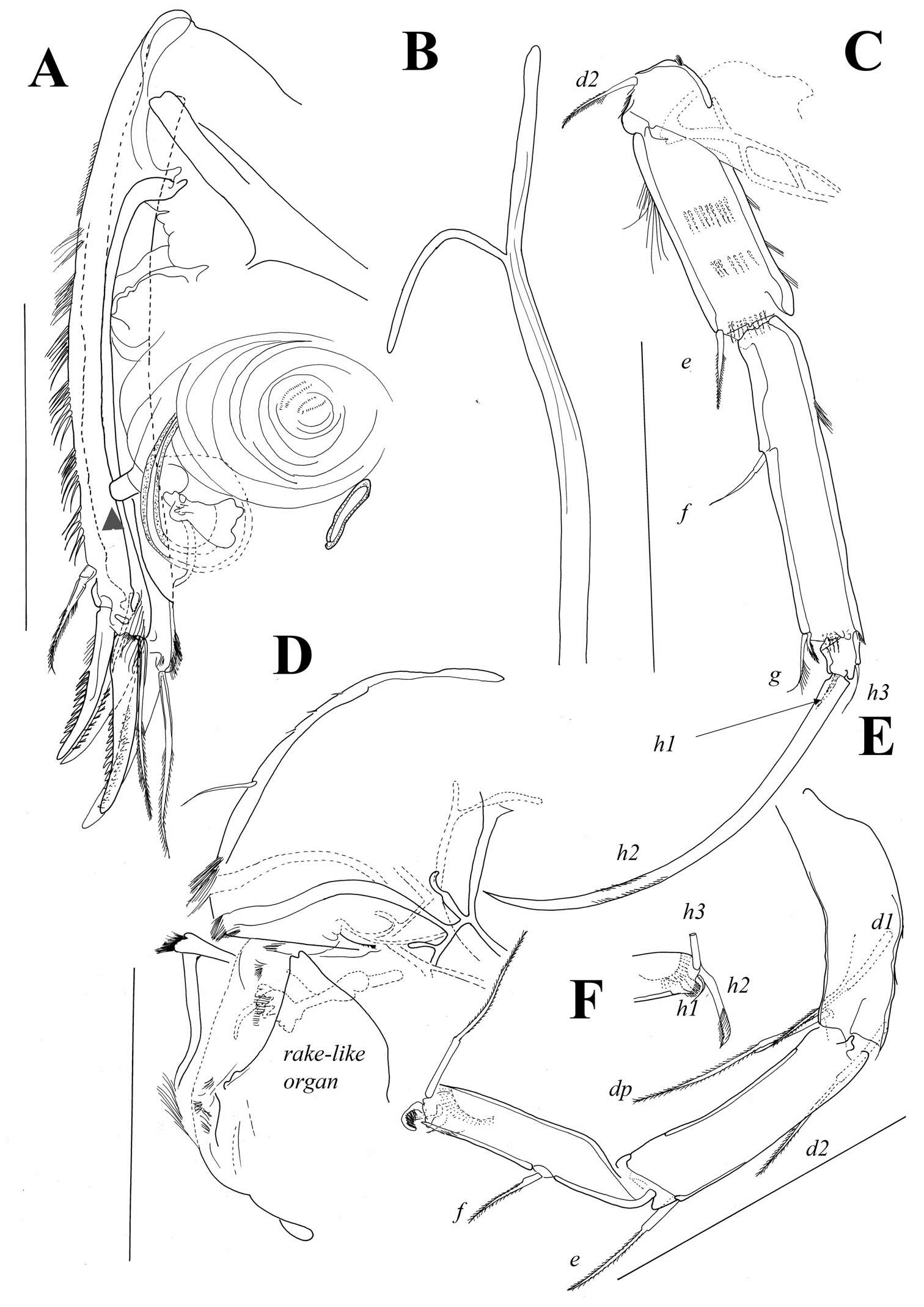
Figure 11.Austromesocypris bluffensis (Holotype): A UR, arrow indicating the genital process B attachment of the UR C L6 D forehead and upper lip E L7 F detail of the distal end of L7. Scales = 0.1 mm.
-
Ricardo L. Pinto, Merlijn Jocqué
Zookeys
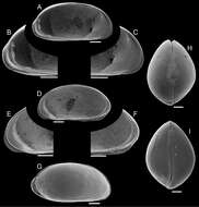
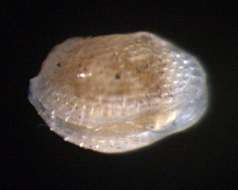
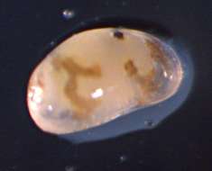
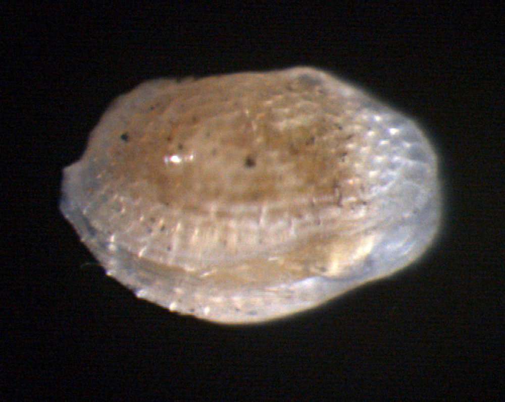
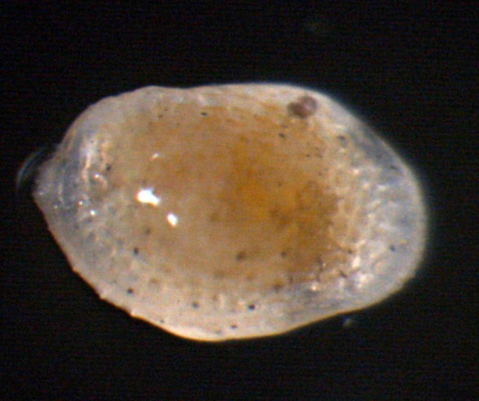
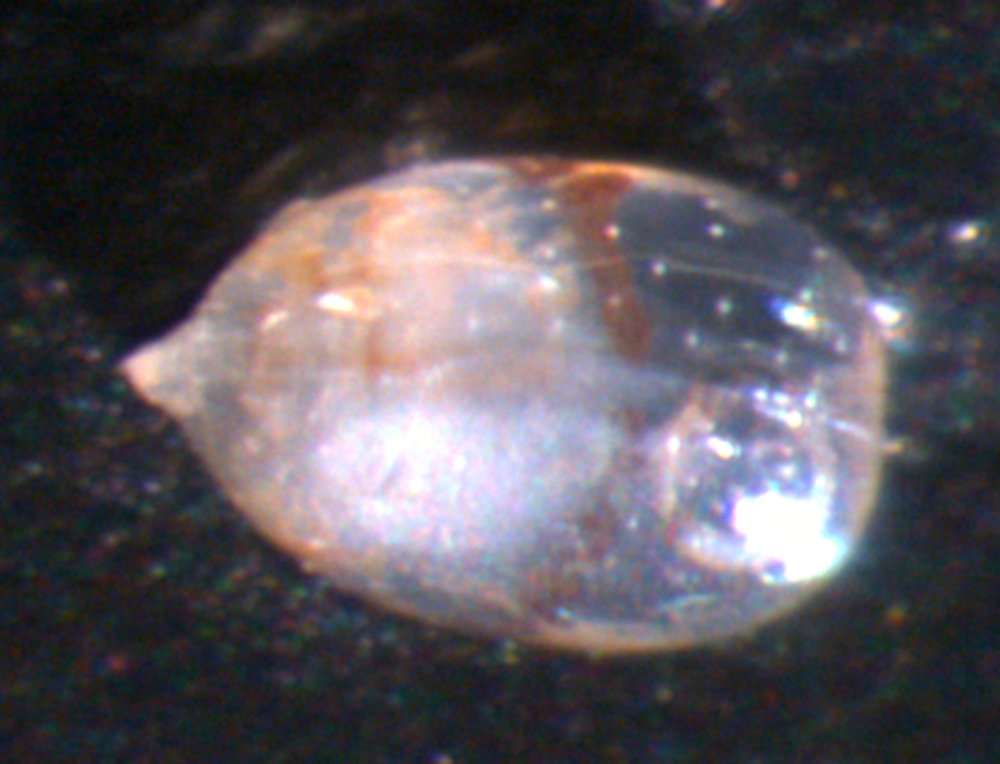
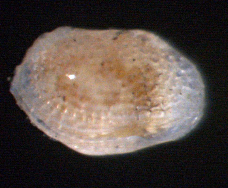
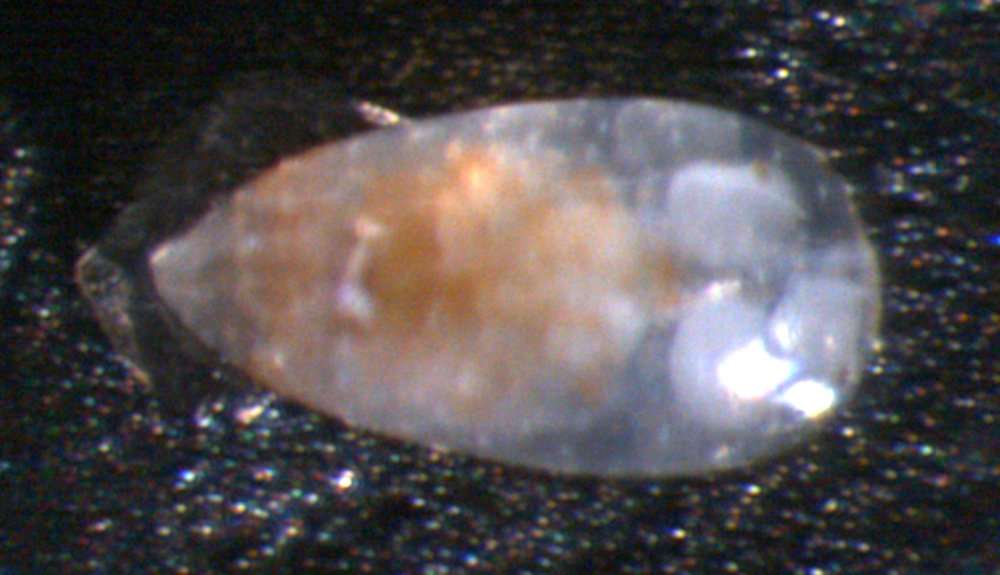
Figure 4.Elpidium merendonense sp. n., female. A Left valve internal view, general B left valve internal view, detail of postero-ventral margin C left valve internal view, detail of antero-ventral margin D right valve internal view, general E right valve internal view, detail of antero-ventral margin F right valve internal view, detail of postero-ventral margin G right lateral view H dorsal view I ventral view. A–F paratype, MZUSP 29076; G paratype, MZUSP 29080; H paratype, MZUSP 29081; I paratype, MZUSP 29082.Scale bars: 100 µm.
-
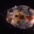
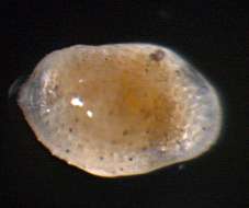
All Biocode files are based on field identifications to the best of the researcher’s ability at the time.
-
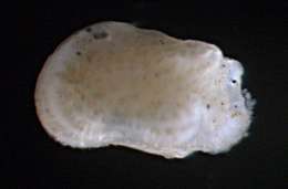
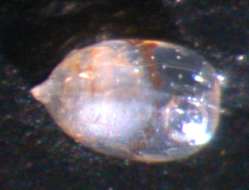
All Biocode files are based on field identifications to the best of the researcher’s ability at the time.
-
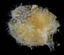
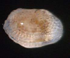
All Biocode files are based on field identifications to the best of the researcher’s ability at the time.
-
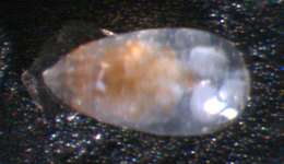
All Biocode files are based on field identifications to the best of the researcher’s ability at the time.
-
Ryouichi Higashi, Akira Tsukagoshi
Zookeys
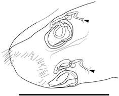
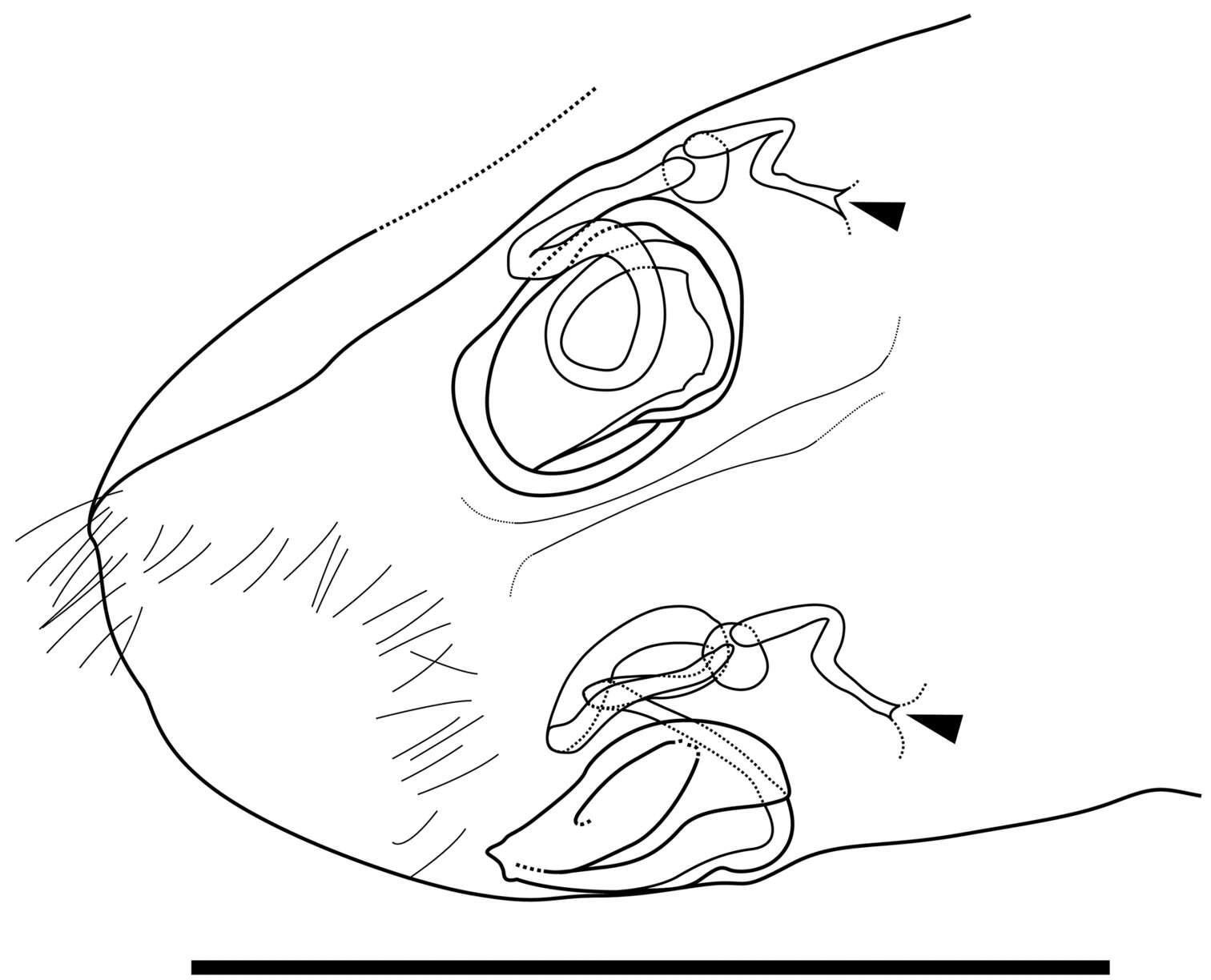
Figure 6. Caudal part of female of Parvocythere gottwaldi sp. n. Dorsal view (paratype, SUM-CO-2038). Arrows indicate openings. Scale bar indicates 50 µm.
-
All Biocode files are based on field identifications to the best of the researcher’s ability at the time.
-
All Biocode files are based on field identifications to the best of the researcher’s ability at the time.
-
Ryouichi Higashi, Akira Tsukagoshi
Zookeys
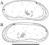
Figure 7. Carapaces of Parvocythere gracilis sp. n. Holotype (SUM-CO-2050). A right external view B left external view. The carapace structures are transmitted images. Scale bar indicates 100 µm.
-
All Biocode files are based on field identifications to the best of the researcher’s ability at the time.
-
All Biocode files are based on field identifications to the best of the researcher’s ability at the time.
-
Ryouichi Higashi, Akira Tsukagoshi
Zookeys
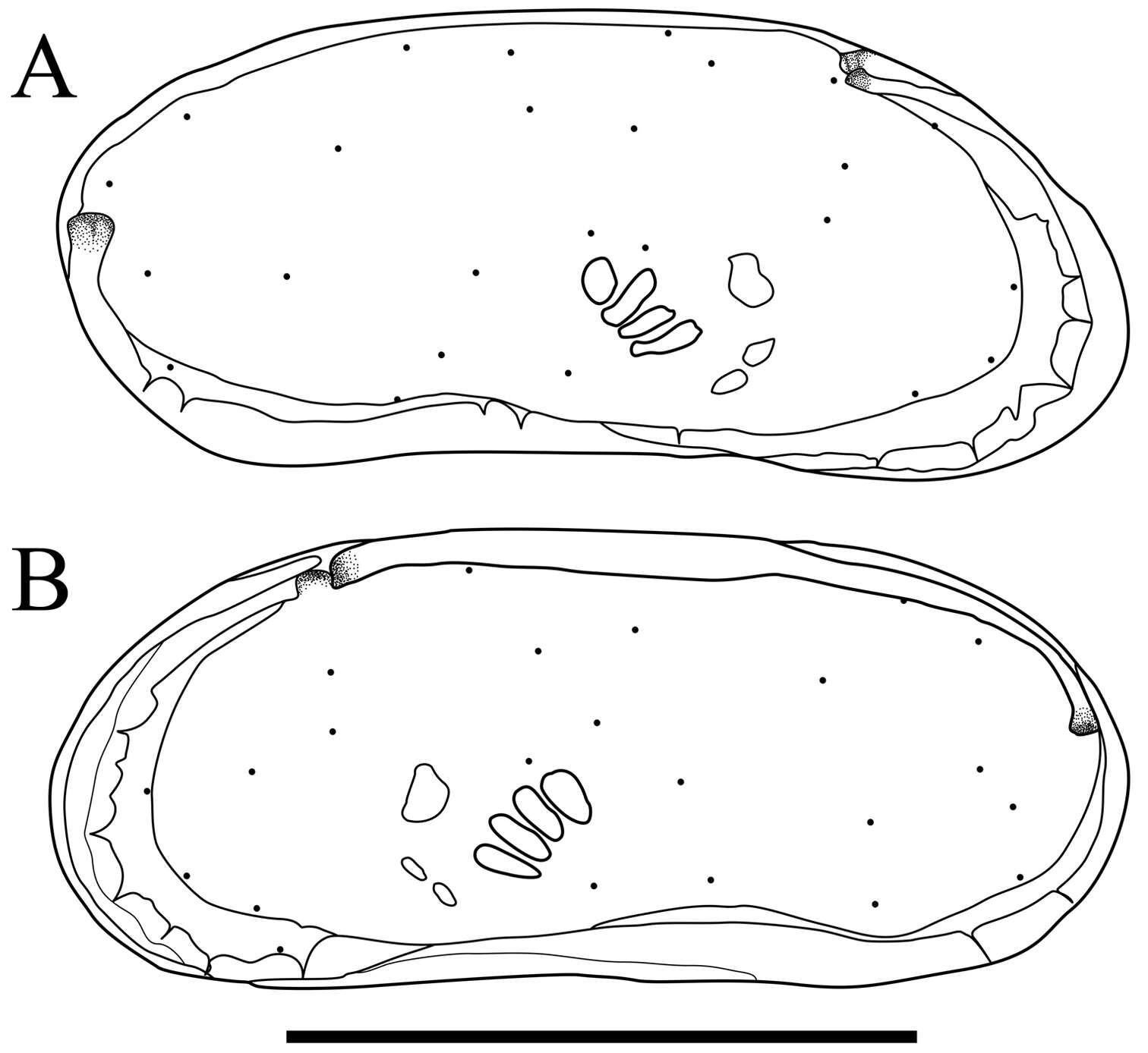
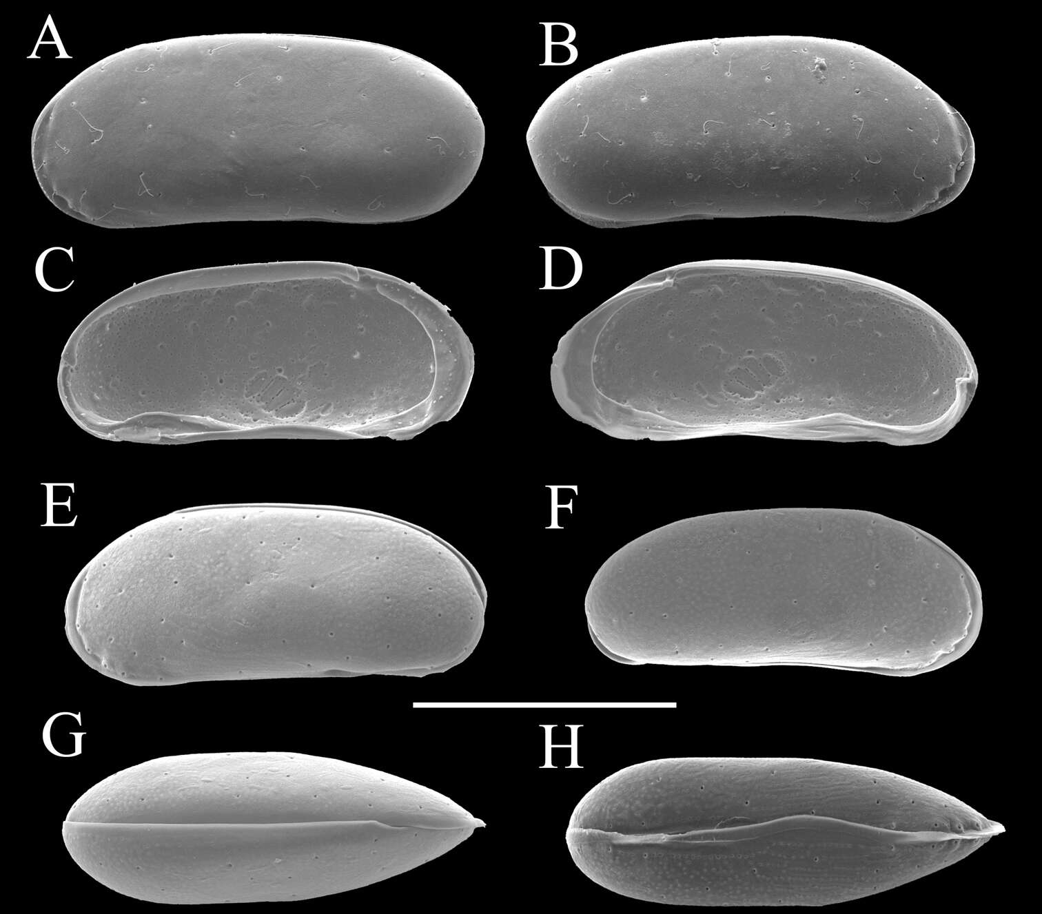
Figure 8. Carapaces of Parvocythere gracilis sp. n. A–D and H male specimens: A paratype (SUM-CO-2051) B paratype (SUM-CO-2052) C and D paratype (SUM-CO-2053) H paratype (SUM-CO-2054). A left external lateral view B right external lateral view C internal view of left valve D internal view of right valve H ventral view. E–G female specimens: E paratype (SUM-CO-2059) F paratype (SUM-CO-2059) G paratype (SUM-CO-2060). E left external view F right external view G dorsal view. Scale bar indicates 100 μm.
-
All Biocode files are based on field identifications to the best of the researcher’s ability at the time.
-
All Biocode files are based on field identifications to the best of the researcher’s ability at the time.
-
Ryouichi Higashi, Akira Tsukagoshi
Zookeys
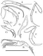
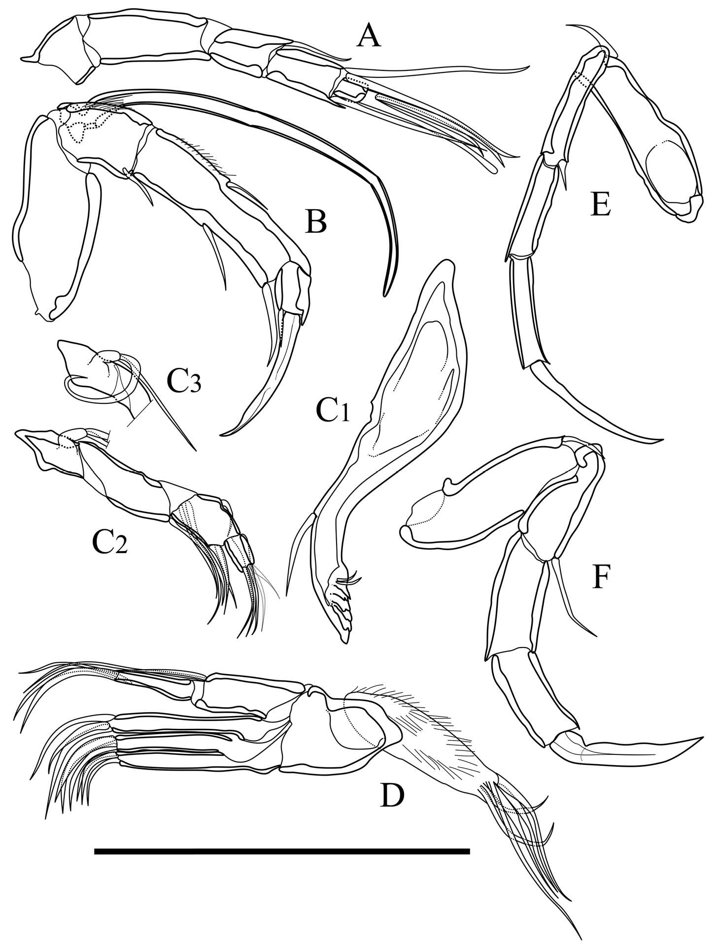
Figure 9. Appendages of Parvocythere gracilis sp. n. Holotype (SUM-CO-2050). A antennula B antenna C1 coxa of mandibula C2 palp of mandibula C3 proximal part of mandibular palp D maxillula E fifth limb F sixth limb. Scale bar indicates 50 µm.
-
All Biocode files are based on field identifications to the best of the researcher’s ability at the time.
-
All Biocode files are based on field identifications to the best of the researcher’s ability at the time.
-
Ryouichi Higashi, Akira Tsukagoshi
Zookeys
Figure 10. Male copulatory organs of Parvocythere gracilis sp. n. Holotype (SUM-CO-2050). A internal view of left organ B external view of right organ. Copulatory ducts are shaded. Abbreviation: Dr dorsal ramus Vr ventral ramus Dl Distal lobe. Scale bar indicates 50 µm.
-
All Biocode files are based on field identifications to the best of the researcher’s ability at the time.
-
All Biocode files are based on field identifications to the best of the researcher’s ability at the time.
-
Ryouichi Higashi, Akira Tsukagoshi
Zookeys
Figure 11. Caudal part of female of Parvocythere gracilis sp. n. Dorsal view (paratype, SUM-CO-2058). Arrows indicate openings. Scale bar indicates 50 µm.
-
All Biocode files are based on field identifications to the best of the researcher’s ability at the time.
-
All Biocode files are based on field identifications to the best of the researcher’s ability at the time.