-
Fig 1a: Balanion comatum Line drawing of protargol stained cell
-
-
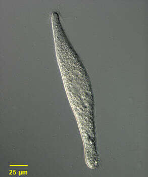
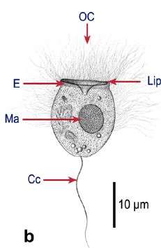
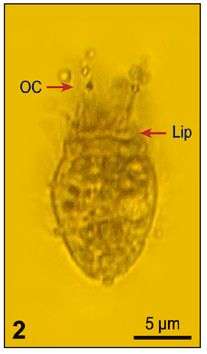
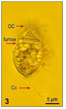
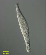
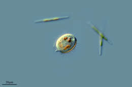
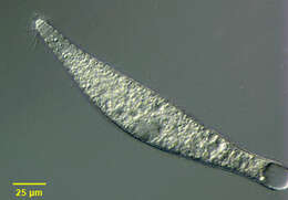
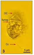
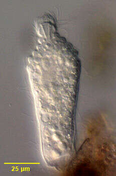
Portrait of extended Lagynus cucumis, a colorless Prostome ciliate found in sapropelic habitats. L. cucumis is synonymous with Lacrymaria cucumis (Penard, 1922) and Lacrymaria putrina (Kahl, 1926). Lagynus cucumis is longer and more slender than L. elegans with about five less pronounced ring-like furrows at the anterior end. The conical head region is much smaller than that of L. elegans. The apical cytostome is supported by fine trichites (seen here). The elongate cell body is flexible, contractile and slightly flattened. Somatic kineties are longitudinal and uniform. Slightly longer cilia surround the anterior end . A bean-shaped macronucleus with a central transverse crease is located in the midportion of the cell (seen well here). There is a single posterior terminal contractile vacuole (not seen in this image). Collected from sapropelic sediments of a freshwater aquaculture tub (pH 7.56) at a Koi farm near Boise, Idaho October 2003. DIC optics.
-
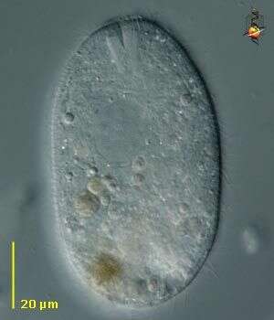
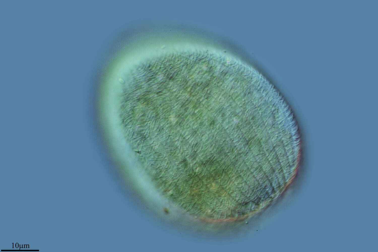
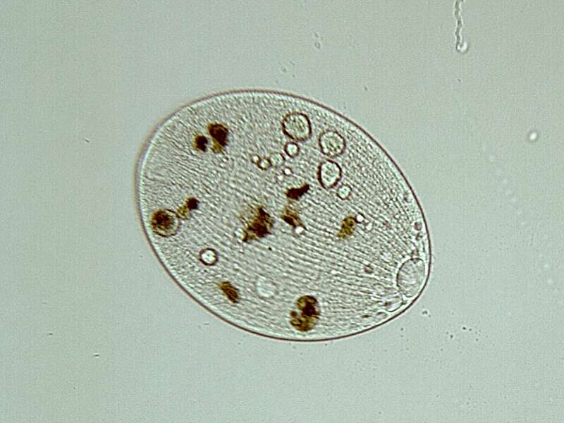
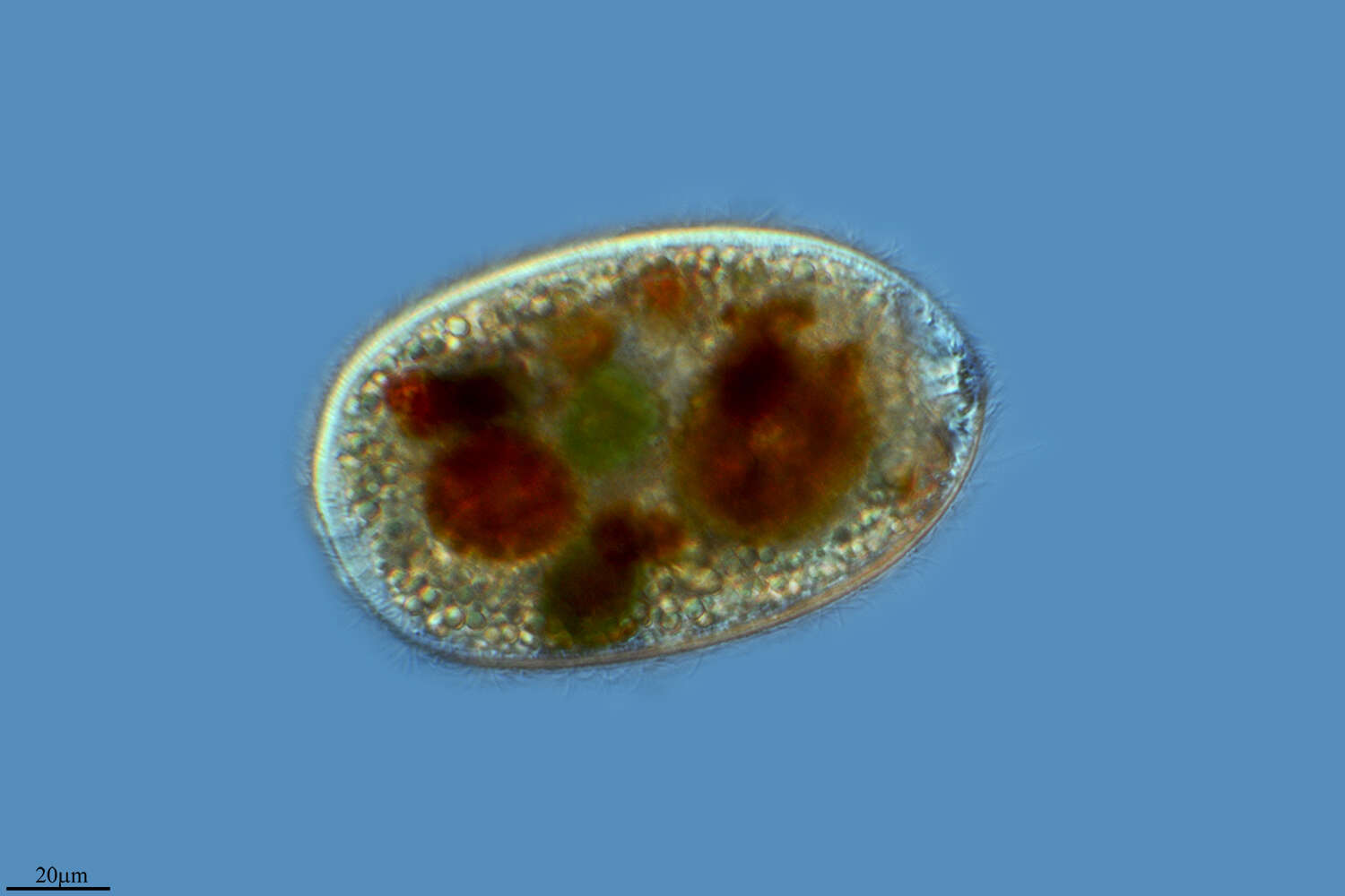
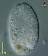
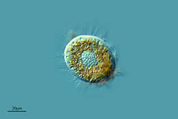
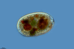
The ciliate Holophrya, with a polar mouth which is surrounded by stiff rods (nemadesmata) that can be used to manipulate food into the body. Normally eats algae and detritus. Differential interference contrast.
-
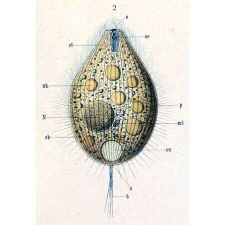
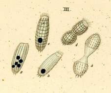
a -- Anus b -- Sensory bristle c.v -- Contractile vacuole ek -- Ectoplasm N -- Macronucleus ncl -- Micronucleus nk -- Food particle o -- Mouth p -- Pellicle st -- Cytopharyngeal basket
-
Lardero, La Rioja, Spain
-
Herrera, Castille and Leon, Spain
-
Villar del Pedroso, Extremadura, Spain
-
Los Cotos, Madrid, Spain
-
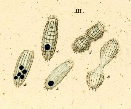
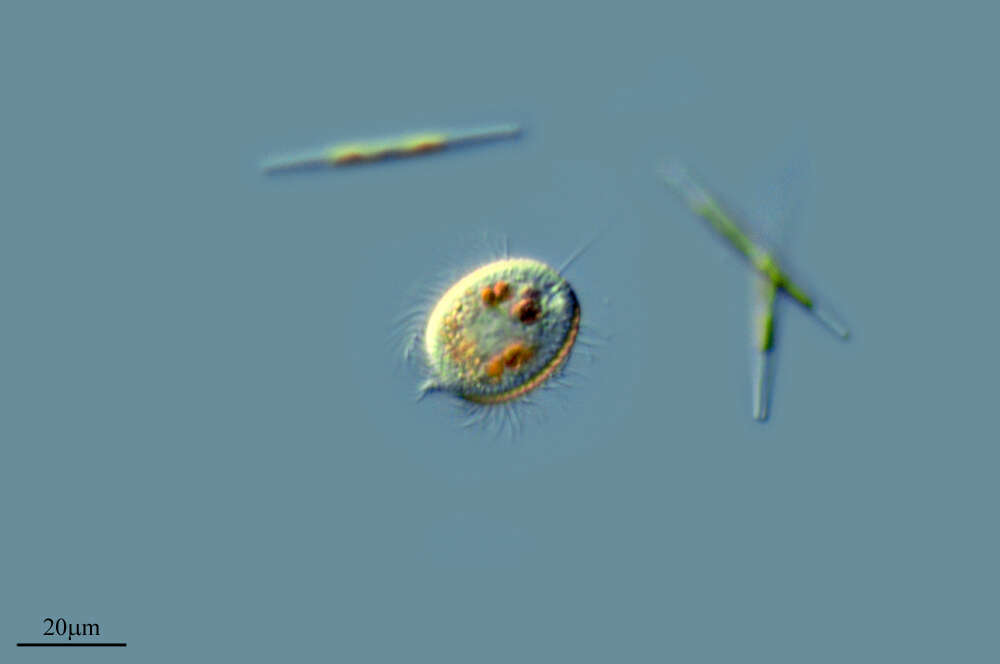
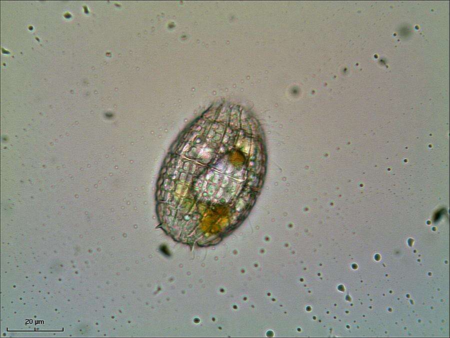
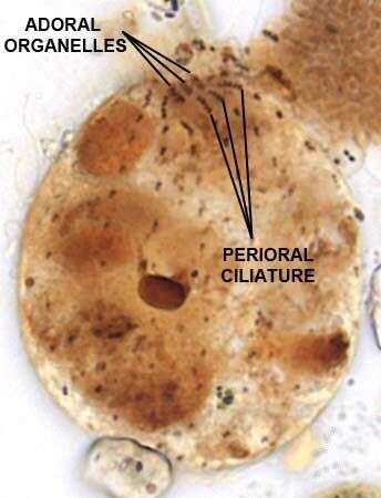
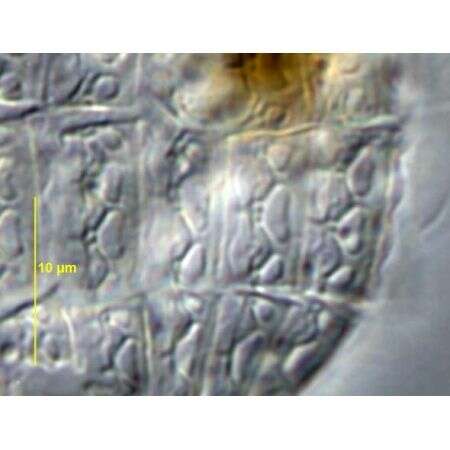
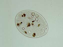
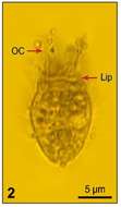
Body with regurarly arranged ectoplasmic plates. Cytostome at anterior end, surrounded by slightly longer cilia . Often spinous projection at or near posterior end.
-
Fig 1b: Balanion comatum Line drawing of live cell (from Wulff, 1919)
-
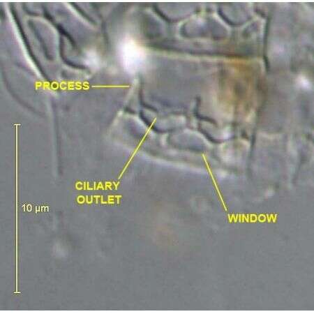
Oral Infraciliature of Nolandia nolandi (KAHL, 1930) SMALL & LYNN, 1985.Collected from a freshwater pond near Boise, Idaho. June 2008.Stained by the Protargol technique (Wilbert modification) (see Foissner, W. Europ. J. Protistol., 27:313-330;1991).Brightfield.
-
Detail view of anterior end of extended Lagynus cucumis, a colorless Prostome ciliate found in sapropelic habitats. L. cucumis is synonymous with Lacrymaria cucumis (Penard, 1922) and Lacrymaria putrina (Kahl, 1926). Lagynus cucumis is longer and more slender than L. elegans with about five less pronounced ring-like furrows at the anterior end. The conical head region is much smaller than that of L. elegans. The apical cytostome is supported by fine trichites (seen here). The elongate cell body is flexible, contractile and slightly flattened. Somatic kineties are longitudinal and uniform. Slightly longer cilia surround the anterior end. A bean-shaped macronucleus with a central transverse crease is located in the midportion of the cell. There is a single posterior terminal contractile vacuole. Collected from sapropelic sediments of a freshwater aquaculture tub (pH 7.56) at a Koi farm near Boise, Idaho October 2003. DIC.
-
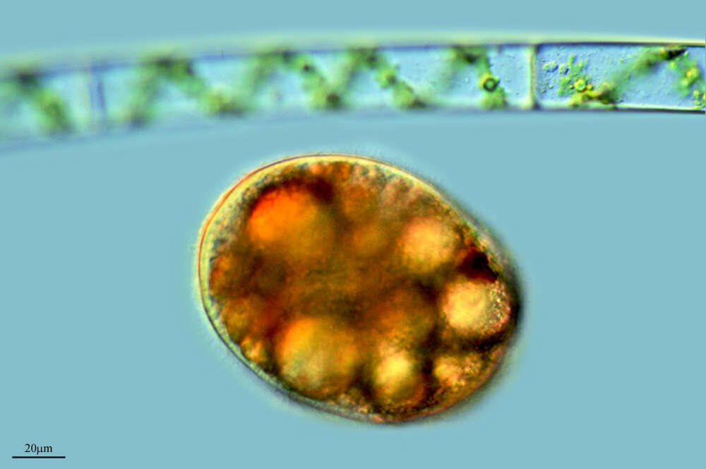
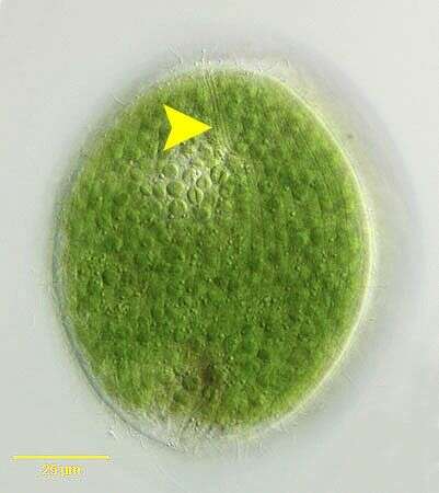
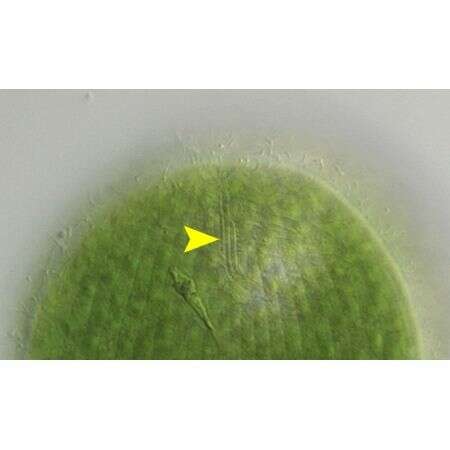
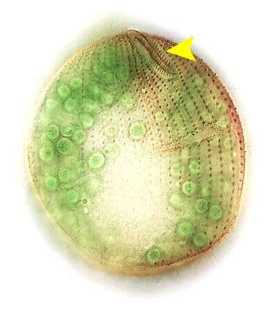
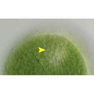
Holophrya ovum (Ehrenberg,1831). The yellow arrowhead indicates the three dorsal brush rows of short cilia. DIC
-
Ovoid to cylindrical; ciliation uniform; oral basket made up of double trichites which end up deep in ectoplasm. Macronucleus ovoid, reniform or ellongate. Cell body 80-200 micron long.
-
Fig. 2: Balanion comatum Lugol's fixed cell, lateral view
-
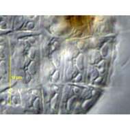
Calcified armor plates of Nolandia nolandi (KAHL, 1930) SMALL & LYNN, 1985.Collected from a freshwater pond near Boise, Idaho. June 2008.DIC.
-
Portrait of extended Lagynus cucumis, a colorless Prostome ciliate found in sapropelic habitats. L. cucumis is synonymous with Lacrymaria cucumis (Penard, 1922) and Lacrymaria putrina (Kahl, 1926). Lagynus cucumis is longer and more slender than L. elegans with about five less pronounced ring-like furrows at the anterior end. The conical head region is much smaller than that of L. elegans. The apical cytostome is supported by fine trichites. The elongate cell body is flexible, contractile and slightly flattened. Somatic kineties are longitudinal and uniform. Slightly longer cilia surround the anterior end. A bean-shaped macronucleus with a central transverse crease is located in the midportion of the cell (seen well here). There is a single posterior terminal contractile vacuole (seen in this image). Collected from sapropelic sediments of a freshwater aquaculture tub (pH 7.56) at a Koi farm near Boise, Idaho October 2003. DIC optics.
-
Holophrya ovum (Ehrenberg,1831). The yellow arrowhead indicates the three dorsal brush rows of short cilia. DIC
-
Cuelgamuros, Madrid, Spain
-
Fig 3: Balanion comatum Lugol's fixed cell, lateral view
-
Calcified armor plates of Nolandia nolandi (KAHL, 1930) SMALL & LYNN, 1985 .Collected from a freshwater pond near Boise, Idaho. June 2008.DIC.
-
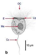
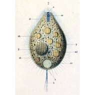
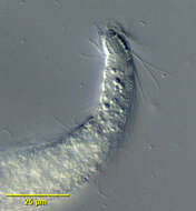
Portrait of contracted Lagynus elegans (Engleman,1862, Quennerstedt,1867), a colorless Prostome ciliate found in sapropelic habitats. The cell body is flask-shaped with a flexible anterior neck marked by concentrically decreasing transverse ciliated furrows (seen well in this contracted individual). A conical head region bears longer cilia and an inconspicuous "dorsal brush" of 3 to 4 groups of kineties between cytopharyngeal nematodesmata (seen well here) and the first perioral ciliary ring. This feature distinguishes Lagynus from Lacrymaria, which has a short longitudinal dorsal brush of cilia on the neck region. Somatic kineties are longitudinal. The silverline system of Lagynus is also different from that of Lacrymaria. The neck region of Lagynus elegans is much less extensible (about 1/3 body length) than that of Lacrymaria olor. The posterior is often flattened. There is a single ellipsoid macronucleus and adjacent micronucleus about mid-body. There is a large posterior terminal contractile vacuole. Multiple food vacuoles are scattered through the cytoplasm. The cytoplasm L. elegans, like that of some other anaerobic ciliates, contains methanogenic bacilli and hydrogenosomes. L. elegans feeds on ciliates, cyanobacteria and other bacteria. Collected from sapropelic sediments of a freshwater aquaculture tub (pH 7.56) at a Koi farm near Boise, Idaho October 2003. DIC optics.
-
Holophrya ovum (Ehrenberg,1831). The yellow arrowhead indicates the three dorsal brush rows of short cilia. Stained by the silver carbonate technique (see Foissner, W. Europ. J. Protistol., 27:313-330;1991).Brightfield.