-
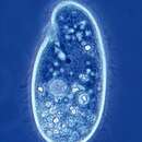
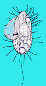
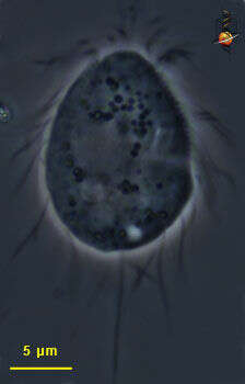
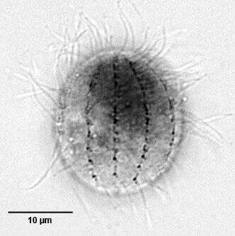
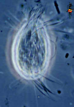
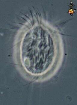
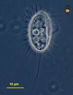
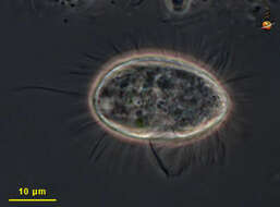
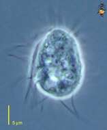
Cyclidium (sigh-clid-ee-um) is a small scuticociliate, a group which usually has a well developed veil of cilia projecting around the mouth region. This undulating membrane is not extended in the cell in this picture. It eats bacteria. May occur in organically enriched habitats with large numbers of bacteria. Common in freshwater and marine habitats. Pleuronema is a larger member of the same group that is often found in marine habitats. Paracyclidium is similar, but there are no locomotor cilia in the centre of the body whereas it is clear from this image, that the kineties are unbroken. Long caudal cilium. Phase contrast.
-
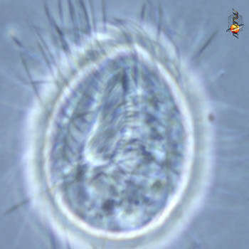
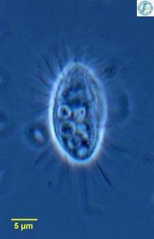
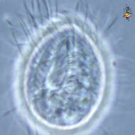
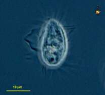
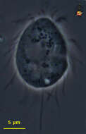
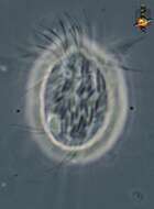
Cyclidium (sigh-clid-ee-um) is a small scuticociliate, a group which usually has a well developed veil of cilia projecting around the mouth region. This image shows the mouth region and the layer of cilia lying over the top of it. Phase contrast.
-
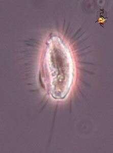
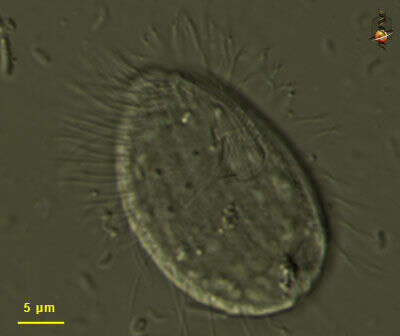
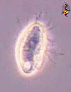
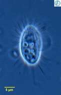
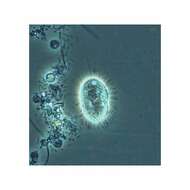
Cyclidium (sigh-clid-ee-um) is a small scuticociliate, a group which usually has a well developed veil of cilia projecting around the mouth region, and these can be see to one side of each cell. It eats bacteria. May occur in organically enriched habitats with large numbers of bacteria. Common in freshwater and marine habitats. Poor image. Phase contrast.
-
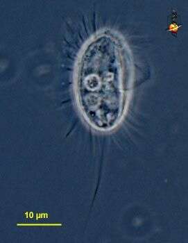
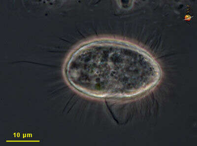
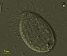
Cyclidium (sigh-clid-ee-um) is a small scuticociliate, a group which usually has a well developed veil of cilia projecting around the mouth region, and these can be see to the right side of this cell. It eats bacteria. May occur in organically enriched habitats with large numbers of bacteria. Common in freshwater and marine habitats. Phase contrast.
-
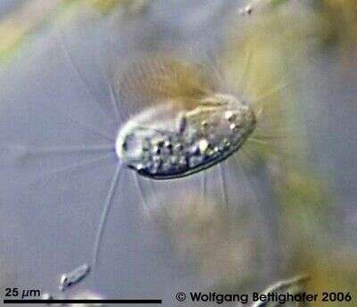
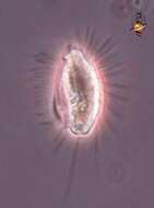
Cyclidium (sigh-clid-ee-um) a common and widespread filter-feeding ciliate. This is a scuticociliate in which one of the aggregates of cilia associated with the mouth forms a veil to one side (here to the left) and this is used to help separate particles of food- bacteria - from the water. Phase contrast. Material from Nymph Creek and Nymph Lake, thermal sites within Yellowstone National Park, photograph by Kathy Sheehan and David Patterson.
-
-
Cyclidium, common bacterivorous ciliate, with undulating membrane extending sideways (here to the right) in feeding cells. From Lake Donghu, China. Phase contrast micrograph.
-
This cell has been slightly compressed, the curving structure upper right is the base of the undulating membrane, showing its path from near the front of the cell to end by curving around the cytosome - the point at which food is ingested. The lines over the surface of the body are where the kineties (rows of cilia) lie. Differential interference contrast optics.
-
The mouth structures are in the upper right part of the cell. Differential interference contrast optics.
-
This image is of a feeding cell. The extensive undulating membrane starts at the front of the cell and gets wider as it gets closer to the mouth - near the middle of the cell. The dark line is an edge of the UM, and the tightly spaced cilia can be seen near the front of the UM. The body has numerous evenly spaced cilia, but there is one unusually long caudal cilium. Phase contrast image.
-
Small ciliate, the bases of the line of cilia that make up the undulating membrane are seen to the upper right of the cell. Phase contrast microscopy.
-
This small ciliat belonging to group of scuticociliates/hymenostomata is not easy to photograph. The picture showes the sail-like undulating membran. Sample collected from Simmelried near Konstanz (Baden-Wuerttemberg, Germany). This image was taken using Zeiss Universal with Olympus C7070 CCD camera.
-
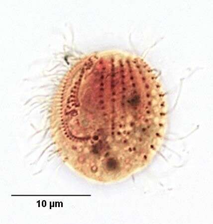
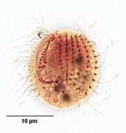
Dorsal view of the small scutociliate, Cyclidium glaucoma (Mueller, 1773). The infraciliature has been stained by the Klein-Foissner silver nitrate technique (see Foissner, W. Europ. J. Protistol.27,313-330;1991). The argyrome or silverline system is revealed. The denser longitudinal argentophilic lines are crossed by finer transverse bridging lines (seen best in the anterior 1/2 here). Collected from stagnant freshwater rich in decomposing matter near Boise, Idaho, August 2004. Brightfield.
-
Ventral view of the small scutociliate, Cyclidium glaucoma (Mueller, 1773). The infraciliature has been stained by the Klein-Foissner silver nitrate technique (see Foissner, W. Europ. J. Protistol.27,313-330;1991). The argyrome or silverline system is revealed. The denser longitudinal argentophilic lines are crossed by finer transverse bridging lines (seen best in the anterior 1/2 here). The reverse "J" shaped paraoral membrane is clearly seen. the line just medial to the paraoral membrane represents the three adoral membranelles. A short dense longitudinal line immediately posterior to the paraoral membrane represents the cytoproct (cell anus). Collected from stagnant freshwater rich in decomposing matter near Boise, Idaho, August 2004. Brightfield.
-
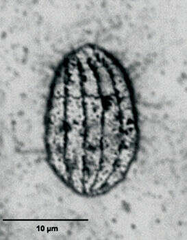
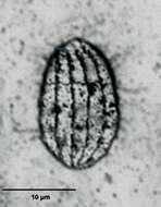
Dorsal view of the small scutociliate, Cyclidium glaucoma (Mueller, 1773). The infraciliature has been stained by the silver carbonate technique (see Foissner, W. Europ. J. Protistol.27,313-330;1991). 15-30 microns in length. The cell is ovoid and laterally compressed. Five of the 10 longitudinal somatic kineties are visible. Collected from stagnant freshwater rich in decomposition products near Boise, Idaho August 2004. Brightfield.
-
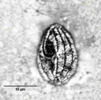
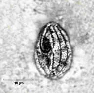
Ventral view of the small scutociliate, Cyclidium glaucoma (Mueller, 1773). The infraciliature has been stained by the silver carbonate technique (see Foissner, W. Europ. J. Protistol.27,313-330;1991). 15-30 microns in length. The cell is ovoid and laterally compressed. A prominent paraoral membrane borders the peristome on the right, curving around it posteriorly to form a pouch. The paraoral membrane extends at least 1/2 the cell length. Just to the left of the paraoral membrane three small polykinetids correspond to adoral membranelles 1, 2 and 3. The nonciliated basal bodies of the scuticovestige at the posterior end of the paraoral membrane are visible in this specimen. Longitudinal somatic kineties are visible to the left of the cytostome. Collected from stagnant organically enriched freshwater near Boise, Idaho August 2004. Brightfield
-
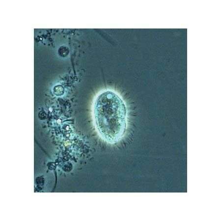
Cyclidium glaucoma: Ciliate protozoa, with long cilia except near the caudal region. A single long caudal cilia. Fast moving, but stationary while feeding. This image was taken by Krishnakumar B. in a sample from an anaerobic bioreactor for organic rich wastewater treatment in Regional Research Laboratory-Trivandrum (CSIR-India).
-
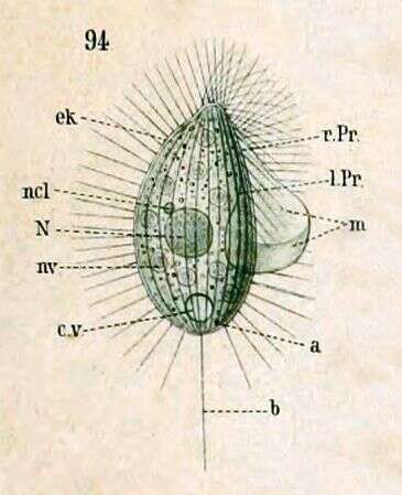
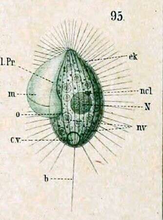
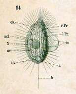
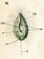
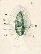
Key to Schewiakoff's abbreviations: a -- Anus b -- Sensory bristle ek -- Ectoplasm l.Pr -- Left peristome N -- Macronucleus ncl -- Micronucleus nv -- Food vacuole r.Pr -- Right peristome
-
Left side view. Key to Schewiakoff's abbreviations: b -- Sensory bristle cv -- Contractile vacuole ek -- Ectoplasm l.Pr -- Left edge of peristome m -- Undulating membrane N -- Macronucleus ncl -- Micronucleus nv -- Food vacuole
-
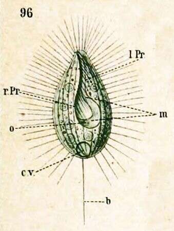
Ventral view. Key to Schewiakoff's abbreviations: b -- Sensory bristle cv -- Contractile vacuole l.Pr -- Left edge of peristome m -- Undulating membrane o -- Mouth r.Pr -- Right edge of peristome
-
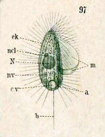
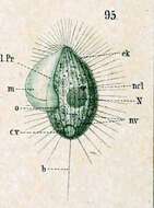
Synononym: Cyclidium glaucoma var. elongatum (Schewiakoff, 1889). Key to Schewiakoff's abbreviations: a -- Anus b -- Sensory bristle cv -- Contractile vacuole ek -- Ectoplasm m -- Unduating membrane N -- Macronucleus ncl -- Micronucleus
-
Paracyclidium (para-sigh-clid-ee-um), one of the cylidiid scuticociliates. Scuticociliates have a curved undulating membrane associated with the mouth, and within the family, this genus is distinguished because the kineties are broken in the centre of the cell. This allows us to see the epibiotic rod-shaped bacteria. Ciliates with external bacteria are often encountered in an anoxic sites suggesting that the symbiosis is linked to respiratory metabolism. Phase contrast.
-
Paracyclidium (para-sigh-clid-ee-um), one of the cylidiid scuticociliates. Scuticociliates have a curved undulating membrane associated with the mouth, and within the family, this genus is distinguished because the kineties are broken in the centre of the cell. This cell is included because of the large numbers of ectosymbiotic bacteria that are attached to the cell surface. Phase contrast.
-
Paracyclidium (para-sigh-clid-ee-um) is a small scuticociliate, a group which usually has a well developed veil of cilia projecting around the mouth region (flopped , and these can be see to one side of each cell. It eats bacteria. May occur in organically enriched habitats with large numbers of bacteria. Rows of cilia (kineties) are interrupted in the central region of the body. Cyclidium is similar, but the kineties are unbroken. Long caudal cilium. Phase contrast.