-
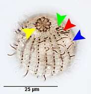
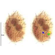
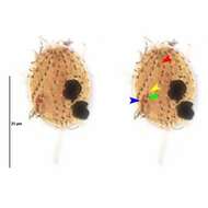
The oblique dikinetids of the circumoral membranelles are indicated by the yellow arrowhed. The anterior row part of the dorsal brush (green arrowhead) and the posterior part (red arrowhead) are seen here.The shortened somatic kinety terminating anteriorly at the dorsal brush is indicated by the blue arrowhead. Stained by the silver carbonate technique (see Foissner, W.Europ. J. Protistol.27:313-330;1991). Brightfield.
-
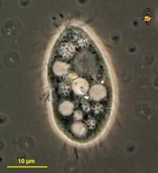
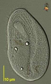
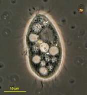
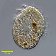
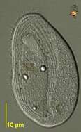
Homalogastra (hom-al-owe-gast-ra) small bacterivorous ciliate. The lighter vacuoles are food vacuoles, the grey area anterior right is the macronucleus. Phase contrast micrograph.
-
Ventral infraciliature of Aspidisca cicada (MUELLER,1786) CLAPARÃDE&LACHMANN,1858. Protargol protocol A. (see Foissner, W. Europ. J. Protistol., 27:313-330;1991).Brightfield.
-
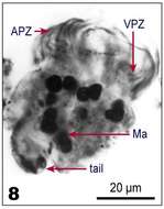
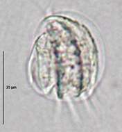
Fig 8: Tontonia gracillima protargol stain, ventral view
-
Left side of Acineria incurvata DUJARDIN,1841, a pleurostomatid ciliate found in heavily polluted freshwater and marine habitats. Collected from effluent of a protein skimmer at a commercial saltwater aquarium in Boise,Idaho. January 2007.
-

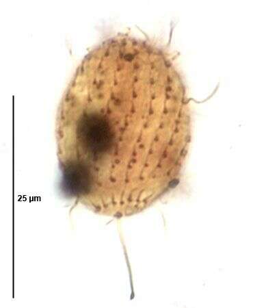
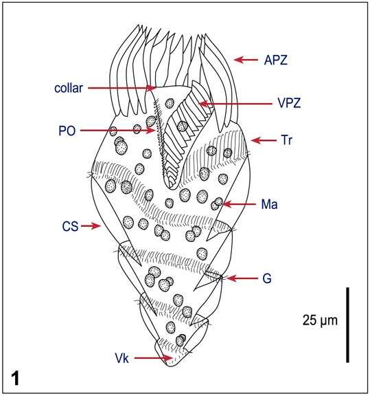
Portrait (dorsal view) of the marine hypotrich ciliate, Kiitricha marina (Nozowa, 1941). The genus is monotypic. The cell has a broadly elliptical outline and is dorsoventrally flattened. The dorsal surface is slightly convex, the ventrum slight concave. The large peristome extends from the anterior end along the left side to terminate in a cytostome in the posterior ¼ of the body. There is a prominent adoral zone of membranelles on the left margin of the peristome and an undulating membrane on its right margin. Uniform dorsal kineties run from left anterior to right posterior on the dorsal surface (seen in this image). On the ventral surface there are approximately 10 longitudinal rows of cirri to the right of and posterior to the cytostome (seen in this image). There are five large transverse cirri lying to the right of the posterior end of the cytostome within the ventral cirral field (not seen here). This feature distinguishes Kiitricha marina from the very similar Caryotricha convexa whose transverse cirri lie outside the ventral cirral field posterior and to the left of the peristome. There is a single ovoid macronucleus (seen in this image) with an adjacent spherical micronucleus in the center. Collected from a commercial saltwater aquarium in Boise, Idaho February 2004. DIC optics.
-
Stained by the silver carbonate technique (see Foissner, W.Europ. J. Protistol.27:313-330;1991).Brightfield.
-
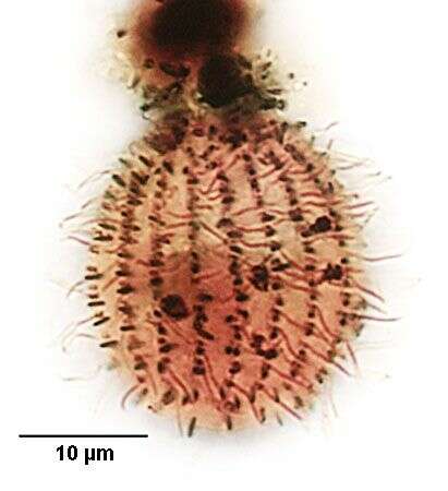
Dorsal infraciliature of the scuticociliate, Homalogastra setosa (KAHL,1926).From non-flooded petri dish culture of topsoil from a public park in Boise,Idaho. Stained by the silver carbonate technique (see Foissner, W.Europ. J. Protistol.27:313-330;1991).Brightfield.
-
Dorsum of Aspidisca cicada (MUELLER,1786) CLAPARÃDE&LACHMANN,1858. Protargol protocol A. (see Foissner, W. Europ. J. Protistol., 27:313-330;1991).Brightfield.
-
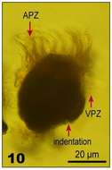
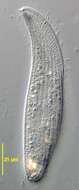
Fig 9: Tontonia gracillima Lugol's fixed cell, ventrolateral view, the tail is lost due to fixation, only the indentation is visible
-
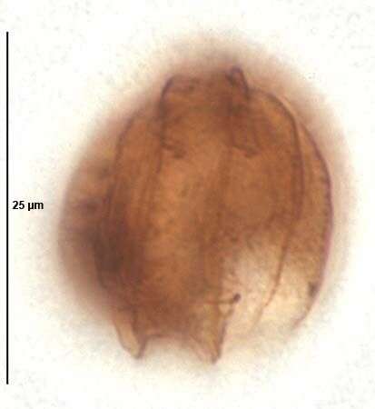
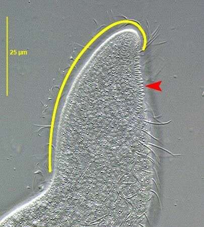
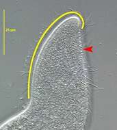
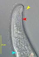
Acineria incurvata DUJARDIN,1841 a pleurostomatid ciliate found in heavily polluted freshwater and marine habitats. The yellow line parallels the distinctive oral bulge which recurves dorsally and to the left at its anterior end. The bulge has a helical structure like a two-stranded string.The pattern is faintly visible here.The dorsal brush consists of a single file of obliquely oriented dikinetids bearing non-motile clavate cilia (red arrowhed). Collected from effluent of a protein skimmer at a commercial saltwater aquarium in Boise,Idaho. January 2007.DIC.
-
Portrait (ventral view) of the marine hypotrich ciliate, Kiitricha marina (Nozowa, 1941). The genus is monotypic. The cell has a broadly elliptical outline and is dorsoventrally flattened. The dorsal surface is slightly convex, the ventrum slight concave. The large peristome extends from the anterior end along the left side to terminate in a cytostome in the posterior ¼ of the body. There is a prominent adoral zone of membranelles on the left margin of the peristome and an undulating membrane on its right margin. Uniform dorsal kineties run from left anterior to right posterior on the dorsal surface. On the ventral surface there are approximately 10 longitudinal rows of cirri to the right of and posterior to the cytostome. There are five large transverse cirri lying to the right of the posterior end of the cytostome within the ventral cirral field. This feature distinguishes Kiitricha marina from the very similar Caryotricha convexa whose transverse cirri lie outside the ventral cirral field posterior and to the left of the peristome. There is a single ovoid macronucleus with an adjacent spherical micronucleus in the center. Collected from a commercial saltwater aquarium in Boise, Idaho February 2004. DIC
-
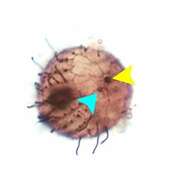
The kinetid of the long posterior cilium is located centrally at the posterior pole (yellow arrohead). The eccentrically located excretory pore of the contractile vacuole is indicated by the light blue arrowhead. Stained by the silver carbonate technique (see Foissner, W.Europ. J. Protistol.27:313-330;1991).Brightfield.
-
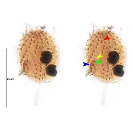
Right ventrolateral view of infraciliature of the scuticociliate, Homalogastra setosa (KAHL,1926).The oral aperture is in the posterior 1/3 of the cell. The blue arrowhead indicates the paraoral membrane to the right of the cytostome. The 2nd and 3rd adoral membranelles are indicated by the yellow and green arrowheads respectively. The small 1st adoral membranelle is not seen here. The two macronuclei indicate that this is an exconjugant specimen.Trophonts have a single spherical macronucleus and a spherical micronucleus. From non-flooded petri dish culture of topsoil from a public park in Boise,Idaho. Stained by the silver carbonate technique (see Foissner, W.Europ. J. Protistol.27:313-330;1991).Brightfield.
-
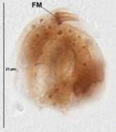
Frontal membranelle of Aspidisca cicada (MUELLER,1786) CLAPARÃDE&LACHMANN,1858. Protargol protocol A. (see Foissner, W. Europ. J. Protistol., 27:313-330;1991).Brightfield.
-
Fig 10: Tontonia gracillima Lugol's fixed cell, ventrolateral view, the tail is lost due to fixation, only the indentation is visible
-
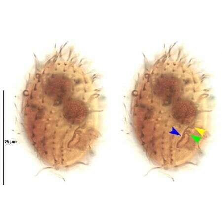
Acineria incurvata DUJARDIN,1841 a pleurostomatid ciliate found in heavily polluted freshwater and marine habitats. The yellow arrowhead indicates cilia of perioral kinety 2 which borders the oral bulge on the right side. The distinctive oral bulge recurves dorsally and to the left at its anterior end. The dorsal brush consists of a single file of obliquely oriented dikinetids bearing non-motile clavate cilia (red arrowhed). The light blue arrowhead indicates the posterior end of the oral bulge.Collected from effluent of a protein skimmer at a commercial saltwater aquarium in Boise,Idaho. January 2007.DIC.
-
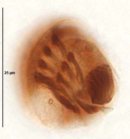
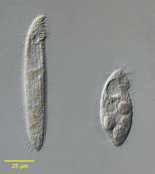
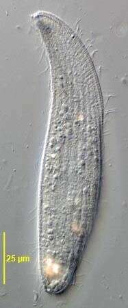
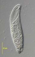
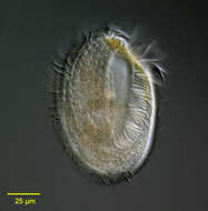
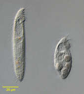
Portrait of the marine spirotrich ciliate Protocruzia granulosa (Kahl,1932) de Faria, da Cunha & Pinto, 1922. The cell is elongate and laterally compressed. The anterior ends in a short rostrum that bends slightly to the left. The posterior is bluntly tapered. The short, subapical anterior periostome has a prominent dense zone of membranelles. The somatic kineties are longitudinal. The cell is highly contractile as seen in this image with an extended individual on the viewer's left and a contracted cell on the right. Large food vacuoles are visible in the individual on the right. Collected from a commercial saltwater aquarium in Boise, Idaho May 2004. DIC optics.
-
Stained by the silver carbonate technique (see Foissner, W.Europ. J. Protistol.27:313-330;1991).Brightfield.
-
Left ventrolateral view of infraciliature of the scuticociliate, Homalogastra setosa (KAHL,1926).The oral aperture is in the posterior 1/3 of the cell. The blue arrowhead indicates the paraoral membrane to the right of the cytostome. The small 1st adoral membranelle is indicated by the red arrowhead. The 2nd and 3rd adoral membranelles are indicated by the yellow and green arrowheads respectively. The two macronuclei indicate that this is an exconjugant specimen.Trophonts have a single spherical macronucleus and a spherical micronucleus. From non-flooded petri dish culture of topsoil from a public park in Boise,Idaho. Stained by the silver carbonate technique (see Foissner, W.Europ. J. Protistol.27:313-330;1991).Brightfield.
-
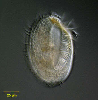
Dorsal view of Aspidisca cicada (MUELLER,1786) CLAPARÃDE&LACHMANN,1858.Brightfield,closed condenser.
-
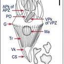
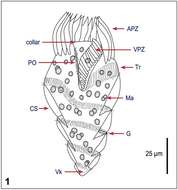
Fig 1 Line drawing of a protargol stained cell.
-
Right side of Acineria incurvata DUJARDIN,1841, a pleurostomatid ciliate found in heavily polluted freshwater and marine habitats. Collected from effluent of a protein skimmer at a commercial saltwater aquarium in Boise,Idaho. January 2007.DIC.
-
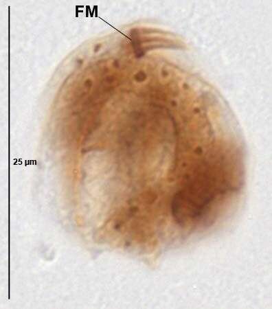
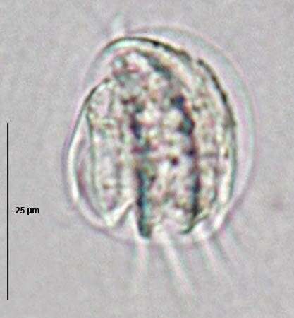
Chilodonella (kai-low-don-ella) is a hypostome ciliate with a mouth stiffened by a palisade of microtubular rods protruding from the ventral surface of the cell. The mouth is used to pick up bacteria and small pieces of detritus and manipulate them into the body. Common in freshwater and marine habitats. Differential Interference Contrast.