-
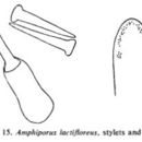
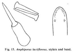
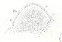
Stylets and head.Brunberg, L. On the Nemertean Fauna of Danish Waters. Ophelia, 1(1), 77-111.
-
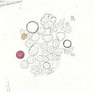
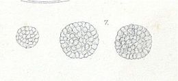
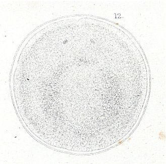
Plate10.9 Contents of the former, with oil-globules
-
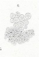
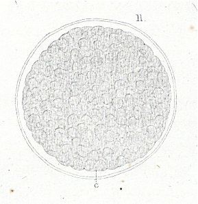
Plate10.8 One of the same slightly compressed glands
-
Plate10.7 Gland-cells from the wall of the digestive cavity of Amphiporus lactifloreus
-
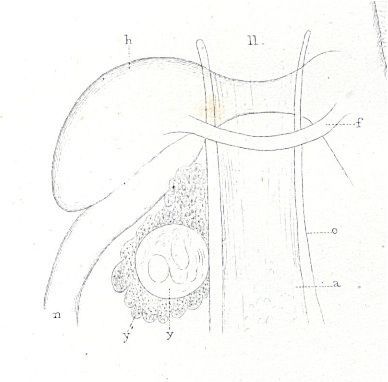
Plate7.14 Parasite extruded from the capsule
-
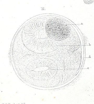
Plate7.13 The same ovum some hours afterwards, showing slight contraction of the discs
-
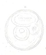
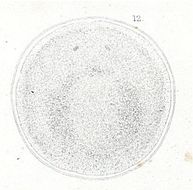
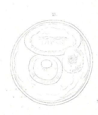
Plate7.12 Parasitic ovum immediately after removal
-
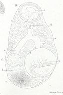
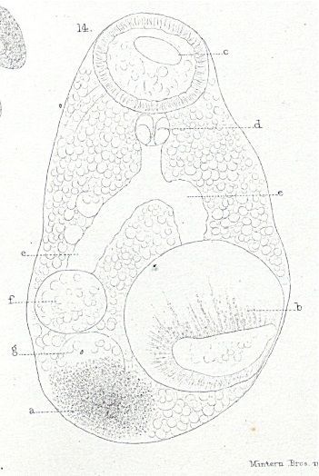
Plate7.11 Magnified view of a ganglionic region of a large one where a parasitic ovum lay imbedded in a granular lobulated mass
-
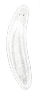
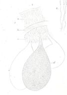
Plate7.2 Another specimen eight days older than the preceding
-
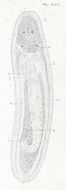
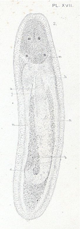
Plate7.1 A young specimen of Amphiporus lactifloreus on extrusion from the egg
-
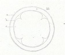
Plate6.12 Ovum just before the extrusion of the embryo
-
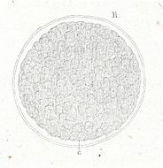
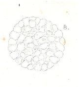
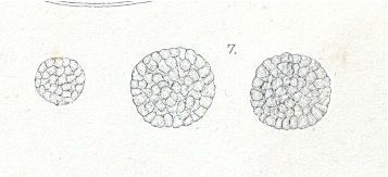
Plate6.11 Ovum of the same species in the mulberry-stage
-
Plate6.10 The same ovum a few hours later
-
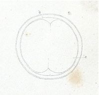
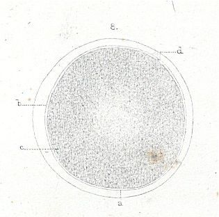
Plate6.9 The same ovum some hours after impregnation
-
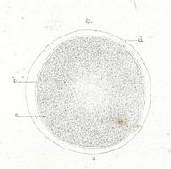
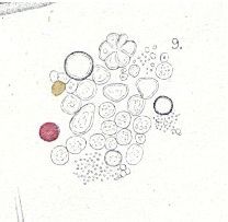
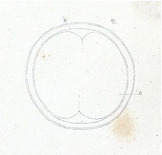
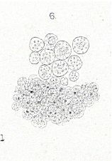
Plate6.8 Unimpregnated ovum of Amphiporus lactifloreus
-
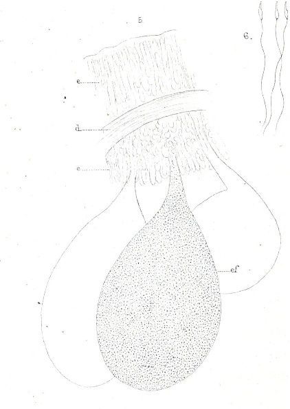
Plate6.5 Three sperm-sacs with a portion of the body-wall of Amphiporus lactifloreus
-
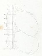
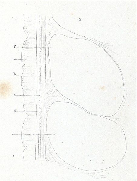
Plate6.2 Longitudinal section of the body-wall of Amphiporus lactfloreus, in a somewhat shriveled condition
-
Plate5.6 Nerve-cells from a cephalic ganglion of Amphiporus lactifloreus
-
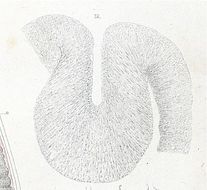
Plate5.4 Portion of the head of the same species considerably flattened
-
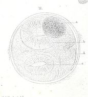
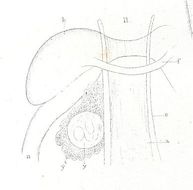
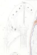
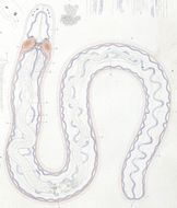
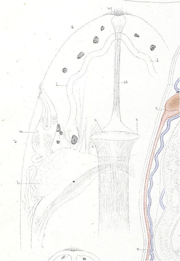
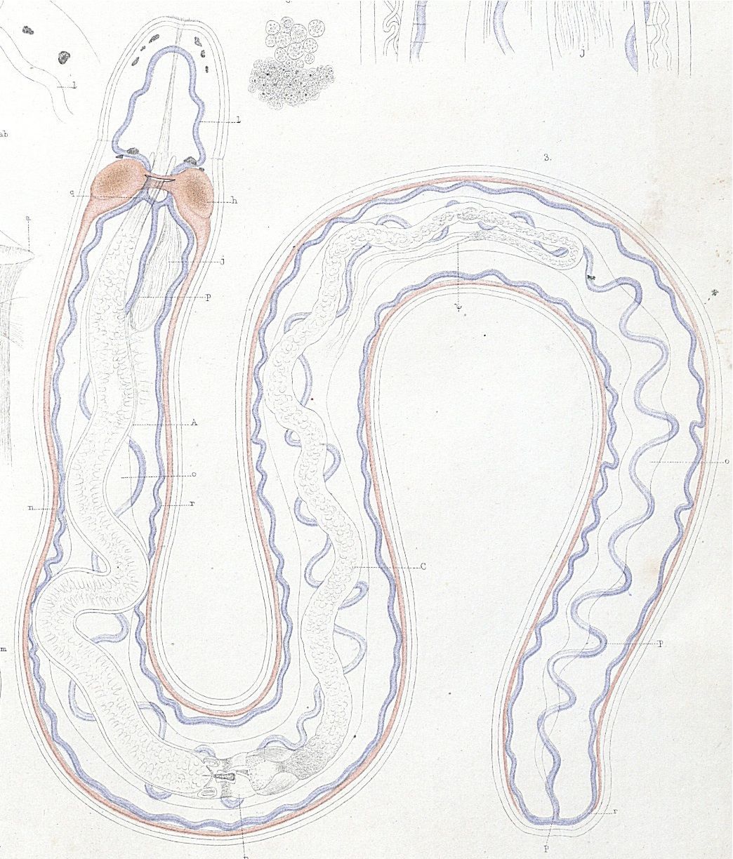
Plate5.3 Arrangement of the circulatory and nervous systems in Amphiporus lactifloreus
-
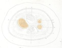
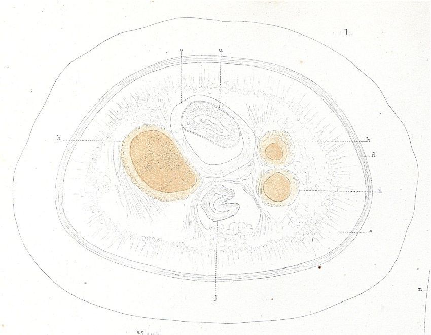
Plate5.1 Transverse section somewhat behind that shown Plate1.1
-
Plate4.18 Spermatozoa of Amphiporus lactifloreus
-
Plate4.12 Transverse section of the wall of the aesophagus of Amphiporus lactifloreus, after mounting in chloride of calcium
-
Plate3.20 Fragment of the aesophageal region of the digestive tract from a living Amphiporus lactifloreus