Thanit Siriboon, Chirasak Sutcharit, Fred Naggs, Somsak Panha
Zookeys
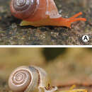
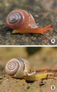
Figure 2.Living snails of A Perrottetia dermapyrrhosa sp. n.(paratype CUMZ 5002) from the type locality (shell width about 7 mm), and B Perrottetia aquilonaria sp. n. (paratype CUMZ 5004) from the type locality (shell width about 6 mm).
Thanit Siriboon, Chirasak Sutcharit, Fred Naggs, Somsak Panha
Zookeys
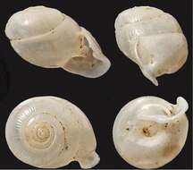
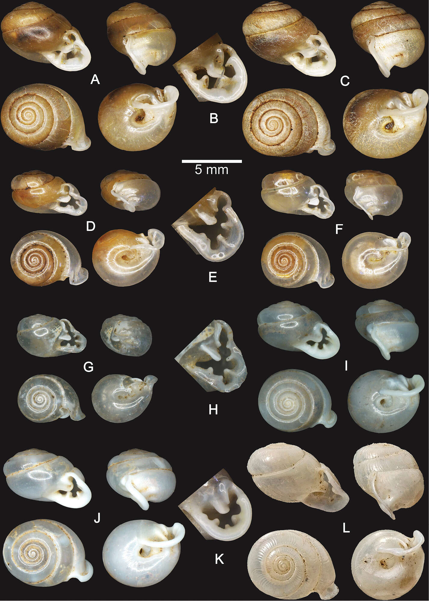
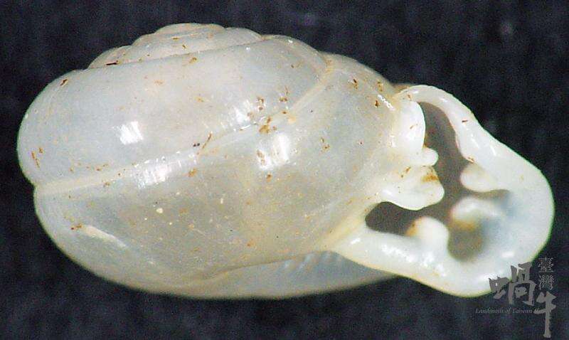
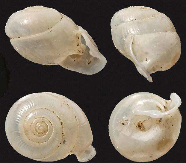
Figure 3.Shells of Perrottetia spp. A–C Perrottetia dermapyrrhosa sp. n. A holotype CUMZ 5001 B apertural dentition of the holotype CUMZ 5001, and C paratype CUMZ 5002 D–H Perrottetia aquilonaria sp. n. D holotype CUMZ 5003 E apertural dentition of the holotype CUMZ 5003 F paratype CUMZ 5004 G specimen from Tam Chiangdao, Chiangmai, CUMZ 5008 and H apertural dentition of the specimen from Tam Chiangdao, Chiangmai CUMZ 5008 I–K Perrottetia phuphamanensis sp. n. I holotype CUMZ 5011 J paratype CUMZ 5012, and K apertural dentition of the holotype CUMZ 5011 L Perrottetia gudei Fulton, 1915, syntype NHMUK 1919.12.31.51.
Thanit Siriboon, Chirasak Sutcharit, Fred Naggs, Somsak Panha
Zookeys
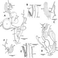
Figure 4.Genitalia of Perrottetia spp. A–C Perrottetia dermapyrrhosa sp. n. (paratype CUMZ 5002) A reproductive system B insertion of vas deferens into penial sheath, and C details of hermaphroditic duct and seminal vesicle D–F Perrottetia aquilonaria sp. n. (paratype CUMZ 5004), D reproductive system E insertion of vas deferens into penis sheath, and F details of hermaphroditic duct and seminal vesicle. Abbreviations: ag, albumen gland; at, atrium; fo, free oviduct; gd, gametolytic duct; gs, gametolytic sac; hd, hermaphroditic duct; ov, oviduct; p, penis; pr, penial retractor muscle; ps, penial sheath; psr, penial sheath retractor muscle; sv, seminal vesicle; ta, talon; v, vagina; vd, vas deferens.
Thanit Siriboon, Chirasak Sutcharit, Fred Naggs, Somsak Panha
Zookeys
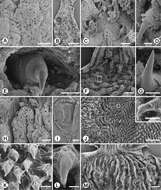
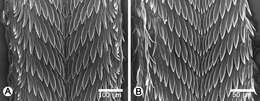
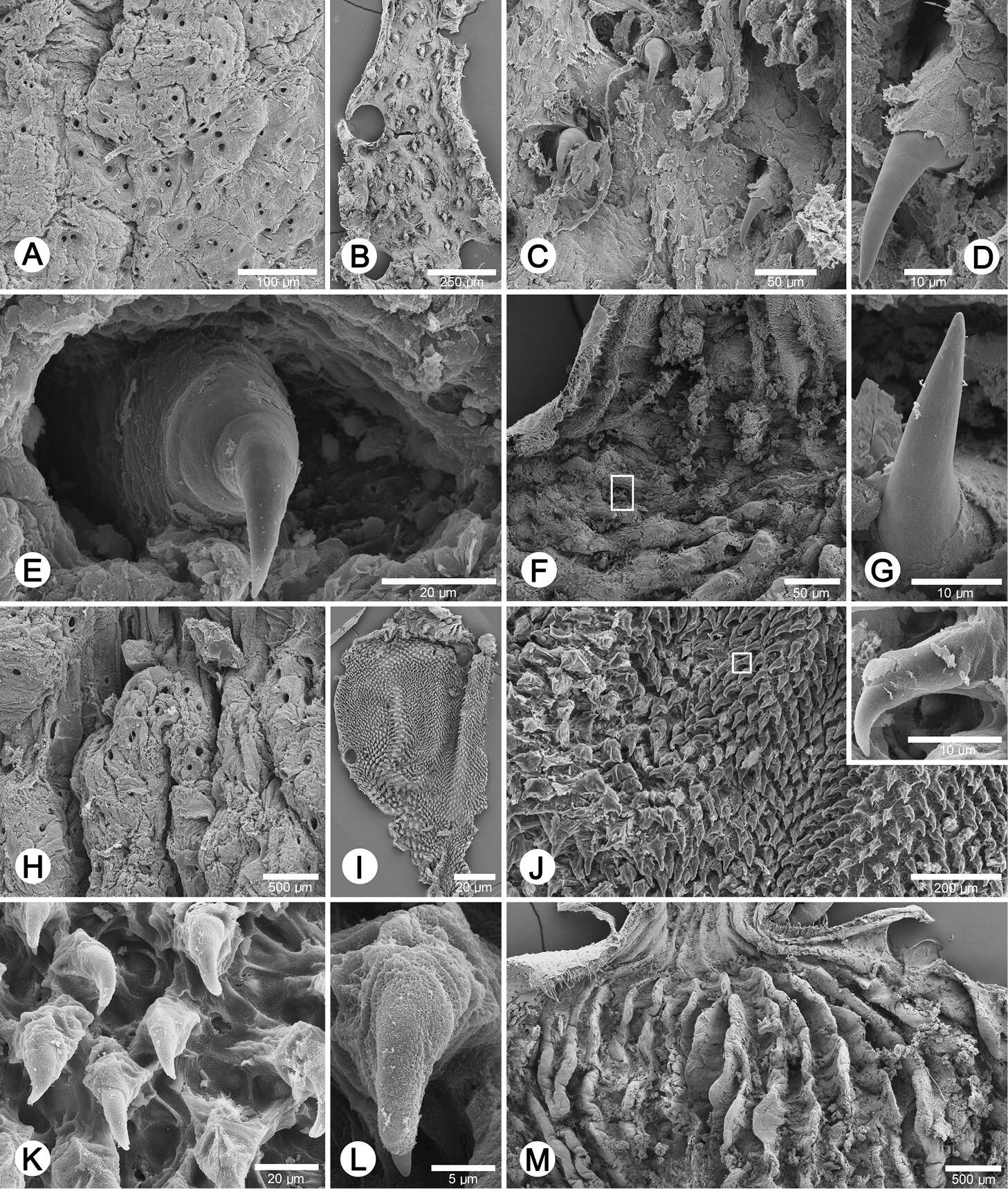
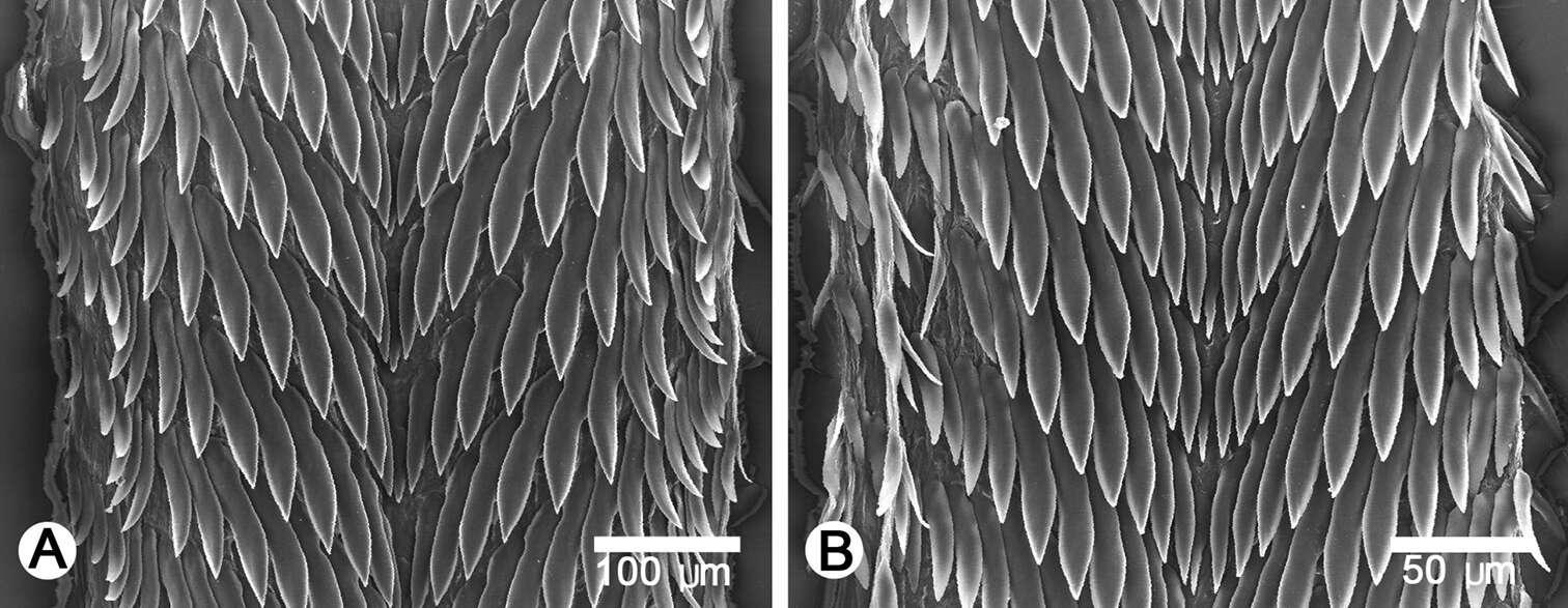
Figure 5.Internal sculpture of genitalia of Perrottetia spp. A–G Perrottetia dermapyrrhosa sp. n. (paratype CUMZ 5002) A details of atrial pore on the atrium surface B low magnification shows arrangement of penial hooks C high magnification of penial hooks D lateral view of penial hook E top view of penial hook situate inside hollow F arrangement of vaginal fold with hook in white square, and G lateral view of vaginal hook (from white square in F) H–M. Perrottetia aquilonaria sp. n. (paratype CUMZ 5004) H details of atrial pore on the atrium surface I low magnification shows dense arrangement of penial hooks J high magnification of penial hooks with (inset) lateral view of penial hook K arrangement of penial hooks L top view of penial hook, and M arrangement of vaginal folds without vaginal hook.
Description: English: ZooKeys - Perrottetia gudei Cut-out from original (Shells of Perrottetia species - ZooKeys-287-041-g003.jpeg) shown below. Date: 11 April 2013, 15:28:51. Source: : This file has been extracted from another file: Shells of Perrottetia species - ZooKeys-287-041-g003.jpeg: . Author: Siriboon T, Sutcharit C, Naggs F, Panha S. Permission (Reusing this file):. : This image is uploaded as part of a collaboration between Wikispecies and ZooKeysবাংলা | català | čeština | Deutsch | English | македонски | polski | português do Brasil | русский | +/−. : This file is licensed under the Creative CommonsAttribution 3.0 Unported license.:. You are free: to share – to copy, distribute and transmit the work to remix – to adapt the work Under the following conditions: attribution – You must attribute the work in the manner specified by the author or licensor (but not in any way that suggests that they endorse you or your use of the work). http://creativecommons.org/licenses/by/3.0 CC BY 3.0 Creative Commons Attribution 3.0 truetrue.. Other versions: .


