-
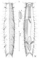
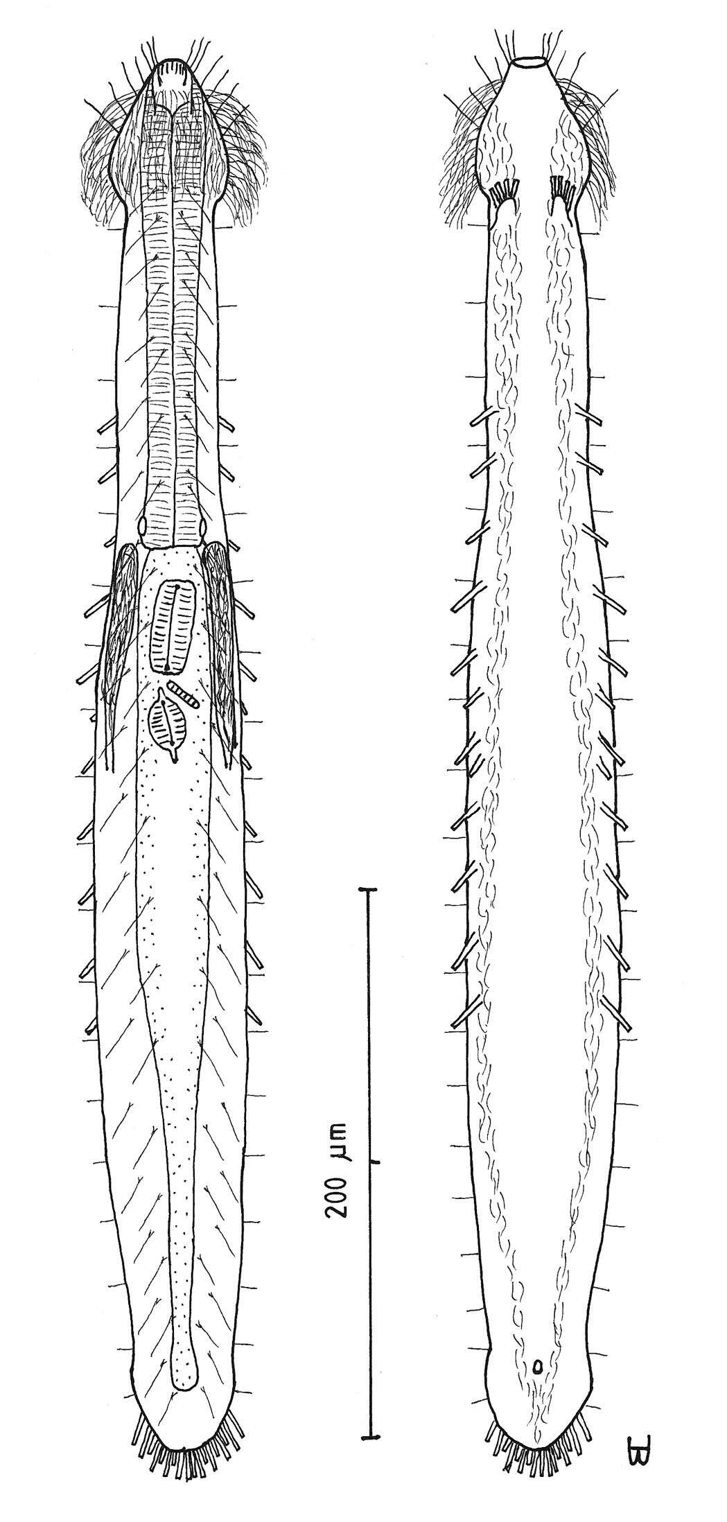
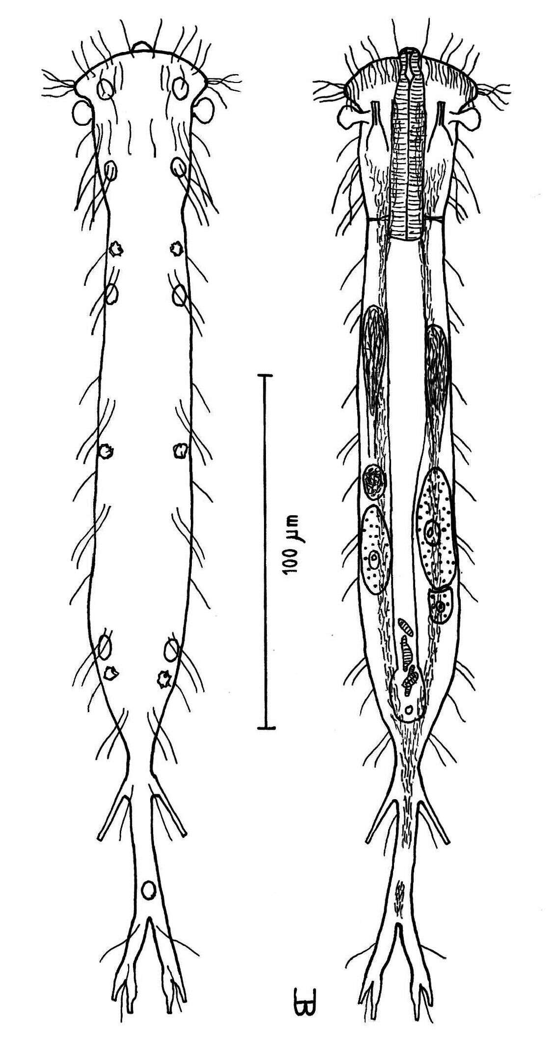
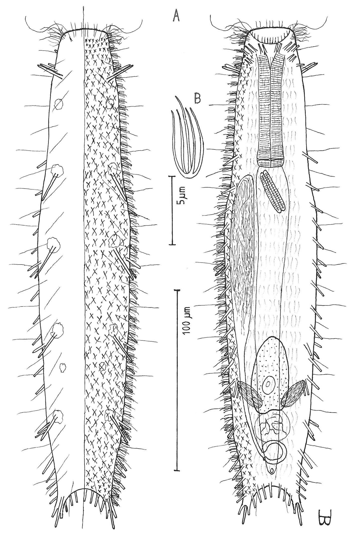
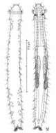
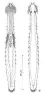
Figure 17.Paraturbanella levantia sp. n. dorsal and ventral views of a mature adult (Lt=657, LPh=163 µm) from Bir Mesud, Alexandria, Egypt; dorsal with pestle organs, pattern of glands, dorsal and lateral body cilia, digestive and reproductive tracts; ventral with adhesive tubes and locomotor ciliary bands.
-
Ribadelago de Franco, Castille and Leon, Spain
-
Aeropuerto de Logroo-Agoncillo, La Rioja, Espaa
-
M. Antonio Todaro, Renzo Perissinotto, Sarah J. Bownes
Zookeys
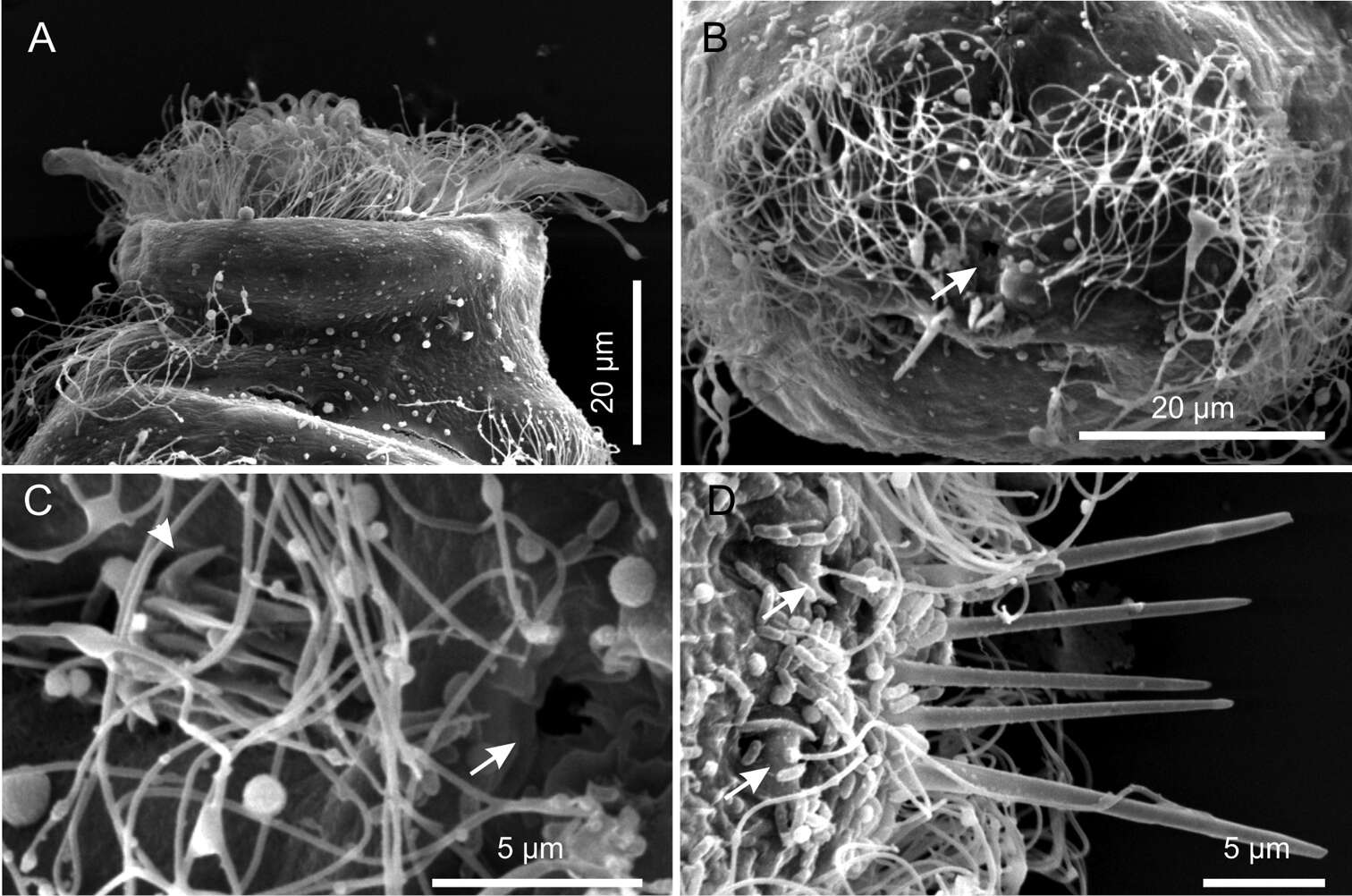
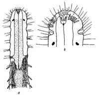
Figure 6.Kijanebalola devestiva sp. n. SEM photomicrographs. A anterior region of specimen with head partially retracted inside the body (ventral view) B posterior trunk region of different specimen, showing the fourth ciliary band and the anus (arrow) C close-up view of the posterior end showing the anus (arrow) and the residual patch of spined scales (arrows) D close-up view of the posterior end (dorsal view), showing theterminal spines and the sensorial bristles (arrows).
-
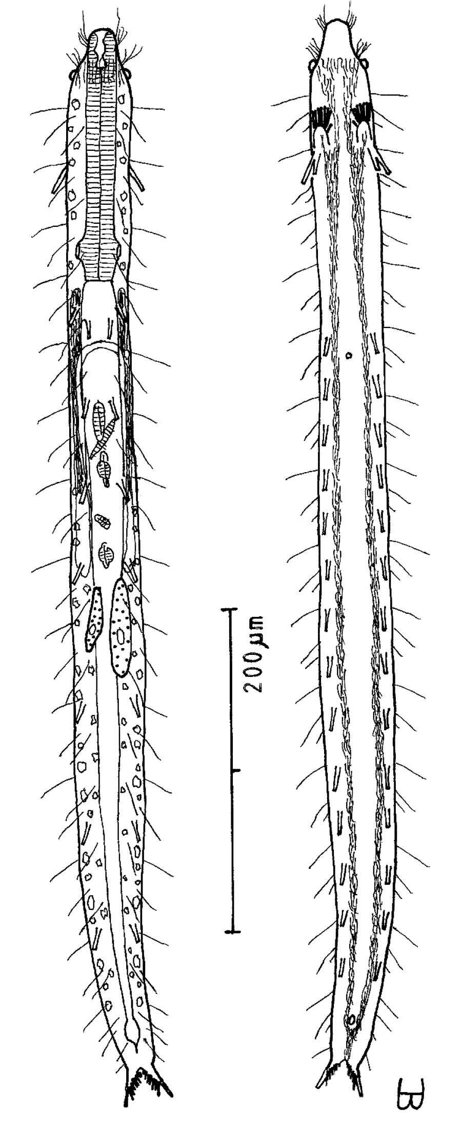
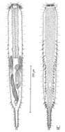
Figure 18.Turbanella erythrothalassia sp. n. dorsal and ventral views of a mature adult (Lt=486, LPh=166 µm) from Moon Valley, Hurghada, Egypt; dorsal with pattern of glands and dorsal and lateral body cilia; ventral with digestive and reproductive tracts, adhesive tubes and locomotor ciliary bands.
-
Neila, Castille and Leon, Spain
-
M. Antonio Todaro, Renzo Perissinotto, Sarah J. Bownes
Zookeys
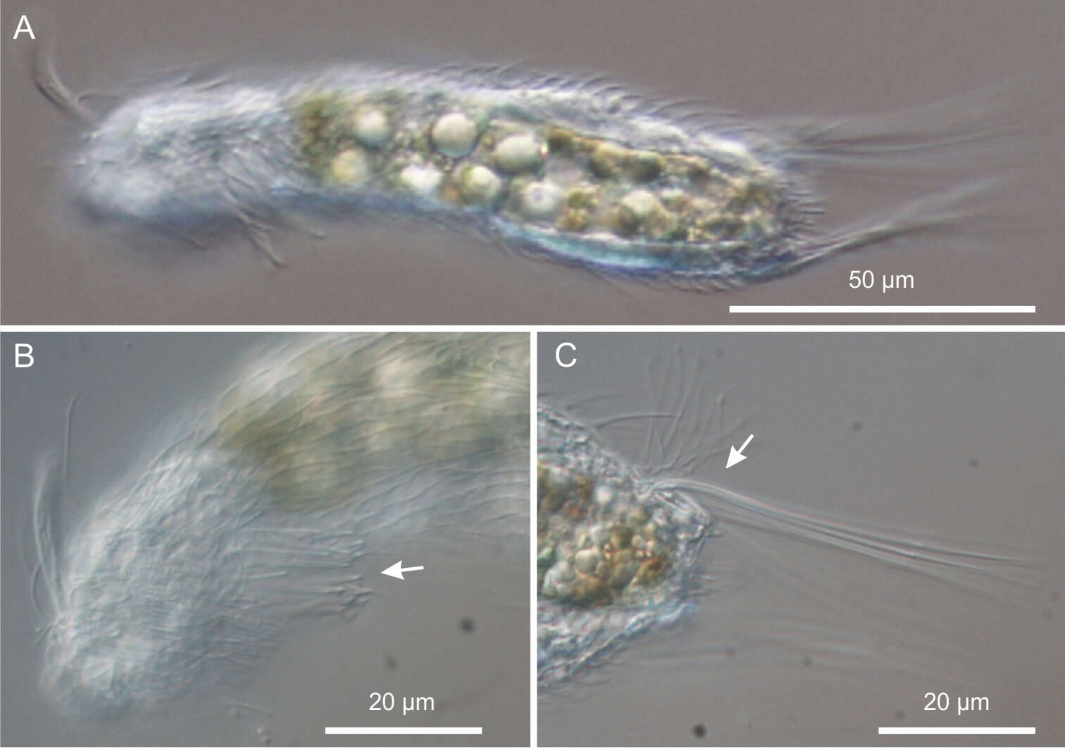
Figure 7.Neogossea acanthocolla. DIC photomicrographs. A habitus B anterior region showing the group of thick spines on the neck (arrow) C close-up of the posterior region of the trunk showing a tuft of long, barbed spines (arrow).
-
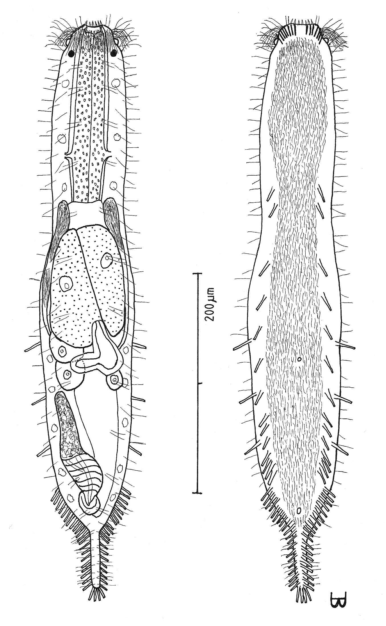
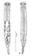
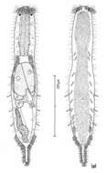
Figure 3.Cephalodasys saegailus sp. n. dorsal and ventral views of a mature adult (Lt=517, LPh=196 µm) from Sidi Abd al-Rahman, Egypt; dorsal with dorsal and lateral body cilia, digestive and reproductive tracts included, ventral with adhesive tubes and locomotor ciliary bands.
-
Figure 5.Dendrodasys rubomarinus sp. n. dorsal and ventral views of a mature adult (Lt=272, LPh=53 µm) from Giftun Island SE, near Hurghada, Egypt; dorsal with body conformation, dorsal and lateral body cilia and pattern of glands; ventral with digestive and reproductive tracts, pestle organs, adhesive tubes and locomotor ciliary bands.
-
Figure 6.Macrodasys imbricatus sp. n. dorsal and ventral views of a mature adult (Lt=544, LPh=232 µm) from Main Beach, Ras Mohamed National Park, S. Sinai, Egypt; dorsal with dorsal and lateral body cilia, digestive and reproductive tracts; ventral with pestle organs, adhesive tubes and locomotor cilia.
-
Figure 7.Macrodasys macrurus sp. n. dorsal and ventral views of a mature adult (Lt=590, LPh=192 µm) from the Giftun Village Spit Outside, near Hurghada, Egypt; dorsal with pestle organs, dorsal and lateral body cilia, digestive and reproductive tracts; ventral with adhesive tubes and locomotor cilia.
-
Figure 8.Macrodasys nigrocellus sp. n. dorsal and ventral views of a mature adult (Lt=300, LPh=161 µm) from Giftun Island SE, near Hurghada, Egypt; dorsal with pestle organs, dorsal and lateral body cilia, pattern of glands, and digestive and reproductive tracts; ventral with adhesive tubes and locomotor cilia.
-
Figure 9.Macrodasys sp. Gerlach 1961 dorso-ventral and ventral views of the fore end of a specimen having black ocelli from Addu-Atol, the Maldive-Archipelago, Indian Ocean.
-
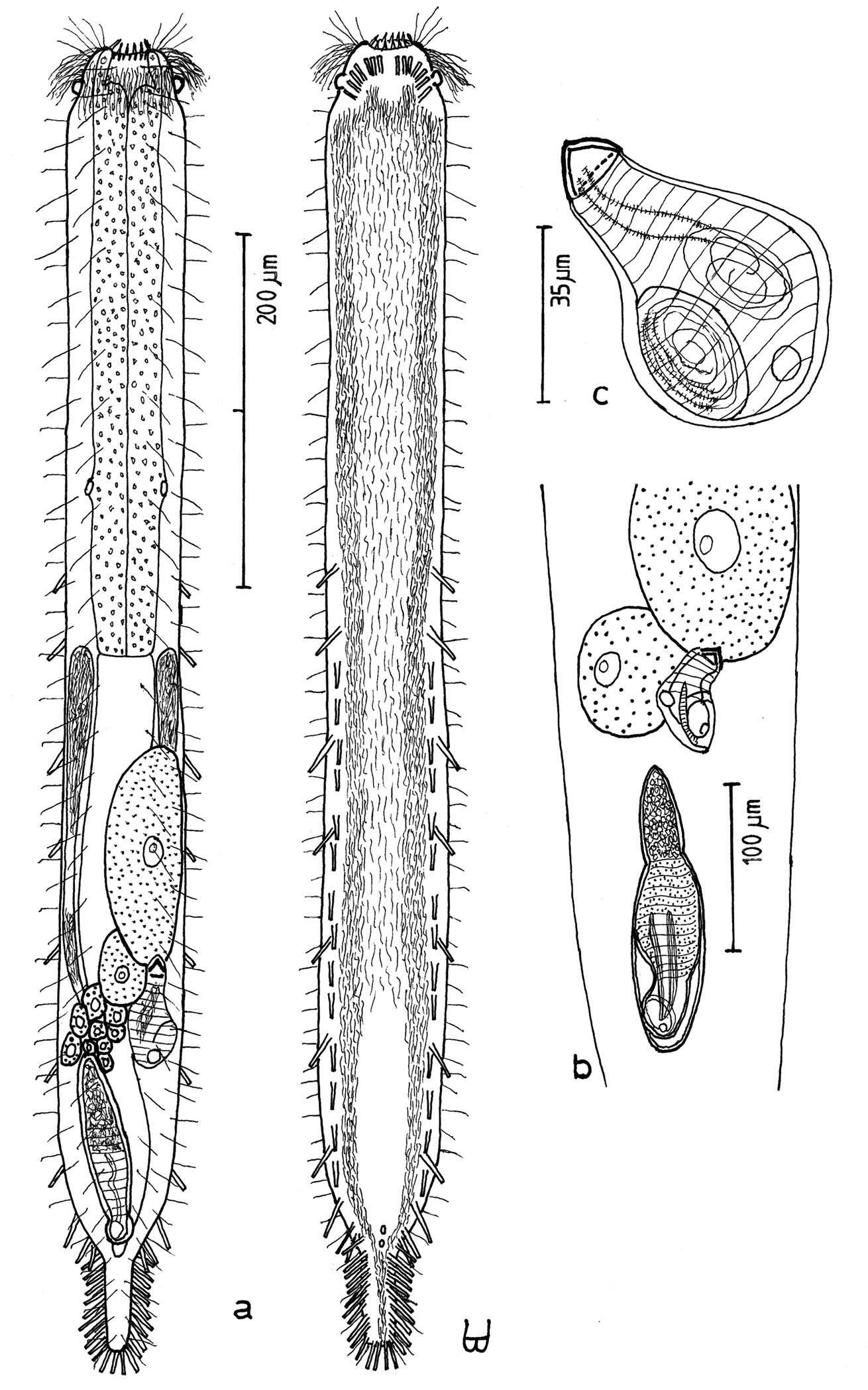
Figure 10.Macrodasys scleracrus sp. n. A dorsal and ventral views of a mature adult (Lt=635, LPh=290 µm) from Main Gate, Ras Mohamed National Park, S. Sinai, Egypt; dorsal with pestle organs, dorsal and lateral body cilia, digestive and reproductive tracts; ventral with adhesive tubes and locomotor ciliary bands B frontal organ with sperm from an animal of Lt=593 µm; C. reproductive organs from another animal of Lt=438 µm.
-
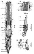
Figure 11.Macrodasys sp. A Schmidt, 1974 A habitus view of a mature adult (Lt=453, LPh=204 µm) from one of the three islands in the Galapagos Islands on which it was found, with pestle organs, body cilia, glands, digestive and reproductive tracts, and adhesive tubes B dorsal view of the fore end C caudal organ; and D two developmental stages of the frontal organ.
-
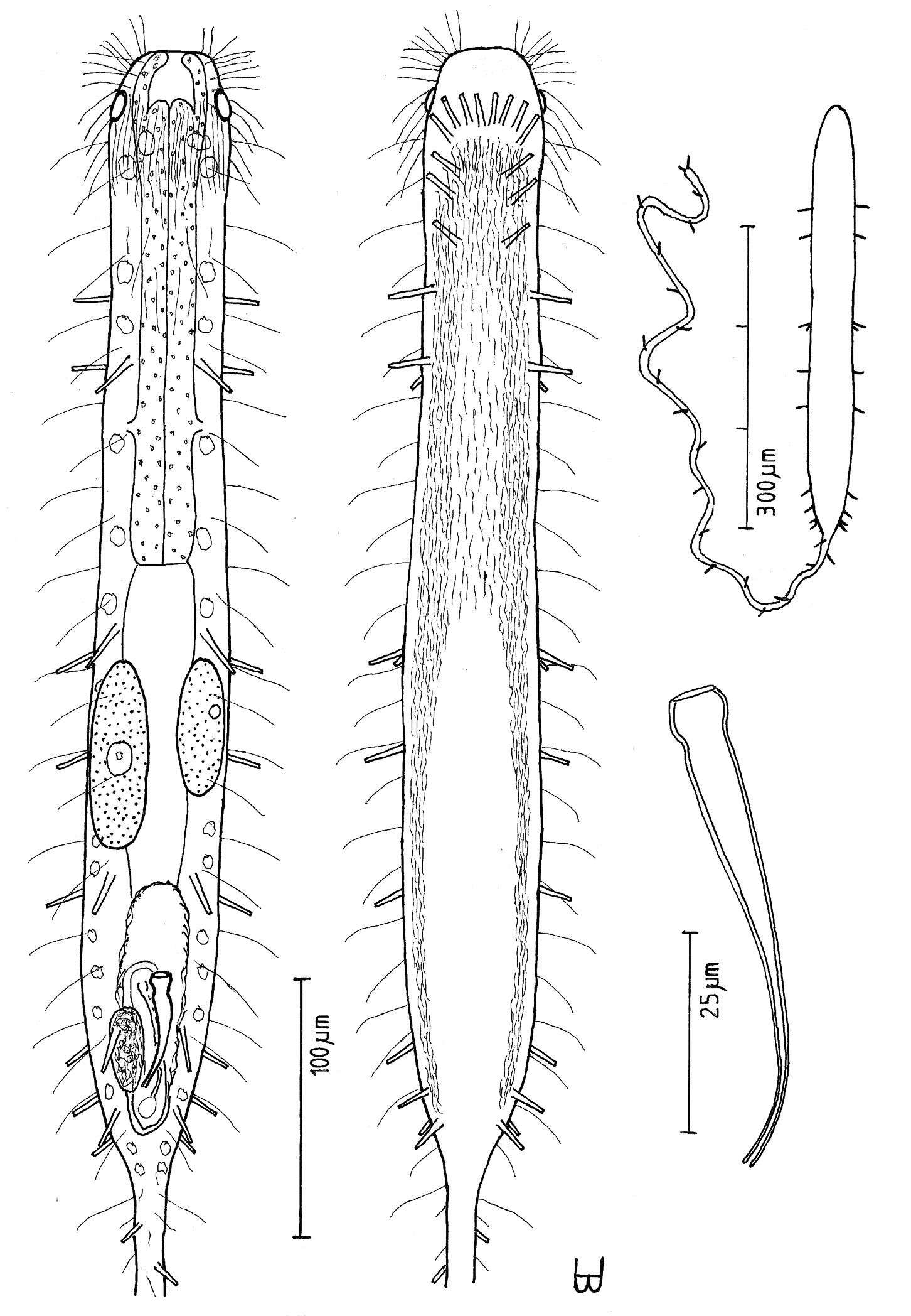
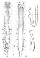
Figure 12.Urodasys toxostylus sp. n. A habitus of a mature adult (L trunk=440, LPh=174, L tail=1100 µm) from Giftun Island SS, near Hurghada, Egypt, showing relative sizes of trunk and tail B dorsal and ventral views of the same specimen; dorsal with pestle organs, dorsal and lateral body cilia, digestive and reproductive tracts, and adhesive tubes; ventral with adhesive tubes and locomotor ciliary bands C the stylet, magnified.
-
Figure 13.Tetranchyroderma corallium sp. n. A dorsal and ventral views of a mature adult (Lt=280, LPh=80 µm) from Middle Garden, S. Sinai, Egypt; dorsal with pentancrous surface (over half of the body), dorsal and lateral body cilia, and cirrata; ventral with digestive and reproductive tracts, adhesive tubes, and the locomotor ciliary band B dorsal pentancre, with a separate scale bar.
-
All Biocode files are based on field identifications to the best of the researcher’s ability at the time.
-
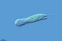
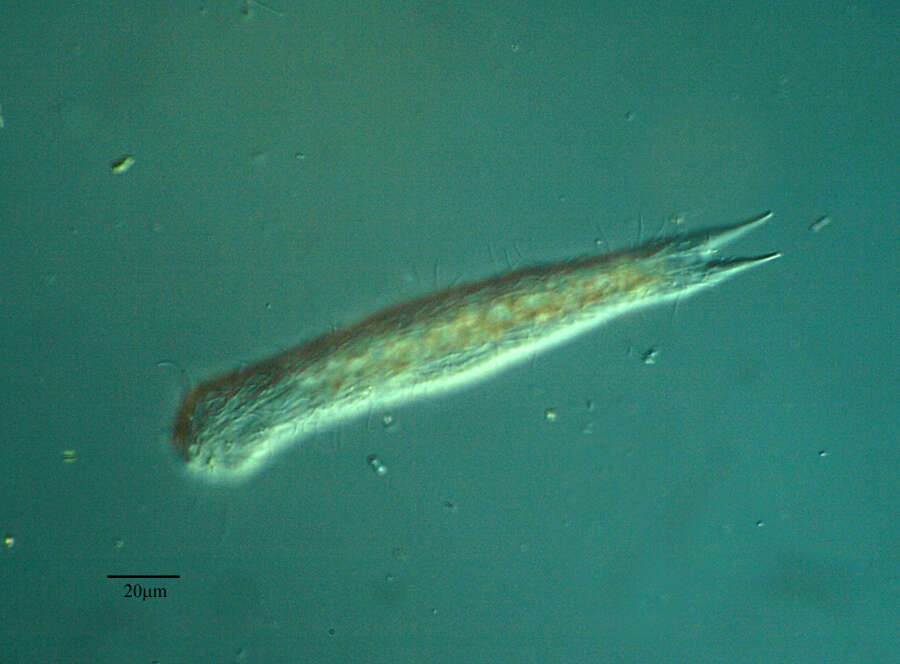
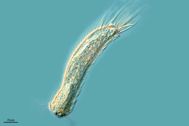
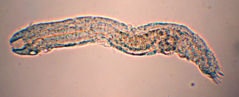
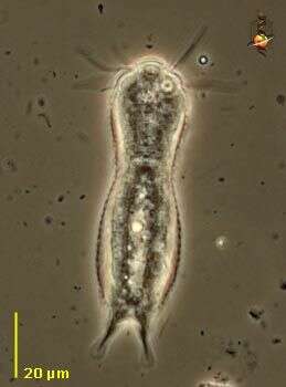
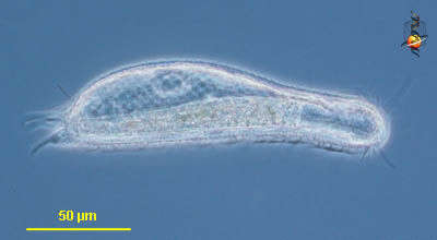
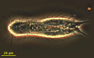
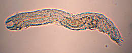
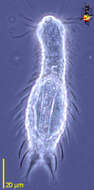
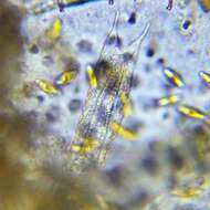
Chaetonotus (keet-a-note-us) a gastrotrich. Perhaps after tardigrades, these are the most inherently cute metazoa likely to be encountered by microscopists. Move by gliding and wriggling over and through the substrate. With an anterior mouth leading to a through gut. Posteriorly with two podites (a bit like rotifers to which they are related, with which they can attach to the substrate. Eat protists, small metazoa and detritus. Phase contrast.
-
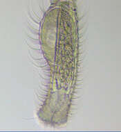
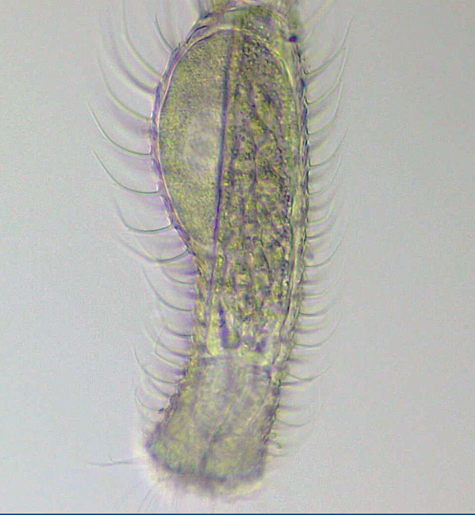
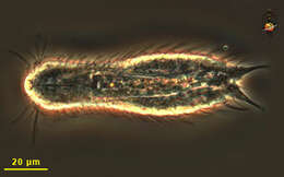
Chaetonotus (keet-a-note-us) a gastrotrich. Perhaps after tardigrades, these are the most inherently cute metazoa likely to be encountered by microscopists. Move by gliding and wriggling over and through the substrate. With an anterior mouth leading to a through gut. Posteriorly with two podites (a bit like rotifers to which they are related, with which they can attach to the substrate. Phase contrast.
-
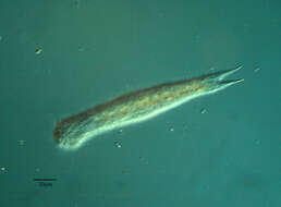
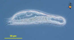
This gastrotrich is about the same size as many ciliates and organisms like this are often encountered in samples, especially from sediments. Phase contrast microscopy.
-
-