-
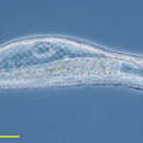
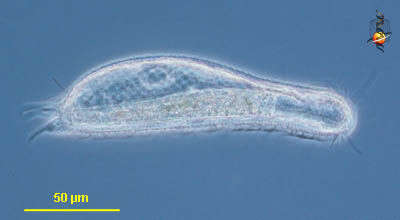
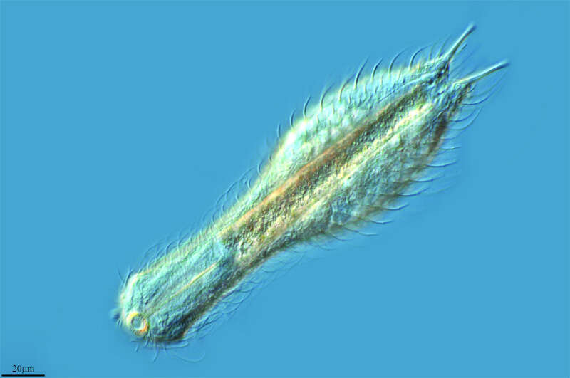
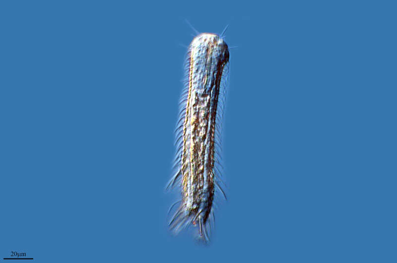
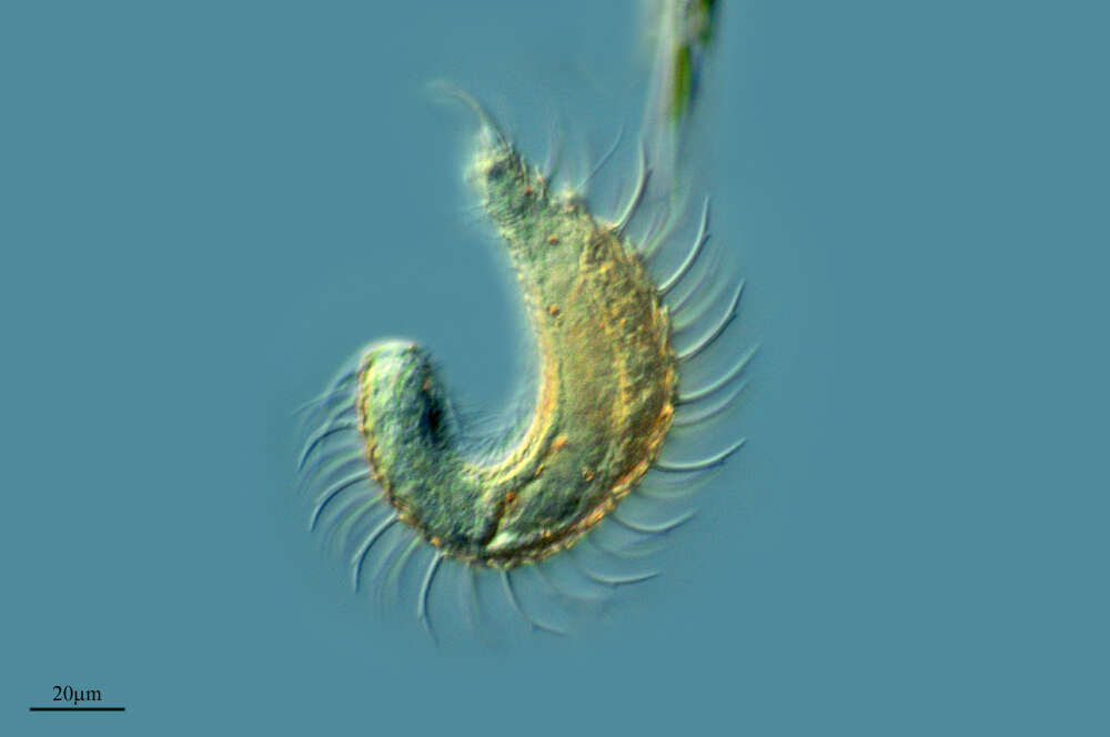
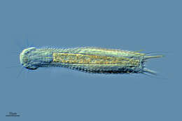
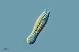
Chaetonotus (keet-a-note-us) a gastrotrich. Perhaps after tardigrades, these are the most inherently cute metazoa likely to be encountered by microscopists. Move by gliding and wriggling over and through the substrate. With an anterior mouth leading to a through gut. Posteriorly with two podites (a bit like rotifers to which they are related, with which they can attach to the substrate. Phase contrast.
-
M. Antonio Todaro, Matteo Dal Zotto, Sarah J. Bownes, Renzo Perissinotto
Zookeys
Figure 2.Gastrotricha from St. Lucia beach, South Africa. Halichaetonotus sanctaeluciae sp. n., schematic drawings A dorsal view B ventral view (locomotor cilia omitted).
-
M. Antonio Todaro, Renzo Perissinotto, Sarah J. Bownes
Zookeys
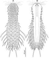
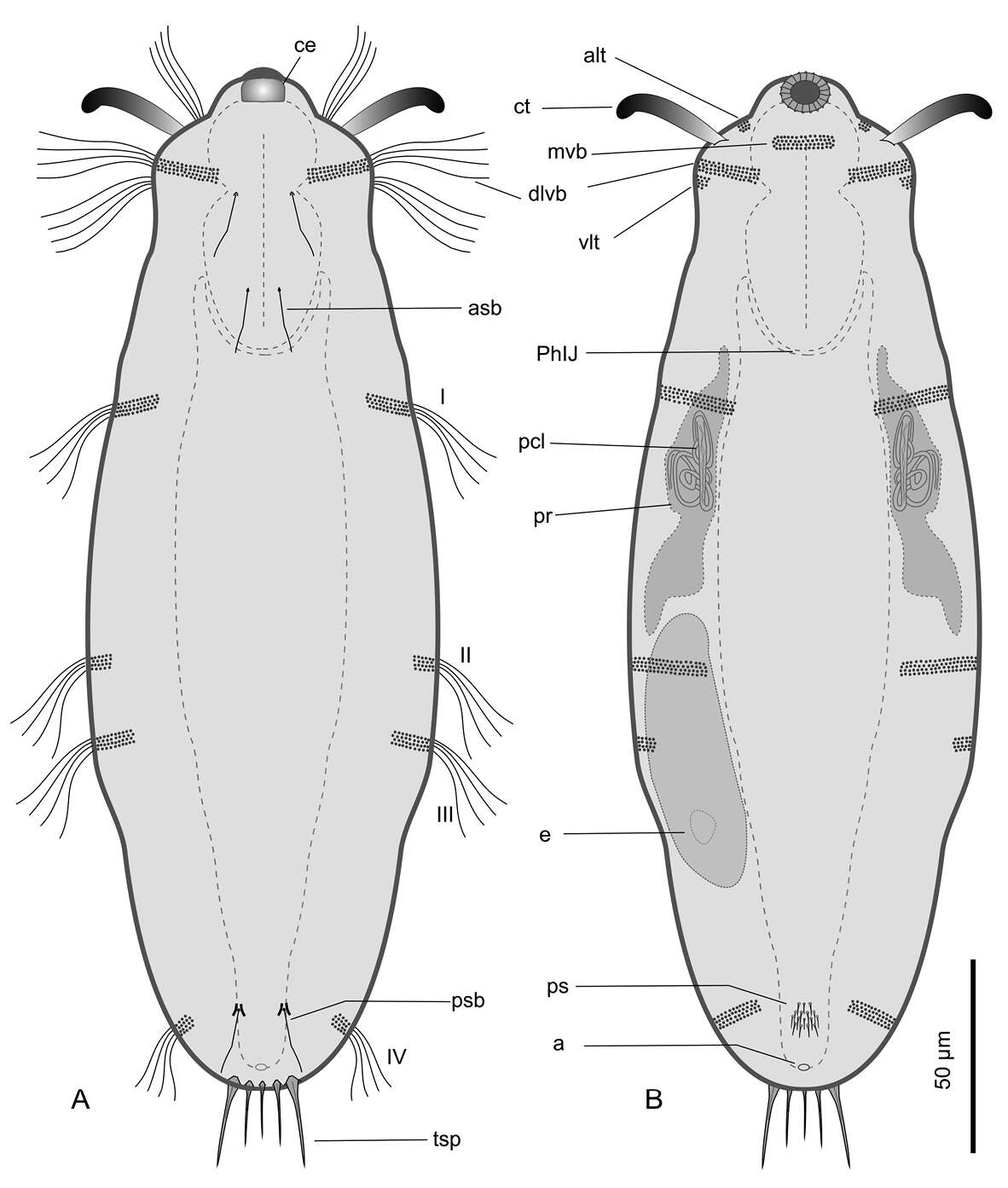
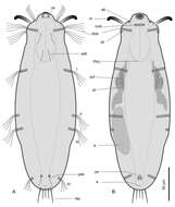
Figure 1.Kijanebalola devestiva sp. n. schematic drawings. A Dorsal habitus B Ventral habitus, showing some internal structures. a anus alt antero-lateral tuft of cephalic cilia asb anterior sensory bristle ce cephalion ct cephalic tentacle dlvb lateral band of cephalic cilia extending dorsally and ventrally e egg I-IV, first to fourth band of trunk ciliature mvb median ventral band of cephalic cilia pcl proximal canal cell lumen PhIJ pharyngeo-intestinal junction ps patch of keeled scales psb posterior sensory bristle tsp terminal spines vlt ventro-lateral band of cephalic cilia.
-
Lumbreras, La Rioja, Spain
-
Rumoroso, Cantabria, Spain
-
-
Lardero, La Rioja, Espaa
-
Ribadelago de Franco, Castille and Leon, Spain
-
Olvega, Castilla y Len, Espaa
-
Ribadelago de Franco, Castille and Leon, Spain
-
Rabanera, La Rioja, Spain
-
Covaleda, Castille and Leon, Spain
-
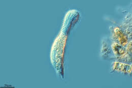
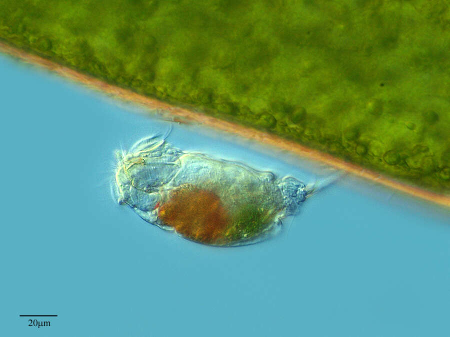
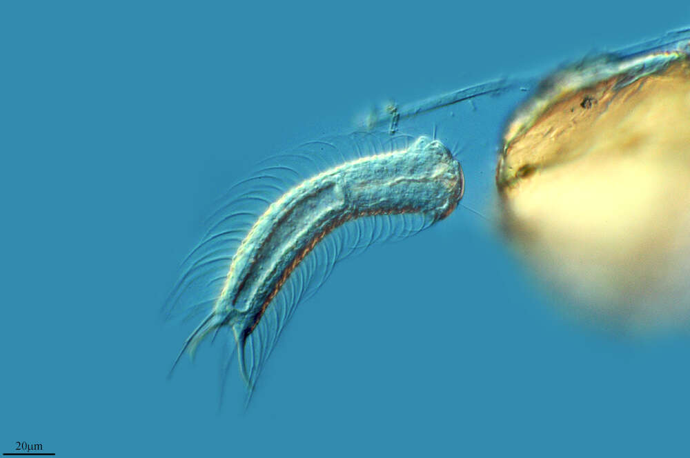
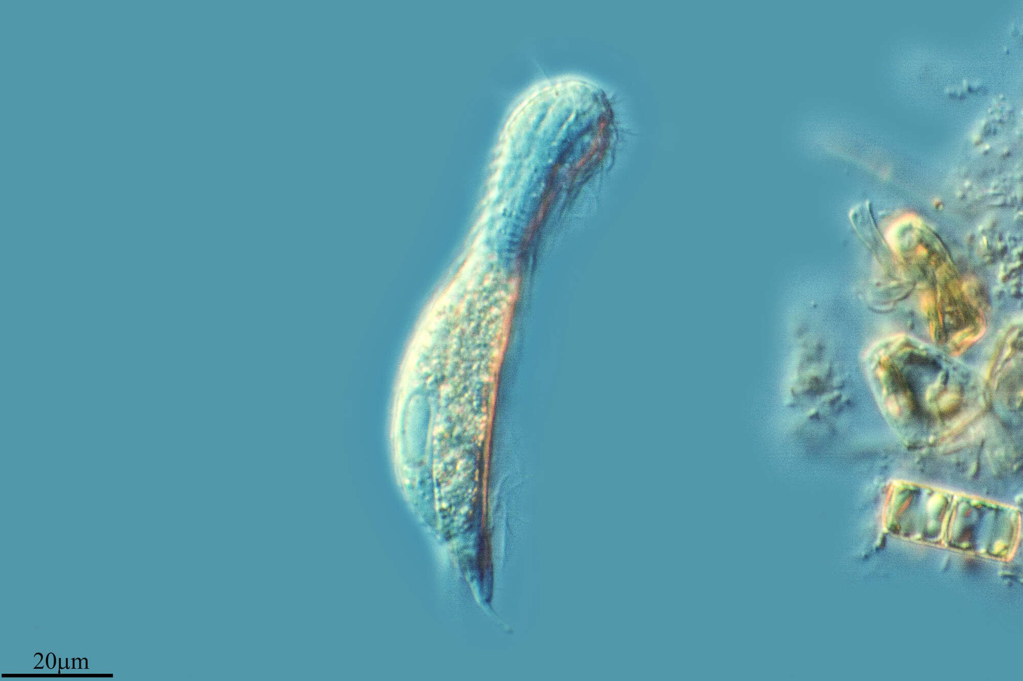
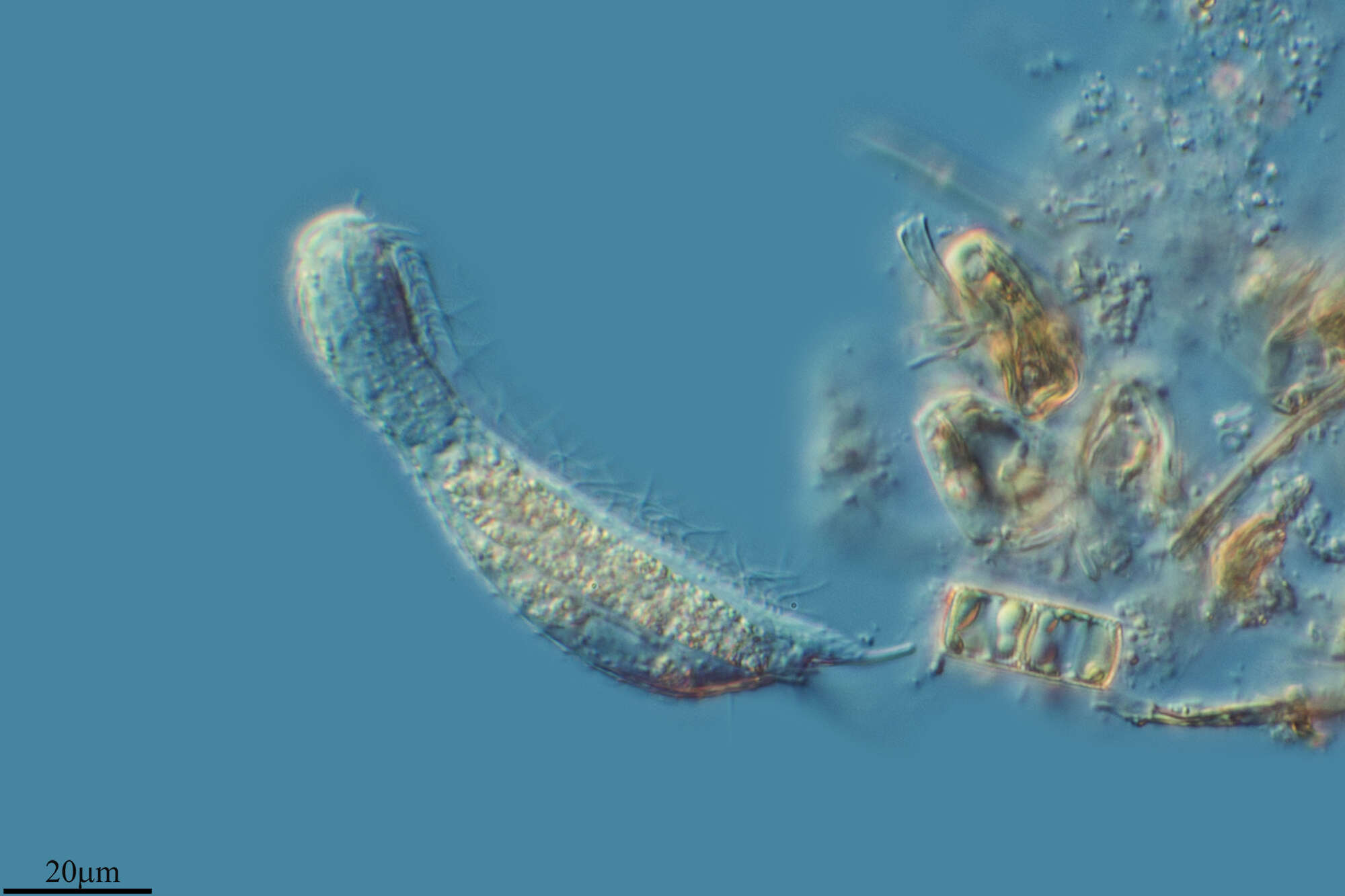
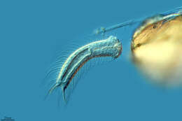
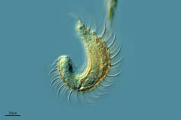
This gastrotrich is about the same size as many ciliates and organisms like this are often encountered in samples, especially from sediments. Phase contrast microscopy.
-
M. Antonio Todaro, Matteo Dal Zotto, Sarah J. Bownes, Renzo Perissinotto
Zookeys
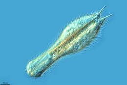
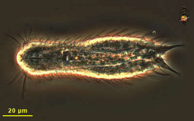
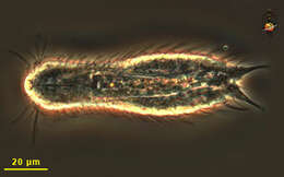
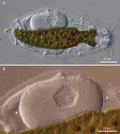
Figure 3.Gastrotricha from St. Lucia beach, South Africa. Halichaetonotus sanctaeluciae sp. n., habitus A dorsal view B ventral view. DIC photomicrographs.
-
M. Antonio Todaro, Renzo Perissinotto, Sarah J. Bownes
Zookeys
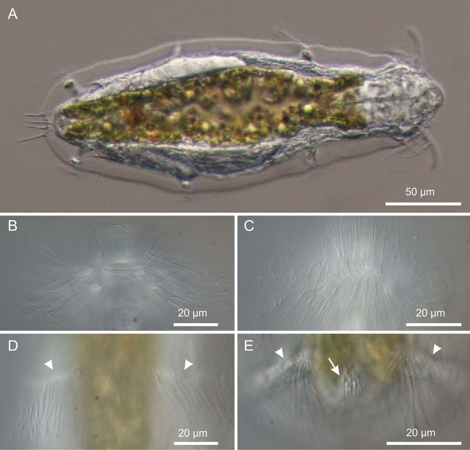
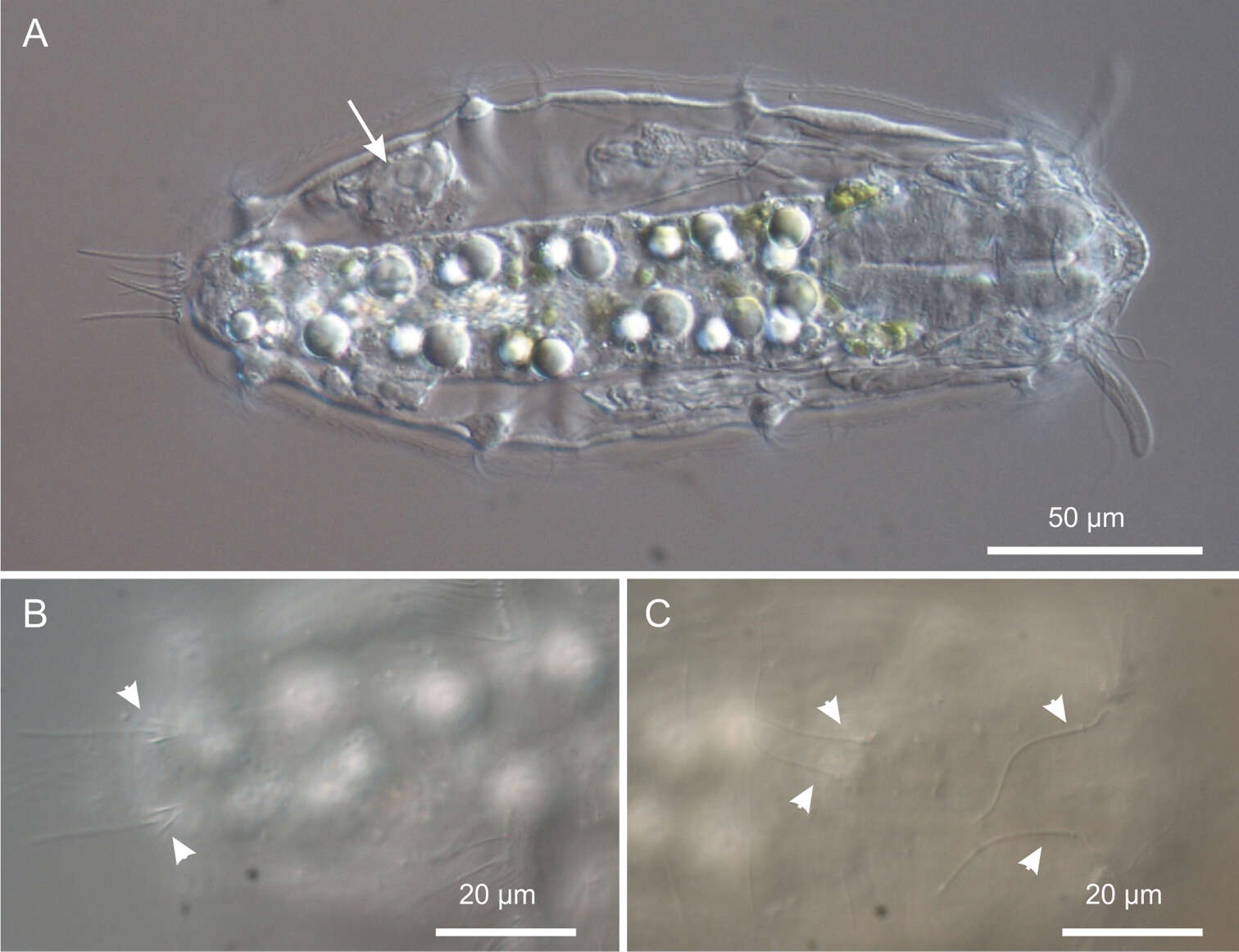
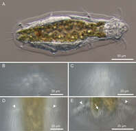
Figure 2.Kijanebalola devestiva sp. n. DIC photomicrographs. A habitus B, C close-up ventral view of theanterior region showing the ciliary bands D close-up ventral view of the mid-trunk region showing the second ciliary bands E close-up ventral view, of the posterior region, showing the fourth ciliary bands (arrowheads) and the residual patch of spined scales (arrow).
-
Lumbreras, La Rioja, Spain
-
Muelas del Pan, Castille and Leon, Spain
-
Torrelles de Foix, Catalonia, Spain
-
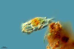
M. Antonio Todaro, Renzo Perissinotto, Sarah J. Bownes
Zookeys
Figure 3.Kijanebalola devestiva sp. n. DIC photomicrographs. A habitus of a gravid specimen B close-up view of the inside egg with the shell bearing spine-like ornamentation (arrowheads).
-
Ribadelago, Castille and Leon, Spain
-
Logrono-Agoncillo Airport, La Rioja, Spain
-
M. Antonio Todaro, Renzo Perissinotto, Sarah J. Bownes
Zookeys
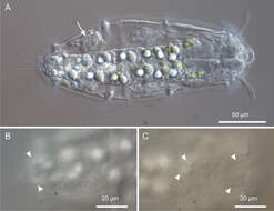
Figure 4.Kijanebalola devestiva sp. n. DIC photomicrographs. A habitus of a subadult specimen showing developing egg (arrow) B close-up dorsal view of the posterior trunk region showing the two sensory bristles (arrow-heads) C close-up dorsal view of the anterior trunk and neck regions, showing two pairs of sensory bristles.
-
Lumbreras, La Rioja, Spain
-
Mahide, Castilla y Len, Espaa