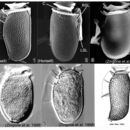
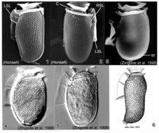
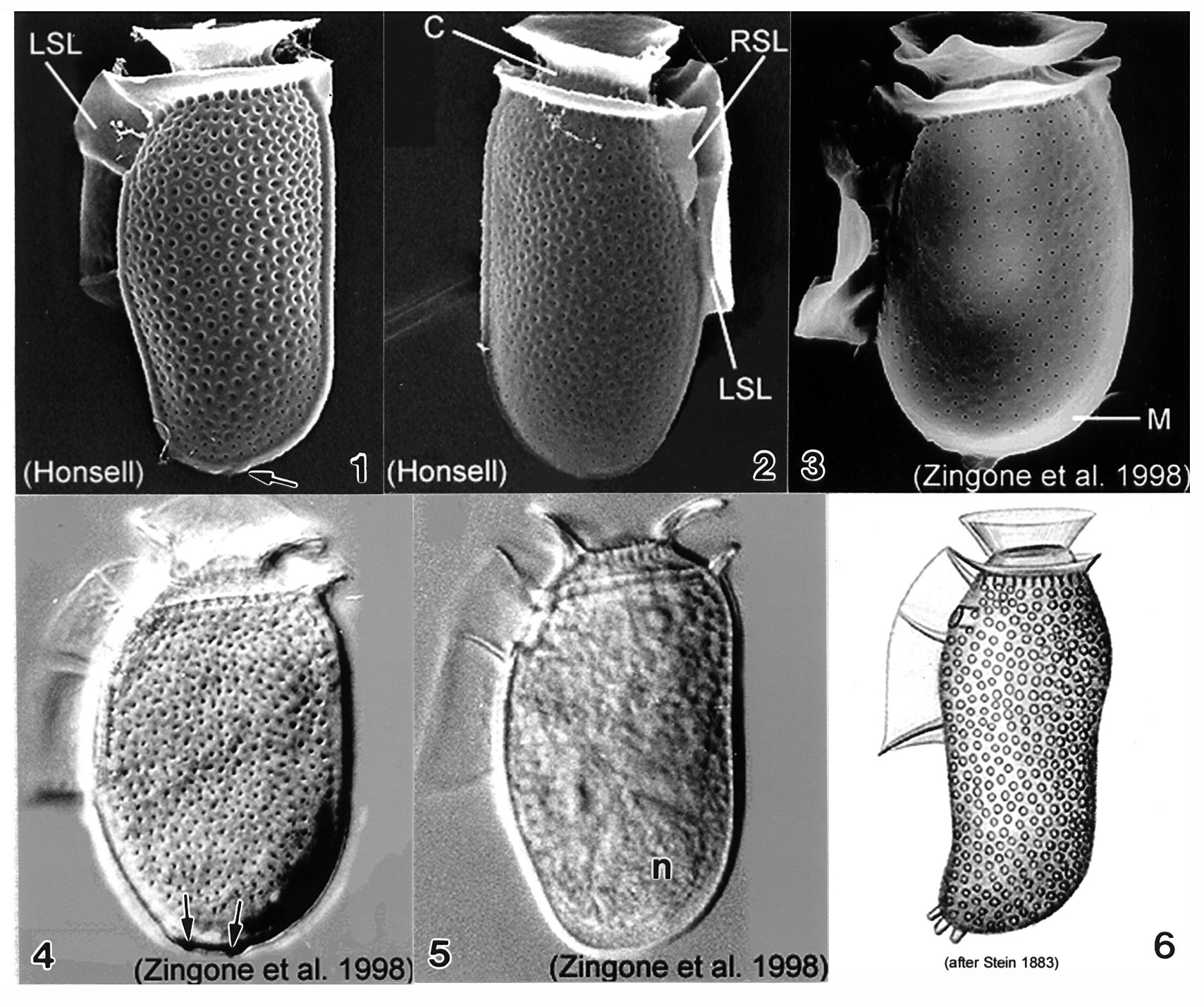
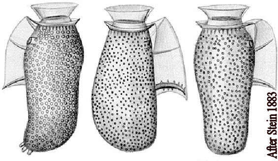
Plate 18. Dinophysis sacculus. Figs. 1-3. SEM: lateral view. Fig. 1. Cell oblong with rounded posterior. Hypotheca long, margins undulate. Thecal surface coarsely areolated. Short left sulcal list (LSL). Cingulum with two well developed lists. Small blunt posterior projections (arrow). Fig. 2. Cingulum lined with pores. Right sulcal list (RSL) visible. Fig. 3. Smooth thecal surface with pores. Metacytic zone (M) devoid of pores. Figs. 4-5. LM: lateral view. Fig. 4. Hypotheca sack-like with deep thecal pores. Posterior end with two blunt projections (arrows). Fig. 5. Large posterior nucleus (n). Fig. 6. Line drawing: morphotype from Stein (1883).