Sivun Keltakuumehyttynen kuva
Kuvaus:
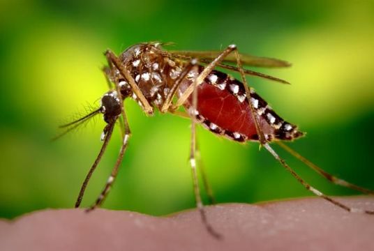
This 2006 image depicted a female Aedes aegypti mosquito as she was completing the activity of obtaining a blood-meal from a human host through her fascicle, which shed begun to resheath in her labium. Both structures are part of her feeding organ known as the proboscis. In this case, what would normally be an unsuspecting host was actually the CDCs biomedical photographers own hand, which hed offered to the hungry mosquito so that shed alight, and be photographed while feeding. After it filled with blood, the abdomen became distended, stretching the exterior exoskeletal surface, thereby, causing it to become transparent, allowing the collecting blood to become visible as an enlarging intra-abdominal red mass.
Created: 2006
Mukana seuraavilla sivuilla:
- Life
- Cellular
- Eukaryota (aitotumaiset)
- Opisthokonta
- Metazoa
- Bilateria (Kaksikylkiset)
- Protostomia (Alkusuiset)
- Ecdysozoa
- Arthropoda (Niveljalkaiset)
- Pancrustacea
- Hexapoda (Kuusijalkaiset)
- Insecta (Hyönteiset)
- Pterygota (Siipikantaiset)
- Neoptera
- Endopterygota
- Diptera (kaksisiipiset)
- Culicomorpha
- Culicidae (hyttyset)
- Aedes
- Aedes aegypti (Keltakuumehyttynen)
- Panarthropoda
Tämä kuva ei ole esillä missään kokoelmassa.
Lähdetiedot
- lisenssi
- cc-publicdomain
- valokuvaaja
- James Gathany
- tarjoaja
- Public Health Image Library
- alkuperäinen
- alkuperäinen mediatiedosto
- käy lähteessä
- kumppanisivusto
- Public Health Image Library
- ID