-
-
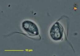
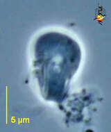
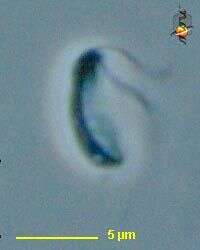
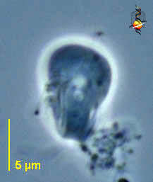
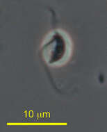
Carpediemonas (car-ped-ee-o-moan-ass), an excavate flagellate - in that there is a ventral groove (to the right of this cell) that is used in feeding. Two flagella insert at the head of the groove. One lies within the groove and beats rapidly, the other extends forward and usually curving back over the anterior end of the cell as in this image. A flap of cytoplasm moves backwards along the groove every few seconds when cells are actively feeding. Nucleus located near the anterior pole of the cell. Phase contrast.
-
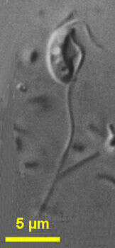
Carpediemonas (car-ped-ee-o-moan-ass), an excavate flagellate - in that there is a ventral groove that is used in feeding. Two flagella insert at the head of the groove. One lies within the groove and beats rapidly, the other extends forward and usually curves back over the anterior end of the cell as in this image. Phase contrast.
-
Centers for Disease Control/Division of Parasitic Diseases and Malaria
EOL staff
Life cycle of Chilomastix mesnili The resistant cyst stage in the life cycle of Chilomastix is responsible for transmission. Both cysts and trophozoites can be found in the feces (diagnostic stages) (1). Infection occurs by the ingestion of cysts in contaminated water or food or by the fecal-oral route (via hands or fomites, i.e., inanimate objects such as towels that transmit infectious organisms to a host) (2). In the large (and possibly small) intestine, excystation releases trophozoites.From
Centers for Disease Control Parasites and Health website.
-

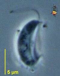
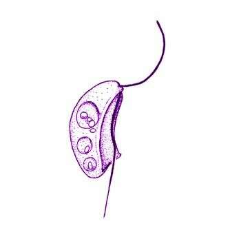
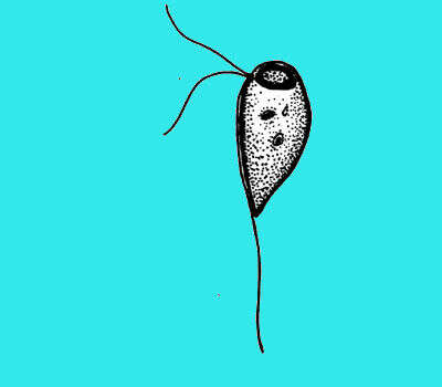
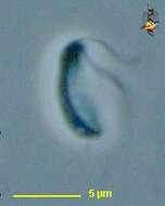
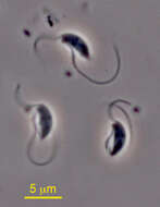
Carpediemonas (carp-ee-dee-a-moan-ass) bialata (Ruinen, 1938) Lee and Patterson, 2000. Cell outline is kidney-shaped. Cells are 6 to 14 microns long (mostly 9 to 12 microns), not rigid, and with a longitudinal ventral groove. A membrane moves down along the groove every 4 - 6 seconds. Two flagella emerge from the anterior part of the cell, the anterior flagellum bends backwards, is about the length of the cell and beats over the cell with a slow sweeping motion. The acronematic posterior flagellum beats asymmetrically and is about 1.5 times cell length. The posterior flagellum may vibrate actively in the groove when not beating. The cells consume bacteria up to 5 microns, and food materials are transferred by the moving membrane to the back of the cell. The cells may have many food vacuoles and attach to the substrate with the tip of the posterior flagellum. The cells move slowly by skidding or gliding with the anterior flagellum beating with a flicking motion. Commonly observed in late cultures.
-
Carpediemonas (car-ped-ee-o-moan-ass), an excavate flagellate - in that there is a ventral groove that is used in feeding. Two flagella insert at the head of the groove. One lies within the groove and beats rapidly, the other extends forward and usually curves back over the anterior end of the cell as in this image. Phase contrast
-
-
Centers for Disease Control/Division of Parasitic Diseases and Malaria
EOL staff
Life cycle of Enteromonas hominisBoth cysts (dormant stage) and trophozoites (active stage) of Enteromonas hominis are shed in feces. Infection occurs after the ingestion of cysts in fecal-contaminated food or water, or on fomites (inanimate objects or substances capable of transferring pathogens). In the large (and possibly small) intestine, excystation releases trophozoites. Enteromonas hominis resides in the large intestine, where it is regarded as a commensal (benefiting from its host but doing no harm) and is not known to cause disease.From
Centers for Disease Control Parasites and Health website.
-
Carpediemonas bialata (Ruinen, 1938) Lee and Patterson, 2000. Cell outline is kidney-shaped. Cells are 6 to 14 microns long, not rigid, and with a longitudinal ventral groove. A membrane moves down along the groove every 4 - 6 seconds. Two flagella emerge from the anterior part of the cell, the anterior flagellum bends backwards, is about the length of the cell and beats over the cell with a slow sweeping motion. The acronematic posterior flagellum beats asymmetrically and is about 1.5 times cell length. The posterior flagellum may vibrate actively in the groove when not beating. The cells consume bacteria up to 5 microns long, and food materials are transferred by the moving membrane to the back of the cell. The cells may have many food vacuoles and attach to the substrate with the tip of the posterior flagellum. The cells move slowly by skidding or gliding with the anterior flagellum beating with a flicking motion.
-
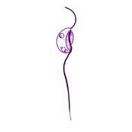
Carpediemonas (carp-ee-dee-a-moan-ass) membranifera Ekebom et al., 1996. Cells are elliptical or obovate and 3 to 6 microns long with a longitudinal ventral groove, which extends most of cell length. When squashed, the cell is pliable. Two flagella unequal in length emerge from the anterior distal part of the cell, the anterior flagellum bent over backwards is as long as the cell and beats stiffly. The acronematic posterior flagellum is about 2.5 - 4 times cell length, beats actively in the ventral depression and usually lies in the depression. The cells usually move by skidding with the anterior flagellum beating with a stiff paddling motion. The cells consume bacteria. Commonly observed in anoxic conditions.
-
-
Carpediemonas membranifera (Larsen and Patterson, 1990) Ekebom et al., 1996. Cells are elliptical or obovate and 3 to 6 microns long with a longitudinal ventral groove, which extends most of cell length. When squashed, the cell is pliable. Two flagella unequal in length emerge from the anterior distal part of the cell, the anterior flagellum bent over backwards is as long as the cell and beats stiffly. The acronematic posterior flagellum is about 2.5 - 4 times cell length, beats actively in the ventral depression and usually lies in the depression. The cells usually move by skidding with the anterior flagellum beating with a stiff paddling motion. The cells consume bacteria.
-
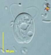
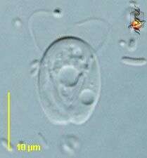
Trepomonas (tree-poe-moan-ass) is one of the free-living diplomonad flagellates. As with almost all diplomonads there are two anterior nuclei and two sets of flagella, one set associated with each nucleus. Different genera are distinguished largely by the numbers and relative lengths of the flagella. his genus has one long anterior flagellum in each group, and three very short ones lying within each of the lateral grooves. The anterior flagella are not evident here. Phase contrast
-
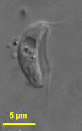
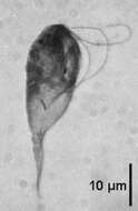
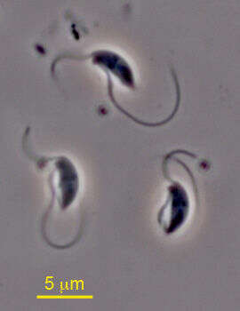
This tiny bean shaped flagellate has a ventral groove almost as long as the cell, from which two flagella emerge. One is longer than the cell and is pointed posteriorly and attached to the substrate, while the other flagellum is pointed anteriorly. The specimen was collected from sediment at Chappaquoit Marsh, Massachusetts, USA. Photo by Banoo Malik.
-
Trepomonas (tree-poe-moan-ass) is one of the free-living diplomonad flagellates. As with almost all diplomonads there are two anterior nuclei and two sets of flagella, one set associated with each nucleus. Different genera are distinguished largely by the numbers and relative lengths of the flagella. This genus has one long anterior flagellum in each group, and three very short ones lying within each of the lateral grooves. Differential interference contrast.
-
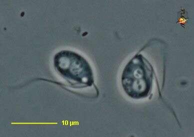
The heterotrophic cells are displayed in three profiles that show an anterior and posterior flagellum each emerging from the anterior end of the ventral feeding groove. The cells here appear to contain a bacterium in a food vacuole. The specimens were collected on coverslips from anoxic sediments from Eel Pond, Woods Hole, MA. Photo by Banoo Malik.
-
Trepomonas (tree-poe-moan-ass) is one of the free-living diplomonad flagellates. As with almost all diplomonads there are two anterior nuclei and two sets of flagella, one set associated with each nucleus. Different genera are distinguished largely by the numbers and relative lengths of the flagella. his genus has one long anterior flagellum in each group, and three very short ones lying within each of the lateral grooves. Phase contrast.
-
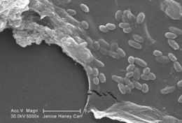
This scanning electron micrograph (SEM) of an untreated water specimen extracted from a wild stream mainly used to control flooding during inclement weather, revealed the presence of unidentified organisms, which included bacteria, protozoa, and algae. In this particular image, a protective biofilm had been inhabited by numbers of what appeared to be unidentified bacterial microorganisms.Created: 2009
-
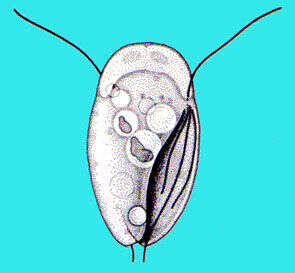
Octomitus is a diplomonad with a broadly pyriform cell body tapered posteriorly, (10-15 µm), bearing six anterior flagella deflected backwards and two posterior trailing flagella. The two anterior nuclei are bean-shaped, they face up and adjoin each other in their anterior part. A large endosome is present in the anterior lobe of the nuclei. The two sets of flagella emerge on each side of the anterior part of the body. The two recurrent flagella, accompanied by a sheath of reticulum, traverse the cell, forming a central axis before emerging as trailing flagella. Two spikes are located at the posterior between the two trailing flagella. There is no cytostomal opening at the emergence of the trailing flagella in contrast to Spironucleus. Anaerobic, parasitic or endocommensal in the intestine of vertebrates such as amphibians, caecum of rodents, rumen. Octomitus intestinalis from mice with two posterior flagella (phase contrast)
-
-
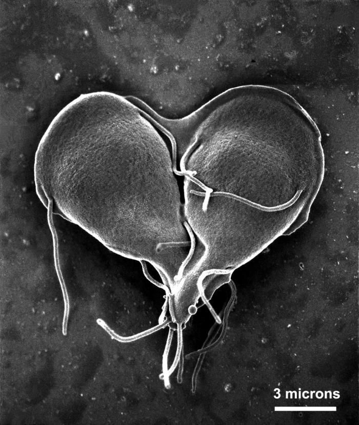
This scanning electron micrograph (SEM) depicted a Giardia lamblia protozoan that was about to become two, separate organisms, as it was caught in a late stage of cell division, producing a heart-shaped form.Created: 1999
-
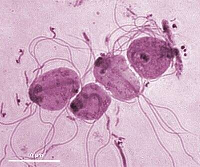
Octomitus is a diplomonad with a broadly pyriform cell body tapered posteriorly, (10-15 µm), bearing six anterior flagella deflected backwards and two posterior trailing flagella. The two anterior nuclei are bean-shaped, they face up and adjoin each other in their anterior part. A large endosome is present in the anterior lobe of the nuclei. The two sets of flagella emerge on each side of the anterior part of the body. The two recurrent flagella, accompanied by a sheath of reticulum, traverse the cell, forming a central axis before emerging as trailing flagella. Two spikes are located at the posterior between the two trailing flagella. There is no cytostomal opening at the emergence of the trailing flagella in contrast to Spironucleus. Anaerobic, parasitic or endocommensal in the intestine of vertebrates such as amphibians, caecum of rodents, rumen. Octomitus intestinalis from mice with two anterior nuclei, six antero-lateral flagella and two posterior flagella which traverse the cell axially (Giemsa staining)
-
-
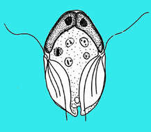
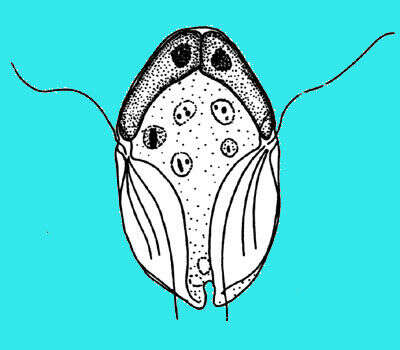
Chilomastix are retortamonad flagellates that have a pyriform and twisted cell body of about 20 µm in length with three anteriorly directed flagella and one short recurrent flagellum beating inside a ventral cytostomal pocket. The right fibril bordering the cytostomal pocket is thicker and forms a hook at its posterior end where food is phagocytosed. Cysts are pyriform and retain the internalised cytostomal fibers. They live in anoxic habitats but one species C. cuspidata is free-living. Among the 29 or so parasitic or endocommensal species described many live in the gut of vertebrates - such as C. mesnili in man and some in the gut of invertebrates such as C. aulastomi from the leech Aulastoma gulo. This species, Chilomastix caulleryi, is from the intestine of amphibia (haematoxylin staining).