-
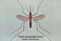
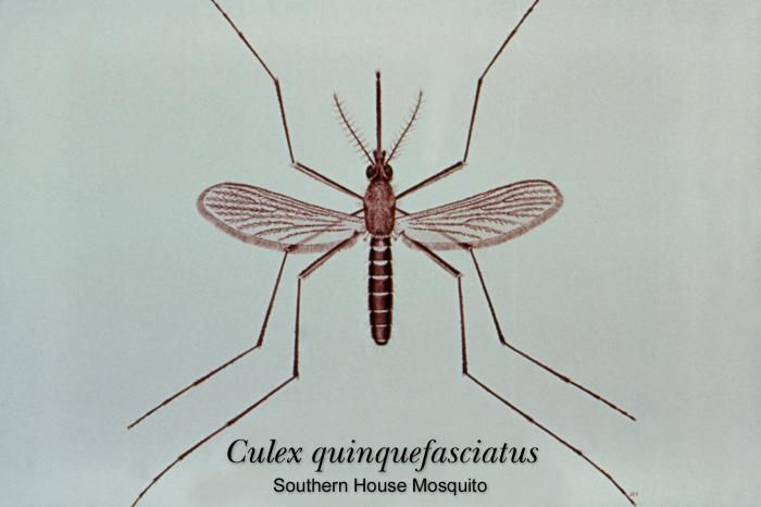
This illustration depicts a typical adult Culex quinquefasciatus mosquito.Created: 1976
-
An illustration of a Culex mosquito larva identifying the terminal abdominal segments.Created: 1975
-
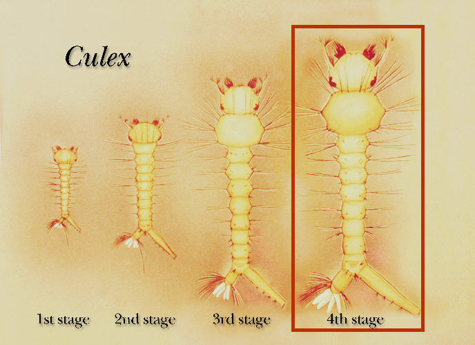
Illustration of the first, second, third and fourth growth stages of a Culex mosquito larva.Created: 1975
-
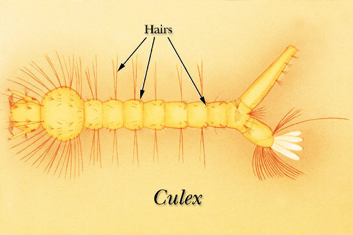
Illustration identifying hairs on the abdominal segments of a Culex mosquito larva.Created: 1975
-
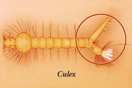
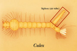
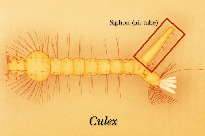
Illustration identifying the siphon, or air tube of a Culex mosquito larva.Created: 1975
-
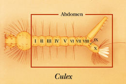
Illustration identifying the abdominal segments of a Culex mosquito larva.Created: 1975
-
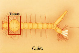
Illustration identifying the thoracic region of a Culex mosquito larva.Created: 1975
-
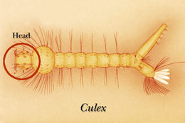
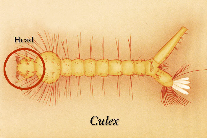
Illustration identifying the head, or cephalic region of a Culex mosquito larva.Created: 1975
-
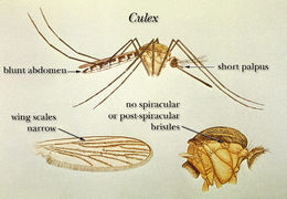
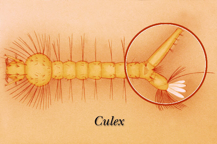
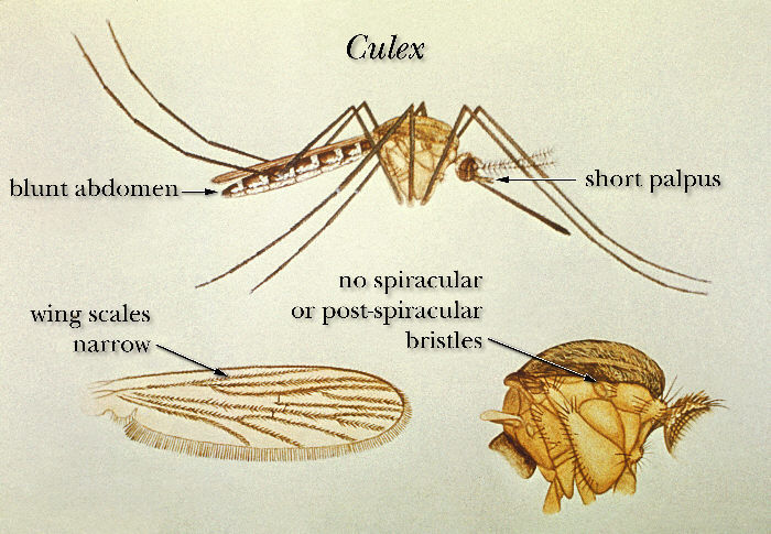
An illustration depicting morphologic characteristics common to Culex mosquitoes.Created:
-
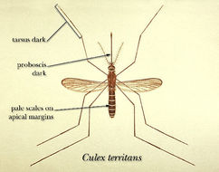
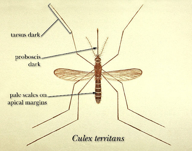
An illustration depicting morphologic characteristics common to Culex territans.Created: 1964
-
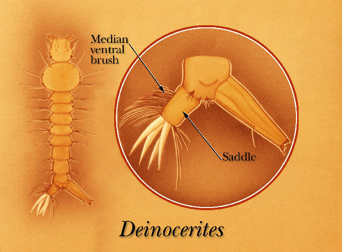
An illustration identifying the saddle and median ventral brush of a Psorophora mosquito larva.Created: 1975
-
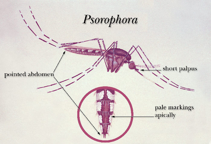
An illustration of a Psorophora mosquito.Created:
-
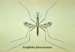
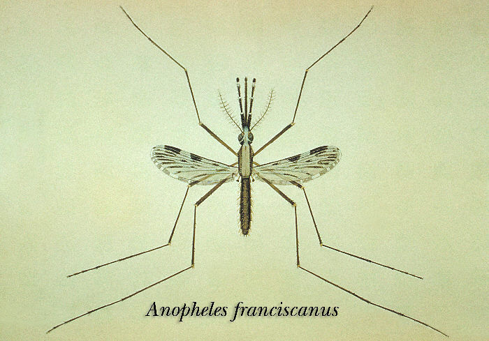
An illustration of the Anopheles franciscanus mosquito.Created:
-
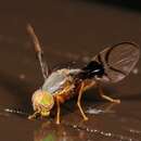
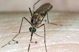
This 2005 photograph depicted a close-up view of a Culex tarsalis mosquito as it was about to begin feeding, after having landed on the skin of what will become its human host. Note the light-colored band wrapped around its dark-scaled proboscis (A), and the multiple similarly light-colored bands wrapped around its distal appendages, i.e., the tibia and femur, of its forelegs and middle pair of legs (B), identifying this as C. tarsalis.Created: 2005
-
This 2005 photograph depicted a close-up anterior view of a Culex tarsalis mosquito as it was about to begin feeding, after having landed on the skin of what will become its human host. Note the light-colored band wrapped around its dark-scaled proboscis, and though not noticeably apparent, if you look closely, the multiple similarly light-colored bands wrapped around its distal appendages, i.e., the tibia and femur, of its forelegs and middle pair of legs, identifying this as C. tarsalis.Created: 2005
-
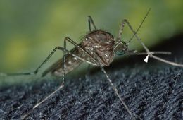
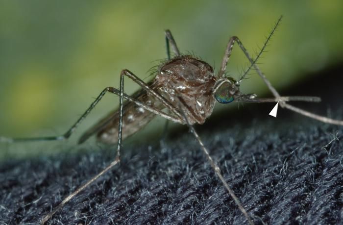
This 2005 photograph shows a close-up view of a Culex Tarsalis mosquito resting on a piece of fabric. Note the light-colored band wrapped around its dark-scaled proboscis (arrowhead), and though not noticeably apparent, if you look closely, the multiple similarly light-colored bands wrapped around its distal appendages, i.e., the tibia and femur, characteristics that identify this as a C. tarsalis.Created: 2005
-
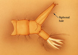
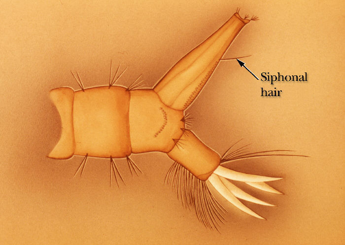
The terminal segment siphonal hairs of either the Aedes, Uranotaenia, or Psorophora mosquito larva.Created: 1975
-
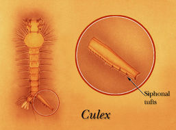
An illustration identifying the row, or scattered siphonal tufts of a Culex mosquito larva.Created: 1975
-
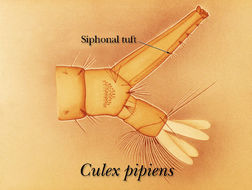
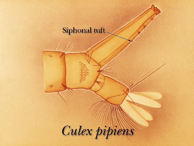
An illustration identifying the siphon tuft on the terminal abdominal segment of a Culex pipiens mosquito larva.Created: 1975
-
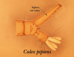
An illustration identifying the siphon on the terminal segment of a Culex pipiens mosquito larva.Created: 1975
-
-
-
-


