portrait
Description:
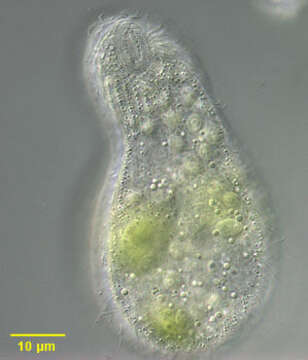
Ventral view of Platyophrya sphagni, a colpodid ciliate. Cells are flask shaped with an indistinct subapical oral aperture (well seen in this image). The cells are very flexible and squirm about rather slowly along the substratum. When swimming they tend to rotate around their long axis. The oral aperture has a right paraoral membrane, and is bordered on the left by a row of adoral organelles and an outer "postoral pseudomembrane". The right lateral surface is more densely ciliated than the left. The cytoplasm contains both large ingested algae and smaller symbiotic zoochlorellae which distinguish this species from other members of the genus. Many small extrusomes are also seen in this image. From freshwater pond near Boise, Idaho. DIC optics.
Included On The Following Pages:
- Life (creatures)
- Cellular (cellular organisms)
- Eukaryota (eukaryotes)
- SAR (Stramenopiles, Alveolates, Rhizaria)
- Alveolata (alveolates)
- Ciliophora (ciliates)
- Intramacronucleata
- Colpodea
- Cyrtolophosidida
- Platyophryidae
- Platyophrya
- Platyophrya sphagni
This image is not featured in any collections.
Source Information
- license
- cc-by-nc
- author
- William Bourland
- provider
- micro*scope
- original
- original media file
- visit source
- partner site
- micro*scope
- ID