ciliature
Description:
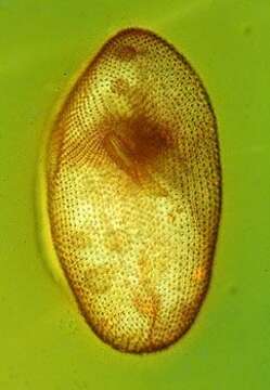
This cell has been fixed and stained with protargol which shows up cilia and other large microtubular structure. The image is of the ventral side, and shows the arrangem,ent of the cilia into kineties, the mpouth with more densely packed rows of cilia within the mouth, and the cytopyge or cell anus through which undigested residues of food will be discharged.
Included On The Following Pages:
- Life (creatures)
- Cellular (cellular organisms)
- Eukaryota (eukaryotes)
- SAR (Stramenopiles, Alveolates, Rhizaria)
- Alveolata (alveolates)
- Ciliophora (ciliates)
- Intramacronucleata
- Oligohymenophorea
- Peniculida (Peniculid)
- Parameciidae
- Paramecium (slipper animalcules)
- Paramecium putrinum
This image is not featured in any collections.
Source Information
- license
- cc-by-nc
- author
- D. J. Patterson.
- provider
- micro*scope
- original
- original media file
- visit source
- partner site
- micro*scope
- ID