Image of Typhochlaena
Description:
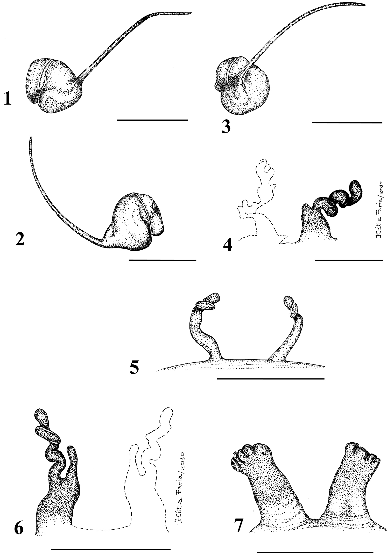
Figures 1–7.1–5 Typhochlaena seladonia C. L. Koch, 1841 1–3 male (IBSP 4551) left palpal bulb 1 prolateral 2 retrolateral 3 frontal 4–5 females, spermathecae 4 exuvium (IBSP 4551) 5 female (IBSP 109718) 6 Typhochlaena curumin sp. n. holotype female (IBSP 8701) spermathecae 7 Typhochlaena paschoali sp. n., paratype female (MNRJ 12928), spermathecae. Scale bar = 1mm.
Included On The Following Pages:
- Life (creatures)
- Cellular (cellular organisms)
- Eukaryota (eukaryotes)
- Opisthokonta (opisthokonts)
- Metazoa (Animal)
- Bilateria
- Protostomia (protostomes)
- Ecdysozoa (ecdysozoans)
- Arthropoda (arthropods)
- Chelicerata (chelicerates)
- Arachnida (arachnids)
- Araneae (spiders)
- Opisthothelae
- Mygalomorphae (mygalomorphs)
- Theraphosidae (tarantulas)
- Typhochlaena
- Typhochlaena curumim
- Panarthropoda
This image is not featured in any collections.
Source Information
- license
- cc-by-3.0
- copyright
- Rogério Bertani
- bibliographic citation
- Bertani R (2012) Revision, cladistic analysis and biogeography of Typhochlaena C. L. Koch, 1850, Pachistopelma Pocock, 1901 and Iridopelma Pocock, 1901 (Araneae, Theraphosidae, Aviculariinae) ZooKeys 230: 1–94
- original
- original media file
- visit source
- partner site
- Zookeys
- ID