Image of Acantholycosa
Description:
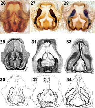
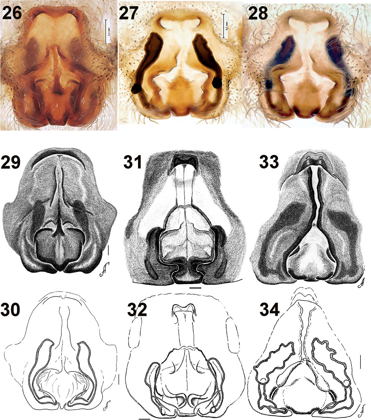
Figures 26–34.Epigyne of Acantholycosa azarkinae sp. n. (26–28), Acantholycosa oligerae (29–30), Acantholycosa norvegica (31–32) and Acantholycosa lignaria (33–34). 26, 28–29, 31, 33 epigyne, ventral 27, 30, 32, 34 vulva, dorsal. 27–28, 30, 32, 34 after maceration. 29–34 after Marusik et al. (2004). Scale = 0.1 mm if not otherwise indicated.
Included On The Following Pages:
- Life (creatures)
- Cellular (cellular organisms)
- Eukaryota (eukaryotes)
- Opisthokonta (opisthokonts)
- Metazoa (Animal)
- Bilateria
- Protostomia (protostomes)
- Ecdysozoa (ecdysozoans)
- Arthropoda (arthropods)
- Chelicerata (chelicerates)
- Arachnida (arachnids)
- Araneae (spiders)
- Opisthothelae
- Araneomorphae
- Entelegynae
- Retrolateral tibial apophysis
- Lycosidae (wolf spiders)
- Acantholycosa
- Acantholycosa azarkinae
- Panarthropoda
This image is not featured in any collections.
Source Information
- license
- cc-by-3.0
- copyright
- Yuri M. Marusik, Mikhail M. Omelko
- bibliographic citation
- Marusik Y, Omelko M (2011) A survey of East Palaearctic Lycosidae (Araneae). 7. A new species of Acantholycosa Dahl, 1908 from the Russian Far East ZooKeys 79: 1–10
- original
- original media file
- visit source
- partner site
- Zookeys
- ID