Images of sensory structures of live Symsagittifera roscoffensis
Description:
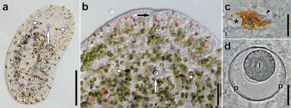
Description: English: Images of sensory structures of live Symsagittifera roscoffensis. a Hatchling. Arrowheads point to eyes, arrow to statocyst. Note absence of symbionts and presence of orange rhabdoids. b Anterior end of adult with symbionts and rhabdoids. White arrowheads point to eyes, white arrow to statocyst, black arrow to frontal organ. c Eye of an adult. Asterisk marks nucleus, arrowhead points to concrements. d Statocyst of an adult. Abbreviations: l lithocyte; p parietal cells. Scale bars: a 100 μm; b 50 μm; c 10 μm; d 10 μm. Date: 30 June 2013. Source: https://link.springer.com/article/10.1007%2Fs13127-012-0112-4. Author: Johannes G. Achatz, Marta Chiodin, Willi Salvenmoser, Seth Tyler , Pedro Martinez.
Included On The Following Pages:
- Life (creatures)
- Cellular (cellular organisms)
- Eukaryota (eukaryotes)
- Opisthokonta (opisthokonts)
- Metazoa (Animal)
- Bilateria
- Xenacoelomorpha
- Acoela (acoel flatworms)
- Aberrantospermata
- Convolutidae
- Symsagittifera
- Symsagittifera roscoffensis
This image is not featured in any collections.
Source Information
- license
- cc-by-sa-3.0
- copyright
- Johannes G. Achatz, Marta Chiodin, Willi Salvenmoser, Seth Tyler , Pedro Martinez
- creator
- Johannes G. Achatz, Marta Chiodin, Willi Salvenmoser, Seth Tyler , Pedro Martinez
- source
- https://link.springer.com/article/10.1007%2Fs13127-012-0112-4
- original
- original media file
- visit source
- partner site
- Wikimedia Commons
- ID