H. pylori urease diagram
Description:
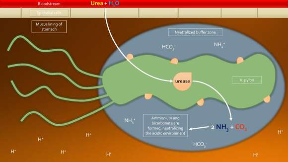
: File:H. pylori urease enzyme diagram.svg is a vector version of this file. File:API File:H. pylori urease enzyme diagram.svg For more information, see Help:SVG. Alemannisch | العربية | Беларуская (тарашкевіца) | Български | বাংলা | Català | Нохчийн | Čeština | Dansk | Deutsch | Ελληνικά | English | Español | Eesti | Euskara | فارسی | Suomi | Français | Frysk | Galego | עברית | Hrvatski | Magyar | Հայերեն | Bahasa Indonesia | Italiano | 日本語 | ქართული | 한국어 | Lietuvių | Македонски | മലയാളം | Bahasa Melayu | Plattdüütsch | Nederlands | Norsk nynorsk | Norsk bokmål | Occitan | Polski | Português | Português do Brasil | Română | Русский | Sicilianu | Scots | Slovenčina | Slovenščina | Српски / srpski | Svenska | ไทย | Türkçe | Татарча | Українська | Vèneto | Tiếng Việt | 中文 | 中文(中国大陆) | 中文(简体) | 中文(繁體) | 中文(马来西亚) | 中文(台灣) | +/− : . Description: English: One mechanism H. pylori has to combat the acidic environment of the stomach is the urease enzyme. This enzyme can be found on the surface of and inside the cell. It takes urea and water from the bloodstream and stomach and converts them to ammonia and carbon dioxide. These products neutralize the strong acid of the stomach, and as a result a buffer zone surrounds the bacteria to protect it from the strong acids in its environment. Date: 20 November 2016. Source: Own work. Author: Olivia Child.
Included On The Following Pages:
- Life (creatures)
- Cellular (cellular organisms)
- Bacteria
- Proteobacteria (Purple Bacteria & relatives)
- deltaepsilon subdivisions
- Epsilonproteobacteria
- Campylobacterales
- Helicobacteraceae
- Helicobacter
- Helicobacter pylori
This image is not featured in any collections.
Source Information
- license
- cc-by-sa-3.0
- copyright
- Olivia Child
- creator
- Olivia Child
- original
- original media file
- visit source
- partner site
- Wikimedia Commons
- ID