Pone.0118415.g001
Description:
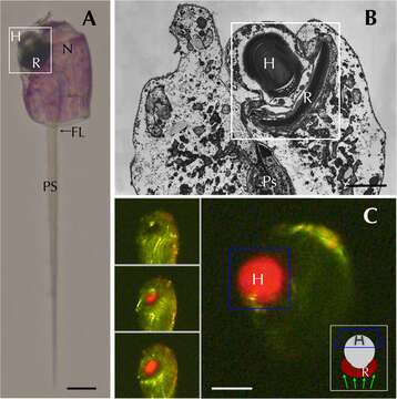
Description: English: Erythropsidinium spp. and its subcellular structure eyespot “ocelloid”. (A) Light micrographs (LM) of Erythropsidinium spp. H = hyalosome (crystallin body), R = retinal body, N = nucleus, FL = flagella, PS = piston. (B) Transmission electron micrographs (TEM) of ocelloid. (C) The refractile nature of the hyalosome under fluorescent microscopy. Bars: 20 μm (A, C), 10 μm (B).The ocelloid is located at the left side of a cell seen in ventral view according to the orientation proposed by Kofoid and Swezy (A). The nucleus was ellipsoidal and at the opposite side of ocelloid, in the anterior of the cell (B). These indices are consistent with the taxonomic criteria of the type specimen that was identified as Erythropsidinium agile. From the serial pictures of autofluorescence in the retinal body (C), lens-effect of the hyalosome can be observed. The front image of the retinal body is larger than side view. Date: 3 March 2015. Source: Fig. 1 at https://journals.plos.org/plosone/article?id=10.1371/journal.pone.0118415 Function and Evolutionary Origin of Unicellular Camera-Type Eye Structure. PLOS ONE 10(3), PMID 25734540, PMC 4348419, doi:10.1371/journal.pone.0118415 . Author: Shiho Hayakawa, Yasuharu Takaku, Jung Shan Hwang, Takeo Horiguchi, Hiroshi Suga, Walter Gehring, Kazuho Ikeo, Takashi Gojobori. Other versions: This file has multiple extracted images: File:Pone.0118415.g001A.tif Plos one hayakawa erythropsidinium ocelloid EM fig1b.png.
Included On The Following Pages:
- Life (creatures)
- Cellular (cellular organisms)
- Eukaryota (eukaryotes)
- SAR (Stramenopiles, Alveolates, Rhizaria)
- Alveolata (alveolates)
- Dinophyceae
- Gymnodiniales
- Warnowiaceae
- Erythropsidinium
This image is not featured in any collections.
Source Information
- license
- cc-by-sa-3.0
- copyright
- Shiho Hayakawa, Yasuharu Takaku, Jung Shan Hwang, Takeo Horiguchi, Hiroshi Suga, Walter Gehring, Kazuho Ikeo, Takashi Gojobori
- creator
- Shiho Hayakawa, Yasuharu Takaku, Jung Shan Hwang, Takeo Horiguchi, Hiroshi Suga, Walter Gehring, Kazuho Ikeo, Takashi Gojobori
- source
- Fig. 1 at https://journals.plos.org/plosone/article?id=10.1371/journal.pone.0118415 Function and Evolutionary Origin of Unicellular Camera-Type Eye Structure. PLOS ONE 10(3), PMID 25734540, PMC 4348419, doi:10.1371/journal.pone.0118415
- original
- original media file
- visit source
- partner site
- Wikimedia Commons
- ID