MSphere.00531-19-f0006
Description:
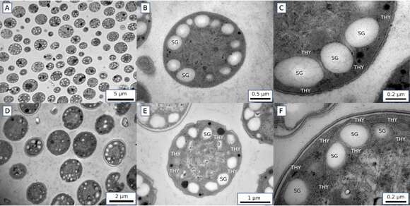
Description: English: Transmission electron micrographs of Crocosphaera cells harvested at the 6-h time point during the light period (A to C) and at the 6-h time point during the dark (D to F). Starch granules (SG) and thylakoid membranes (THY) are observed mostly on the edge of the cytosol. More-detailed images (C and F) show that SG are observed mostly between THY. Date: 11 December 2019. Source: Fig. 6 at https://europepmc.org/article/PMC/PMC6908418 Quantifying Oxygen Management and Temperature and Light Dependencies of Nitrogen Fixation by Crocosphaera watsonii. In: Msphere 4(6), doi:10.1128/msphere.00531-19, PMID 31826967, PMC 6908418. Author: Inomura K, Deutsch C, Wilson ST, Masuda T, Lawrenz E, Lenka B, Sobotka R, Gauglitz JM, Saito MA, Prášil O, Follows MJ.
Included On The Following Pages:
- Life (creatures)
- Cellular (cellular organisms)
- Bacteria
- Cyanobacteria
- Oscillatoriophycideae
- Chroococcales
- Aphanothecaceae
This image is not featured in any collections.
Source Information
- license
- cc-by-sa-3.0
- copyright
- Inomura K, Deutsch C, Wilson ST, Masuda T, Lawrenz E, Lenka B, Sobotka R, Gauglitz JM, Saito MA, Prášil O, Follows MJ
- creator
- Inomura K, Deutsch C, Wilson ST, Masuda T, Lawrenz E, Lenka B, Sobotka R, Gauglitz JM, Saito MA, Prášil O, Follows MJ
- source
- Fig. 6 at https://europepmc.org/article/PMC/PMC6908418 Quantifying Oxygen Management and Temperature and Light Dependencies of Nitrogen Fixation by Crocosphaera watsonii. In: Msphere 4(6), doi:10.1128/msphere.00531-19, PMID 31826967, PMC 6908418
- original
- original media file
- visit source
- partner site
- Wikimedia Commons
- ID