Elife-25940-fig8-v2
Description:
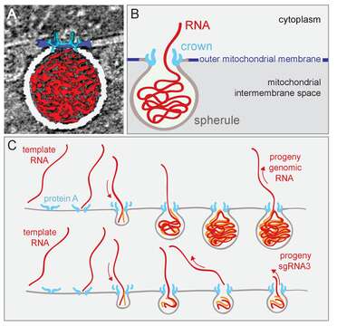
Description: English: Model of Flock house nodavirus (FHV) replication complex structure and function. (A) Amira-assisted 3D segmentation of a single virus replication compartment in FHV-infected cells. The spherule membrane (white) encloses the viral RNA (red) and is anchored to the mitochondrial outer membrane (dark blue), by the FHV protein A crown (light blue, derived from the subtomogram average). (B) Cartoon rendering of the image in (A). Viral RNA is shown as exiting the spherule to include the observation of fibrillary extrusions in many other tomograms. (C) Model of the FHV RNA complex and RNA synthesis, where positive-strand RNA (red) associates with protein A on the mitochondrial membrane to initiate the synthesis of negative-strand RNA (orange) and progeny positive-strand RNA accommodated by increasing volume of the spherule as if blowing up a balloon. In this model, subgenomic RNA3 results from attenuated negative strand synthesis and subse- quent positive-strand production in small spherules. Date: 27 June 2017. Source: eLife 2017;6:e25940 DOI: 10.7554/eLife.25940 https://elifesciences.org/articles/25940. Author: Kenneth J Ertel, Desirée Benefield, Daniel Castaño-Diez, Janice G Pennington, Mark Horswill, Johan A den Boon, Marisa S Otegui, and Paul Ahlquist.
Included On The Following Pages:
This image is not featured in any collections.
Source Information
- license
- cc-by-3.0
- copyright
- Kenneth J Ertel, Desirée Benefield, Daniel Castaño-Diez, Janice G Pennington, Mark Horswill, Johan A den Boon, Marisa S Otegui, and Paul Ahlquist
- creator
- Kenneth J Ertel, Desirée Benefield, Daniel Castaño-Diez, Janice G Pennington, Mark Horswill, Johan A den Boon, Marisa S Otegui, and Paul Ahlquist
- source
- eLife 2017;6:e25940 DOI: 10.7554/eLife.25940
- original
- original media file
- visit source
- partner site
- Wikimedia Commons
- ID