Image of Orientia tsutsugamushi
Description:
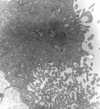
This 1976 transmission electron micrograph (TEM) depicted a hypertrophic peritoneal mesothelial cell of mouse that had been experimentally infected intraperitoneally with Orientia tsutsugamushi rickettsial micro-organisms. In this TEM, several organisms were visible, free within the host cell's cytoplasm. One O. tsutsugamushi appeared within a phagocytic vacuole, still bearing a third outer membrane layer of probable host cell origin.
Created: 1976
Included On The Following Pages:
- Life (creatures)
- Cellular (cellular organisms)
- Bacteria
- Proteobacteria (Purple Bacteria & relatives)
- Alphaproteobacteria (alphaproteobacterium; alphaproteobacteria)
- Rickettsiales
- Rickettsiaceae
- Orientia
- Orientia tsutsugamushi
This image is not featured in any collections.
Source Information
- license
- cc-publicdomain
- provider
- Public Health Image Library
- original
- original media file
- visit source
- partner site
- Public Health Image Library
- ID