Image of Borrelia burgdorferi Johnson et al. 1984
Description:
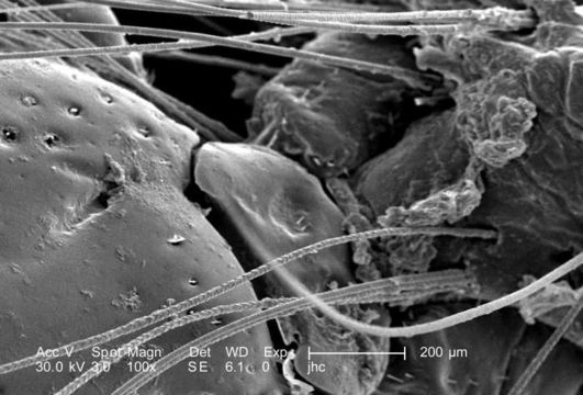
Under a low magnification of 100X, this scanning electron micrographic (SEM) image depicted a dorsal view of an unidentified engorged female tick, which had been extracted from the skin of a pet cat while in the process of obtaining its blood meal. Note the presence of some of the cats fur, along with some of its skin tissue in which the ticks gnathosoma were still embedded. See PHIL 9972 and 9973 for additional, less magnified views of this scenario. It is from the basis capituli that the two spread pedipalps, and hidden skin-piercing hypostome and chelicerae emanate. On the dorsal surface of the basis capituli youll see two depressed areas known as the porose areas, through which secretions produced by dermal glands are released.
Created: 2006
Included On The Following Pages:
- Borrelia burgdorferi
- Life (biota)
- Cellular
- Bacteria (bacteria)
- Spirochaetes
- Spirochaetales
- Borrelia
- Spirochaetaceae
- Spirochaetes
This image is not featured in any collections.
Source Information
- license
- cc-publicdomain
- photographer
- Janice Carr
- provider
- Public Health Image Library
- original
- original media file
- visit source
- partner site
- Public Health Image Library
- ID