Image of Hartmannella vermiformis Page 1967
Description:
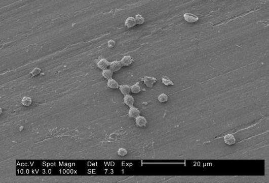
Under a moderately-high magnification of 1000X, this 2002 scanning electron micrograph (SEM) revealed some of the ultrastructural morphology exhibited by a number of Hartmannella vermiformis amoebae cysts.
As part of a study to determine whether Legionella pneumophila bacteria can colonize and grow in biofilms both with, and without the presence of H. vermiformis, here these protozoa were situated on the surface of a stainless steel coupon. See PHIL 11165, which depicted numbers of these amoebae atop a base biofilm, within which Pseudomonas aeruginosa, Klebsiella pneumoniae and a Flavobacterium sp. bacteria had been suspended. By preying upon these bacteria, these amoebae had scoured the stainless steel coupon, thereby, exposing its surface.
Created: 2002
Included On The Following Pages:
- Hartmannella vermiformis
- Life (creatures)
- Cellular (cellular organisms)
- Eukaryota (eukaryotes)
- Amoebozoa (amoeboid protists)
- Tubulinea
- Euamoebida
- Hartmannellidae
- Hartmannella
This image is not featured in any collections.
Source Information
- license
- cc-publicdomain
- photographer
- Janice Haney Carr
- provider
- Public Health Image Library
- original
- original media file
- visit source
- partner site
- Public Health Image Library
- ID