Plate 13
Description:
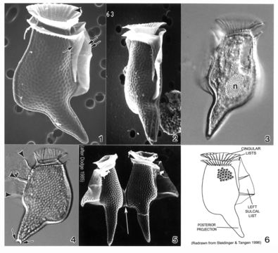
Plate 13. Dinophysis caudata. Figs. 1-2. SEM. Fig. 1. Large, long and distinctive cell with extended ventral hypothecal process. Cingulum narrow; lists supported by ribs (arrowhead). Strong left sulcal list (double arrows). Right sulcal list present (single arrow). Fig. 2. Ventral view: cell compressed laterally. Figs. 3-4. LM. Fig. 3. Large posterior nucleus (n). Fig. 4. Left sulcal list with three supporting ribs (arrowheads); posterior projection with small knob-like spines (arrows). Surface areolae evident. Fig. 5. SEM. Paired cells joined at dorsal expansion (arrow). Fig. 6. Line drawing.
Included On The Following Pages:
- Life (biota)
- Cellular
- Eukaryota (eukaryotes)
- SAR (Stramenopiles, Alveolates, Rhizaria)
- Alveolata (alveolates)
- Dinophyceae (dinoflagellates)
- Dinophysiales
- Dinophysiaceae
- Dinophysis
- Dinophysis caudata
- Dinoflagellata (Dinoflagellate)
This image is not featured in any collections.
Source Information
- license
- cc-publicdomain
- bibliographic citation
- Faust, Maria A. and Rose A. Gulledge. Identifying Harmful Marine Dinoflagellates. Smithsonian Contributions from the United States National Herbarium, volume 42: 1-144 (including 48 plates, 1 figure and 1 table).
- original
- original media file
- visit source
- partner site
- NMNH Marine Dinoflagellates
- ID