Plate 19
Description:
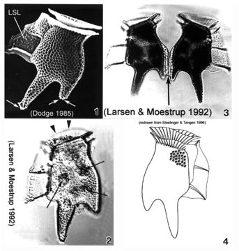
Plate 19. Dinophysis tripos. Fig. 1. SEM: lateral view. Cell large, oblong and heavily areolated. Hypothecal projections with toothed posterior ends (arrows). Left sulcal list (LSL) large, wide and reticulated. Figs. 2,3. LM: lateral view. Fig. 2. Anterior cingular list (ACL) projected anteriorly obscuring low epitheca (arrowheads). Narrow cingulum. Chloroplasts visible (arrows). Fig. 3. Paired cells. Hypothecal projection on dorsal margin sometimes seen with a narrow list (arrow) connecting two daughter cells during cell division. Fig. 4. Line drawing.
Included On The Following Pages:
- Life (creatures)
- Cellular (cellular organisms)
- Eukaryota (eukaryotes)
- SAR (Stramenopiles, Alveolates, Rhizaria)
- Alveolata (alveolates)
- Dinophyceae
- Dinophysiales
- Dinophysiaceae
- Dinophysis
- Dinophysis tripos
- Dinoflagellata (dinoflagellates)
This image is not featured in any collections.
Source Information
- license
- cc-publicdomain
- bibliographic citation
- Faust, Maria A. and Rose A. Gulledge. Identifying Harmful Marine Dinoflagellates. Smithsonian Contributions from the United States National Herbarium, volume 42: 1-144 (including 48 plates, 1 figure and 1 table).
- original
- original media file
- visit source
- partner site
- NMNH Marine Dinoflagellates
- ID