Plate 44
Description:
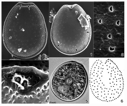
Plate 44. Prorocentrum maculosum. Figs. 1-4. SEM. Fig. 1. Right valve: cell broadly ovate, narrowing apically. Valve surface rugose with scattered poroids; valve center devoid of poroids. Marginal pores evenly spaced (arrows). Fig. 2. Left valve: anterior end flat to slightly concave with raised apical ridge (arrows). Valve margins appear as a flange around cell. Fig. 3. Valve poroids: unevenly distributed on valve surface; circular to oblong or kidney-shaped. Fig. 4. Periflagellar area: broad V-shaped depression on right valve. Apical ridge (raised margin) on left valve. Flagellar (f) and auxiliary (a) pores surrounded by protuberant periflagellar collar (arrowheads); equal in size. Fig. 5. LM. Right valve: central pyrenoid (P) and large posterior nucleus (N) (M.A. Faust). Fig. 6. Line drawing: valve poroid and marginal pore arrangement (Figs. 1-4,6 after Faust 1993b)
Included On The Following Pages:
- Life (biota)
- Cellular
- Eukaryota (eukaryotes)
- SAR (Stramenopiles, Alveolates, Rhizaria)
- Alveolata (alveolates)
- Dinophyceae (dinoflagellates)
- Prorocentrales
- Prorocentraceae
- Prorocentrum
- Prorocentrum maculosum
- Dinoflagellata (Dinoflagellate)
This image is not featured in any collections.
Source Information
- license
- cc-publicdomain
- bibliographic citation
- Faust, Maria A. and Rose A. Gulledge. Identifying Harmful Marine Dinoflagellates. Smithsonian Contributions from the United States National Herbarium, volume 42: 1-144 (including 48 plates, 1 figure and 1 table).
- original
- original media file
- visit source
- partner site
- NMNH Marine Dinoflagellates
- ID