-
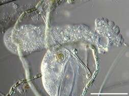
The posterior part of a actively moving naked amoebae usually adopts a distinctive appearance. The cytoplasm is often more rigid in this area, and the surface of the cell is thrown into folds or is the source of numerous filaments. In this amoeba, the uroid has a rumpled appearance as a result of folds forming in the cell surface. Phase contrast micrograph.
-
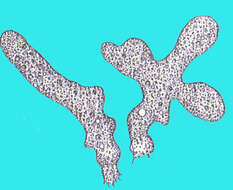
There are only a few genera of free-living amoebae that have lobose pseudopodia and which measure over half a millimetre. The genus Chaos is distinguished in part because the cytoplasm contains many nuclei. This cell is lysing, and the nuclei are spilling out of it. Many more remains inside the cell. Phase contrast micrograph.
-
-
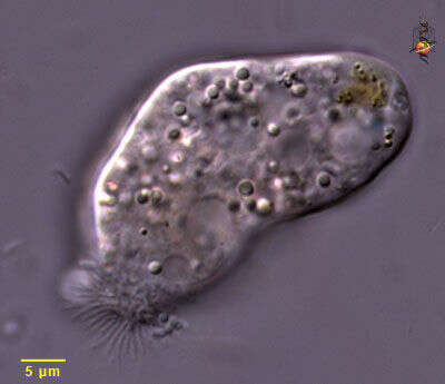
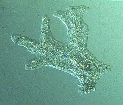
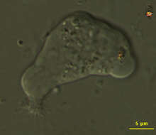
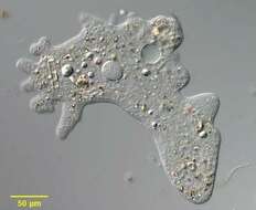
Free-living amoeba, moving towards right, a small hyaline cap indicates the direction of movement. The threads at the lower left are emerging from the uroid of the cell. Differential interference contrast optics.
-
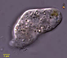
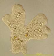
Free-living amoeba, moving towards upper right. The threads at the lower left are emerging from the uroid of the cell. Differential interference contrast optics.
-
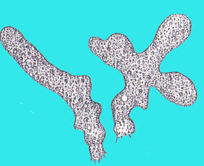
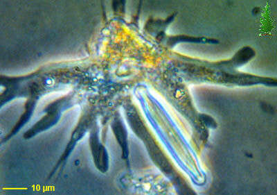
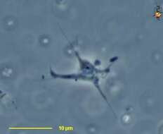
The identity of this amoeba is uncertain. It is a amoeba that forms lobose pseudopodia and has a large nucleus. This cell is squashed and has taken on this star-like form. Phase contrast microscopy.
-
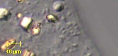
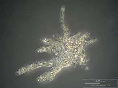
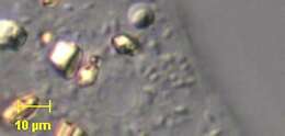
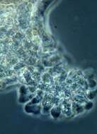
Detail of Polychaos dubium, a large polypodial naked amoeba. P. dubium is the type species. Palmate form. Pseudopodia lack the longitudinal ridges seen in Amoeba species. The posterior is composed of a collection of fused remnant pseudopodia. May become monopodial during rapid locomotion. The granular nucleus is spherical to ovoid in shape. The cytoplasm contains ingested algae and numerous refractile crystals. This image shows a large bipyramidal cytoplasmic crystal, probably composed of urea. From freshwater pond near Boise, Idaho. DIC optics.
-
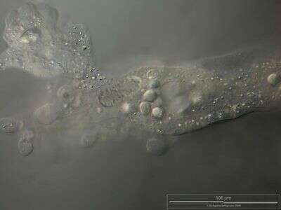
Portrait of Polychaos dubium, a large polypodial naked amoeba. P. dubium is the type species. Palmate form. Pseudopodia lack the longitudinal ridges seen in species of Amoeba. The posterior (left side of image) is composed of a collection of fused remnant pseudopodia. May become monopodial during rapid locomotion. The granular nucleus is spherical to ovoid in shape. A large contractile vacuole is visible. The cytoplasm contains ingested algae and numerous refractile crystals. From freshwater pond near Boise, Idaho. DIC optics.
-
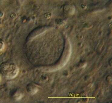
Portrait of the polypoidal amoeba. The single nucleus with itâs distinct nucleolar material in narrow peripheral lobes well visible in the central part of the cell.
-
The nucleus of Polychaos fasciculatum with it's nucleolar material in typically peripheral located narrow lobes
-
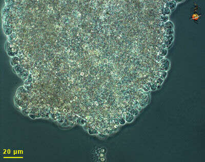
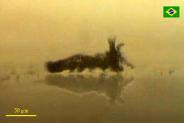
Scale bar indicates 50 µm. Sample from the pond Hegne Moor situated in the vicinity of Lake Constance. The image was built up using several photomicrographic frames with manual stacking technique. Images were taken using Zeiss Universal with Olympus C7070 CCD camera.Image under Creative Commons License V 3.0 (CC BY-NC-SA).
-
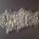
Collected from Cumloden Swamp December 23, 2003.
-
Collected from Cumloden Swamp on December 23, 2003.
-
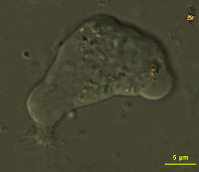
Amoeba crawling on a glass slide. This picture was taken with a light microscope laid sideways in the table, and the amoeba was inside a very tiny handcrafted fishbowl (amoebabowl?)
-
-
Single cell, living, moving to upper right. The uroid is the cumpled region at the posterior end of the cell. Actively progressing pseudopodia (such as the one pointing north) have a hyaline cap. Cell surface with folds. Contractile vacuole and nucleus not visible.
-
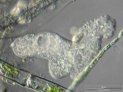
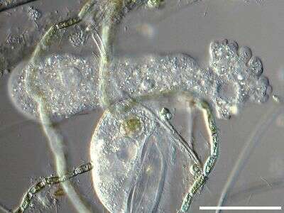
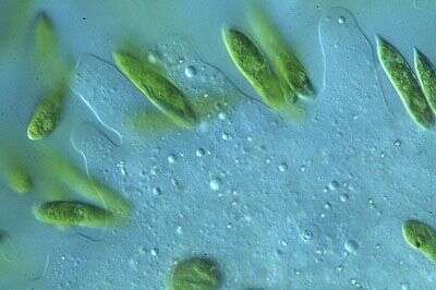
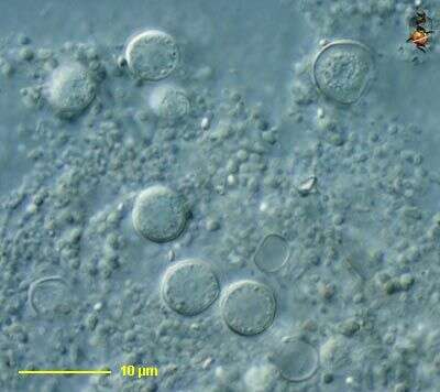
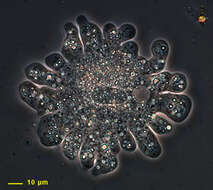
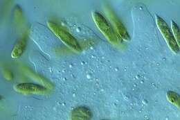
Image of live cell capturing Euglena gracilis cells. Even though the amoebae move considerably slower than the flagellates, the flagellates cluster around the amoeba and are ingested.
-
The posterior end of most moving lobose amoebae has a different appearance to the rest of the cell. In the case of Amoeba proteus, it looks crumpled. This is in part due to the interactions of actin and myosin that cause the forward movement of the cell.
-
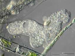
Amoeba proteus with numerous crystals showing typical polypodial movement. The specimen was gathered in a tiny freshwater pond at the island of Hiddensee (Baltic Sea, Germany) which shows a fascinating biodiversity of naked and testate amoebae. Images were taken using Zeiss Standard with Olympus C7070 CCD camera.
-
Nucleus (center) and trailing uroid (upper left) of Amoeba proteus. To the right of the nucleus there are several food vacuoles. In addition many tiny crystals are visible. The specimen was gathered in a tiny freshwater pond at the island of Hiddensee (Baltic Sea, Germany) which shows a fascinating biodiversity of naked and testate amoebae. Images were taken using Zeiss Standard with Olympus C7070 CCD camera.
-
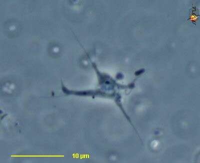
Gephyramoeba (ge-fire-a-me-ba) a naked amoeba of uncertain affinities. Often with cytoplasm drawn out into long pseudopodia. Nucleus with nucleolus also visible. Phase contrast.
-
Gephyramoeba (ge-fire-a-me-ba) a naked amoeba of uncertain affinities. Often with cytoplasm drawn out into long pseudopodia. Nucleus with nucleolus also visible. Cysts. Differential interference contrast.
-