-
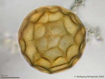
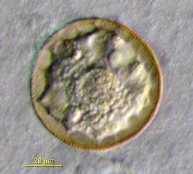
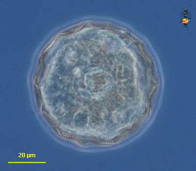
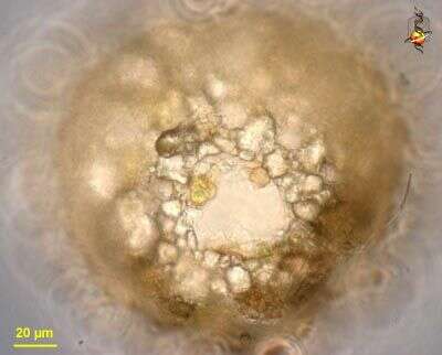
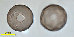
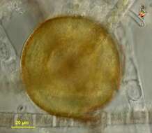
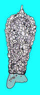
Large species of Arcella with high dome and an internal collar. Surface has typical honeycomb pattern, often with dented surface, and gets darker with age.
-
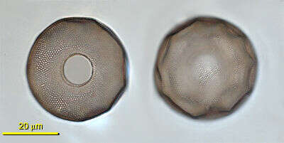
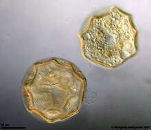
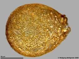
This testate amoeba constructs a proteinaceous shell with numerous tiny alveoli reinforcing the test. The picture shows the test from dorsal and ventral. On the ventral side the aperture, two contractile vacoules and one nucleus with centric nucleolus is visible. Arcellae have two nuclei. Multi layer image using 25 DIC frames with manual stacking technique using Corel Photopaint. The scale bar indicates 25 µm. Collected from bottom sediments of a rain storage reservoir in Kiel (Schleswig-Holstein, Germany). Images were taken using Zeiss Universal with Olympus C7070 CCD camera.
-
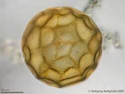
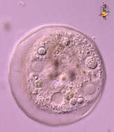
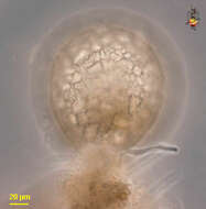
Multi layer image using 35 frames generating depth of focus, stacked manualle using Corel Photopaint. Scale bar indicates 25 µm. Sample from a pond on the island of Hiddensee (Baltic Sea, Germany). This image was taken using Zeiss Universal with Olympus C7070 CCD camera.
-
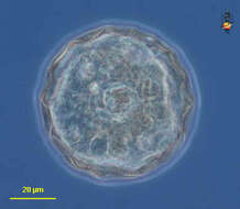
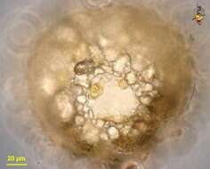
Scale bar indicates 25 µm. Sample from the pond Hegne Moor situated in the vicinity of Lake Constance. The image was built up using several photomicrographic frames with manual stacking technique. Images were taken using Zeiss Universal with Olympus C7070 CCD camera.Image under Creative Commons License V 3.0 (CC BY-NC-SA).
-
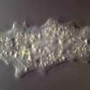
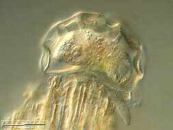
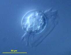
Differential interference contrast image showing an extensive pseudopodia veil.
-
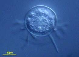
Differential interference contrast image showing pseudpodia of living cell.
-
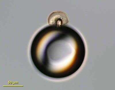
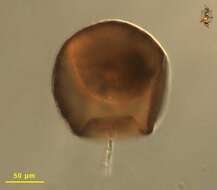
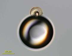
Image of Arcella cell sitting stop an air bubble.
-
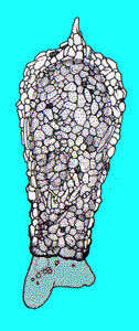
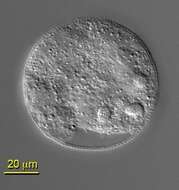
This image was taken with a scanning light microscope. The texture of the test is visible around the edge of the cell. Three nuclei are also visible.
-
This is a DIC image that shows the central position of the aperture.
-
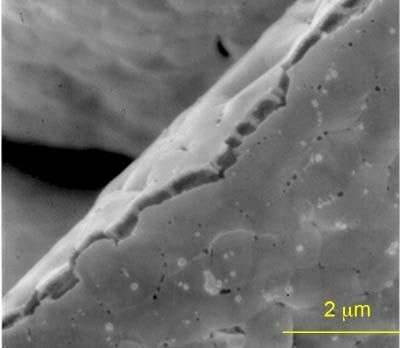
Detail of test structure captured by SEM. Notice honeycomb.
-
This testate amoebae was collected from a White Cedar Swamp in Woods Hole, MA. Note the areolate pattern on the test. Image by Dan Lahr.
-
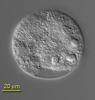
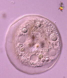
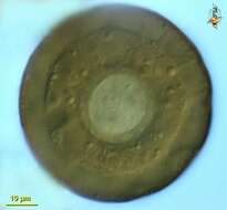
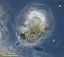
Arcella (our-sell-a), one of the more common testate amoebae, in which the amoeba lives within a lorica. The lorica is organic and in older cells usually becomes brown. The dorsal surface of the lorica of this genus is usually domed, and the ventral surface has a single round aperture or hole through which the pseudopodia emerge. These organisms usually eat detritus (the food vacuoles contain bacteria). There are two nuclei (4 and 10 o clock) (distinguished by the large rounded nucleoli) and a single contractile vacuole. Differential interference contrast.
-
Arcella (our-sell-a), one of the more common testate amoebae, in which the amoeba lives within a lorica. The lorica is organic and in older cells usually becomes brown. Phase contrast
-
Arcella (our-sell-a), one of the more common testate amoebae, in which the amoeba lives within a lorica. The lorica is organic and in older cells usually becomes brown. The dorsal surface of the lorica of this genus is usually domed, and the ventral surface has a single round aperture or hole through which the pseudopodia emerge. These organisms usually eat detritus (the food vacuoles contain bacteria). Cytoplasm evident within the lorica. Differential interference contrast.
-
Arcella (are-sell-ah) a testate amoeba in which the cell is located within a domed lorica that has a single round opening (aperture) on the ventral side. Found occasionally in cooler waters. Differential interference contrast. Material from heated thermal sites of Nymph Creek and Nymph Lake in Yellowstone National Park, photograph by Kathy Sheehan and David Patterson.
-
Test of a freshwater Difflugina. Multi layer image using 28 frames generating depth of focus, stacked manually using Corel Photopaint. Collected from a creek sediment. This image was taken using Zeiss Universal with Olympus C7070 CCD camera.
-
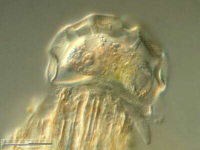
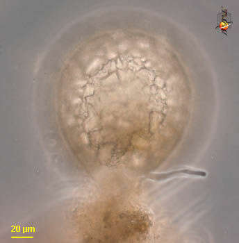
Difflugia (dif-flew-gee-a) is a testate amoeba, in which the amoeba is enclosed within a shell that is comprised of an organic cement and variously sized inorganic granules. This individual has produced one pseudopodium from the opening or aperture which is at the bottom of the test and is surrounded by accumulated debris. Phase contrast.
-
Difflugia (dif-flew-gee-a) is a testate amoeba, in which the amoeba is enclosed within a shell that is comprised of an organic cement and variously sized inorganic granules. This image shows the aperture through which the pseudopodia emerge. Phase contrast.
-
-
Difflugia (diff-flew-gee-a) a testate amoeba. The test is comprised of cemented inorganic granules. There is an aperture at one end of the cell, from which lobose pseudopodia emerge. Phase contrast.
-
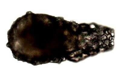
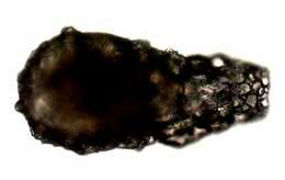
Difflugia is an amoeba that lives within a shell or 'test' comprised of small particles of sand and grit. Pseudopodia, used for movement or to capture food, if present would extend from a hole or aperture at the right end of the test.
-
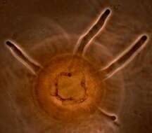
The aperture of the shell of Difflugia from which four pseudopodia are emerging.
-
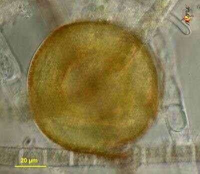
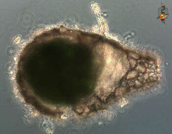
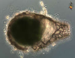
This shelled amoeba has a test that includes small sand granules that have been glued together. The cytoplasm of the enclosed amoeba is darh green because the cytoplasm includes large numbers of symbiotic algae. The aperture is to the right in this image. No pseudopodia are visible. Phase contrast microscopy.
-
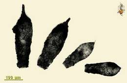
Small collection of Difflugia from one site illustrating the variation in shape and size within a population. Image two of two, this one assembled using Mr Zeiss' nice software - extended focus option.