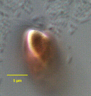
Surface detail of the cryptomonad flagellate, Rhodomonas (Karsten,1898). The inner layer of the periplast is composed of overlapping rounded or square proteinaceous organic plates about 0.4 um in diameter. This specimen was collected from a commercial saltwater aquarium in Boise, Idaho, September 2004. DIC.
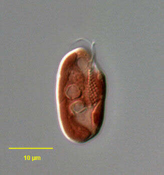
Portrait (lateral view) of the cryptomonad flagellate, Rhodomonas (Karsten,1898). The cells are laterally flattened. The anterior end is obliquely truncate and the posterior rounded. Two subequal flagella insert into a ventral furrow-gullet complex. A single contractile vacuole is seen adjacent to the anterior opening of the ventral furrow. The inner layer of the periplast is composed of overlapping rounded or square proteinaceous organic plates about 0.4 um in diameter. There is a single large boat-shaped chloroplast with a small pyrenoid. Although not always this color, this species is red due to a chloroplast containing Cr-phycoerythrin 545. Like other cryptomonads, ultrastructural studies of Rhodomonas reveal a nucleomorph associated with the plastid. The nucleomorph is thought to represent a nuclear remnant of an ancestral endosymbiotic red alga. The function, if any, of the nucleomorph is unknown. Large ejectosomes are seen here lining the ventral furrow-gullet. Rhodomonas is phototrophic. This genus is found in both freshwater and marine habitats. This specimen was collected from a commercial saltwater aquarium in Boise, Idaho, September 2004. DIC.
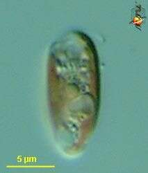
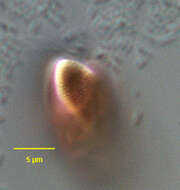
Rhodomonas (row-doe-moan-ass) salina, a cryptomonad / cryptophyte alga, with red coloured plastids, two flagella (not well imaged here) arise in a wide channel which opens near the front of the cell. Differential interference microscopy. data on this strain.
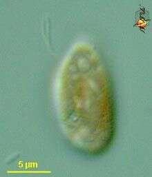
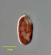
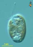
Rhodomonas (row-doe-moan-ass) salina, a cryptomonad / cryptophyte alga, with red coloured plastids, two flagella, visible upper left, arise in a wide channel which opens near the front of the cell. With starch inclusions. "" Differential interference microscopy.
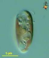
Rhodomonas (row-doe-moan-ass) salina, a cryptomonad / cryptophyte alga, with red coloured plastids, two flagella arise in a wide channel which opens near the front of the cell. The cell surface is underlain by a layer of stiffening plates, and the texture of these can be seen in places in this micrograph. Differential interference microscopy. data on this strain.
George B. McManus, Weiwei Liu, Rachel A. Cole, Daniel Biemesderfer and Jennifer L. Mydosh
Wikimedia Commons
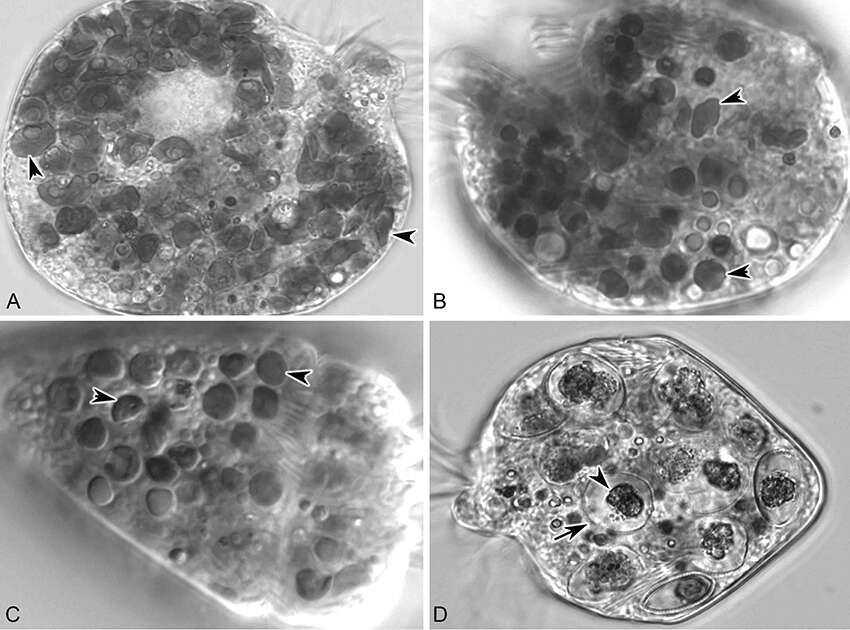
Description: English: Strombidium rassoulzadegani fed four food algae that supported positive growth in the light (12:12 L:D). (A) S. rassoulzadegani fed Tetraselmis chui PLY429; arrowheads indicate different shapes and sizes of retained chloroplasts; (B) S. rassoulzadegani fed Rhodomonas lens RHODO; arrowheads as in A; (C) S. rassoulzadegani fed Isochrysis sp. ISO SP; arrowheads as in A; (D) S. rassoulzadegani fed Prorocentrum minimum JA; note that no retained plastids appear in the cell. Arrow marks the rigid wall of the algal cell and arrowhead marks the partially-digested algal cytoplasm. Date: 12 June 2018. Source: Fig. 6 at https://www.frontiersin.org/articles/10.3389/fmars.2018.00205/full Strombidium rassoulzadegani: A Model Species for Chloroplast Retention in Oligotrich Ciliates. In: Front. Mar. Sci., doi:10.3389/fmars.2018.00205. Author: George B. McManus, Weiwei Liu, Rachel A. Cole, Daniel Biemesderfer and Jennifer L. Mydosh. Other versions: .


