Didymella bryoniae (anamorph: Phoma cucrubitacearum) is the causal agent of gummy stem blight (GSB), a disease affecting members of the family Cucurbitaceae. The cucurbit family includes economically important crops such as watermelon (Citrullus lantanus), muskmelon (Cucumis melo), cucumber (Cucumis sativa), and pumpkin (Cucurbita pepo and Cucurbita maxima). Infection can occur on the leaves, the stems, and the fruit. It is also suspected to infect the seed as well. When infection occurs on the fruit, the disease is often called black rot (Zitter, 1992).
Taxonomy and phyologeny
The original name, Sphaeria bryoniae was first described by Fuckel in 1870. Since then, there have been many additional classifications of this ascomycete (Index fungorum 2013). Didymella bryoniae was first observed as a pathogen in France in 1891 on a chinese variety of cucumber. The pathogen was reported in the US the same year in Delaware on watermelon, squash, muskmelon, and pumpkin (Chui, W.F. and Walker, J.C. 1949a). F.B. Chester described the fungus found on infected watermelon in Delaware. After isolating the culture and re-infecting new plants, he concluded it was in the genus Phyllostica and named it Phyllostica citrullina (Chester 1892). In 1949, W.F. Chui and J.C. Walker released two publications looking at the morphology and variability (1949a) along with a paper describing the physiology and pathogenicity of the pathogen collected from infected fruit (1949b). Chui and Walker classified the fungus as Mycosphaerella bryoniae. (1949a)
The imperfect fungal stage has been described as Phoma cucurbitacearum. This was first described as Sphaeria cucurbitacearum by Lundae in 1823. (Index Fungorum, 2013) Didymella bryoniae is in the phylum, Ascomycota Class, Dothideomycotes, Order, Pleosporomycetidae Family, Pleosporale. It should be noted that the latin term “Incertae sedis” is often included in the classification. This term suggests that the placement of the fungus in the classification is uncertain. (Index Fungorum, 2013).
General classification scheme:
Kingdom: Fungi
Phylum: Ascomycota
Class: Dothideomycetes
Subclass: pleospormycetidae
Order: Pleosporales
Family: Didymella
Genus: D. bryoniae
Species:
The genome of Didymella bryoniae has been sequenced in several regions including the Internal Transcribed Spacer (ITS) region to identify the species. Additionally, the species has been separated by a region for virulence, named the Phoma I and Phoma II groups. (Somai et al, 2002). There has also been some molecular characterization of the resistant isolates for polymorphisms of importance to fungicide resistance. (Arvenot et al, 2012).
Morphology
Didymella bryoniae (anamorph: Phoma cucrubitacearum) is a homothallic ascomycete. There are two stages of spore formation, sexual and asexual. Sexual spores are produced by structures called perithecia, which give rise to ascospores. Another form of ascocarp is possible, known as a pseudothecia, although this is rarely observed. Ascospores are believed to serve as the primary inoculum and are better suited to wind dispersal. Asexual spores, called conidia, are produced in structures called pycnidia. Often, both types of spores can be found in the same lesion. Conidia are typically single-celled which tend to be 3 times longer than they are wide. Conidia are also believed to be better adapted to shorter distance transmission, such as rainsplash. (Wehner, 2008) Most experts agree that the conidia serve as a secondary spread of the disease. (Paret, Dufault, and Olson, 2011).
In culture
In culture, hyphae may appear brown, white, gray or green. The culture will form concentric circles with the altering of day and night light conditions. Hyphae are septate and black pycnidia are often found in the culture. The genome of Didymella is known to mutate quickly. (Wehner, 2008). Spore morphology appears to depend on the structure that formed the aforementioned spores. Perithecia diameter can be as large as 200 µm with a narrower osteole. Ascospores arising from Perithecia tend to be 5-8 µm wide with an overall length ranging from 15 to 21 µm. Spores are monoseptate and hyaline in color. Pseudothecia diameter vary between 125 to 215 µm in diameter and appear dark and have only been observed on the stems of infected cucurbits. Ascospores arising from pseudothecia are smaller, with widths ranging from 4-6 µm and a length of 14-18 µm. Spores are monoseptate and hyaline in color. One distinctive characteristic of these ascospores is the top cell above the septum tends to be larger than the bottom. Both perithecia and pseudothecia produce spores in a sac or asci which contain eight genetically distinct spores (Chiu and Walker 1949a).
In the case of asexual spore formation (Phoma), the pycnidia also appear dark, with a smaller diameter of 120-180 µm. These fruiting bodies have been observed on the leaves, stems and on the fruit. Conidia also appear hyaline in color and range in length from 6 to 13 µm. Conidia may be monoseptate or nonseptate. (Paret, Dufault, and Olson, 2011).
In field (Signs and symptoms)
Symptoms of gummy stem blight can span a wide range, depending on the species of cucurbit. Leaf symptoms are the most common, which may be round, water-soaked lesions and sometimes are surrounded by a yellow halo. Necrotic scorching around the interveinal region is also possible. Stem symptomology includes water-soaked lesions, which may turn tan over tie. A classical symptom is the red or black beads of exudate found along the stem, hence the disease name (Zitter, 1992). This is caused by a pectolytic enzyme called polygalacturonase. This is an important virulence tool for the pathogen; this enzyme allows for the cell walls of the host to be degraded. (Chilosi, G and P Magro 2002). When the disease is present on the fruit, it is sometimes called black rot. This becomes economically important especially postharvest. As with leaf symptomology, the symptoms may differ depending on the species. Lesions on butternut squash (Cucurbita moschata) appear bronze and in irregular shapes. Lesions may also be raised or corklike. The characteristic black rot becomes evident during the storage process. Pycnidia may be present, which will appear as black specks on a ring pattern. This can appear on leaves, stems, or fruit. Perithecia and pycnidia can be found readily, sometimes within the same lesion. (Zitter, 1992).
Ecology
Many isolates of Didymella bryoniae are pathogenic to plants. (Wehner, 2008). This organism is often associated with the production of cucurbit crops, which include economically important vegetables such as cucumber, watermelon, squash, pumpkin, luffa, and muskmelon. (The plant list 2010). D. bryoniae gets its carbon by parasitizing the aforementioned crops as hosts.
In the United States, Didymella bryoniae tends to be found more often in the southern US compared to the northern counterpart. The major range-limiting factor is the overwintering period. This fungus has been reported in other countries, including major cucurbit producing countries. D. bryoniae is considered a facultative necrotroph. While there is no evidence that D. bryoniae can be saprophytic, crop debris can harbor this pathogen. Infected debris can be a source of inoculum from one year to the next. In many fields, infected vines may harbor D. bryoniae for up to 24 months (Keinath 2008). However, with good crop rotation, appropriate tillage and elimination of volunteer plants, this is a rare source of D. bryoniae. (Keinath 2013)
More often, D. bryoniae inoculum enters the field is through infected transplants. This entails a grower buying infected transplants and planting them into the field. Additionally, transplants are at a greater risk of infection since their cotyledons tend to be highly susceptible in watermelon (Sitterly et al 1996). Furthermore, seedborne infection has been observed in watermelon (Hopkins, 1996) and muskmelon (Sudisha ,Niranjana, Umesha, Prakash, and Shetty 2006). This could prove to be a major source of infection without proper seed treatment.
Moisture and temperature are important factors in infection, as is the case with many plant pathogens. These factors are also important in the biology of the pathogen and play a role germination, colonization, and sporulation of the conidia. Optimum infection temperatures range from 61-75o F with a high relative humidity (>85%). Symptoms tend to occur 7-12 days after spore germination. Additionally, wounding, either mechanical or biological, can lead to increased incidence of Gummy Stem Blight infections (Paret, Dufault, and Olson, 2011).
Relevance to humans
Commercial cucurbit production in the US was estimated at 109 million metric tons on nearly 230,000 hectares in 2007. The cucurbit production in 2007 was estimated at $1.43 billion. In fact, in 2004, Watermelon, muskmelon and cucumber are in the top 15 vegetables produced (total production), ranked at 4th, 11th, and 13th respectively (Cantliffe, Shaw, and Stoffella, 2007). With such a large production scale and a high value, it becomes clear that any threat to the industry should be taken seriously.
Growers have limited management options depending on the crop. Watermelon is the most susceptible crop. (Paret, Dufault, and Olson, 2011). While there is an array of resistant varieties available resistant to other pathogens on watermelon, such as anthracnose (Colletotrichum orbicularaea) and downy mildew (Pseudperonospora cubensis), there is no commercially available variety which has a resistance package to D. bryoniae. Breeders have identified some resistance factors present in some populations. There is an ongoing breeding trial at the University of Georgia addressing this problem (Langston and McGregor, 2010). As a result, D. bryoniae has emerged as a major pathogen in watermelon. (Sitterly et al, 1996).
It is believed that the pathogen can also be transmitted via infected seed (sitterly et al, 1996). Growers should always start with clean seed. Most commercial seed companies will test each lot for contamination. Wet seed treatments, such as fermentation, peroxyacetic acid, and hydrochloric acid washes were found to significantly reduce incidence of both Acidivorax (bacterial fruit blotch) and gummy stem blight (GSB) (Hopkins 1996).
Without a resistant variety available for growers, the main tool in their disease management is chemical control through the use of fungicides. One major challenge with all fungicides is the pathogen development of resistance. This particular pathogen has been through several cycles of resistance. The first recorded resistant isolate of Didymella was found in Greece in 1981 and subsequently confirmed to be in the US in 1995. These strains were equally virulent to their sensitive counterparts. (Keinath and Zitter, 1998). There is widespread resistance to modern fungicides among many classes of chemistries. There is resistance recorded in three of the four major fungicide classes labeled for control. (Keinath, 2012).
To conclude, the management of this disease remains to be a challenge. Growers are limited to fungicide application, which is costly and becomes ineffective over time. Further research is needed on the fungicide resistance and how to best manage resistant strains. Additionally, cultural practices are important to implement to reduce inoculum loads from season to season. Commercial varieties with resistance to D. bryoniae would also reduce disease severity for growers.
Didymella bryoniae ist ein mikroskopisch kleiner Schlauchpilz (Ascomycota) aus der Familie der Didymellaceae. Der pflanzenpathogene Pilz ruft bei verschiedenen Kulturpflanzen die sogenannte Gummistängelkrankheit hervor. Mitunter wird die Krankheit auch als Stängelbrand bezeichnet. Die heute weltweit verbreitete Art befällt vor allem Kürbisgewächse (Cucurbitaceae) wie Kürbisse, Gurken und Melonen und richtet dadurch bedeutenden ökonomischen Schaden an.
Didymella bryoniae hat einen pleomorpher Entwicklungszyklus, das heißt, die Art hat eine Hauptfruchtform, bei der die Ascosporen für die sexuelle Vermehrung gebildet werden, und eine Nebenfruchtform, bei der asexuelle Konidiosporen gebildet werden.
Bei Didymella bryoniae kommt es wie bei vielen Schlauchpilzen im Zuge der geschlechtlichen Vermehrung zur Bildung eines Fruchtkörpers aus sehr eng ineinander verflochtenen Zellfäden, dem Ascokarp. Genauer handelt es sich beim Ascokarp um ein Pseudothecium; das ist ein Fruchtkörper, an dessen Oberseite sich eine kleine Pore, die Ostiole, befindet.
Das Pseudothecium ist kugelig, dunkel bis schwarz, in das Gewebe der Wirtspflanze eingesunken und dann hervorbrechend. Es findet sich häufig auf Sprossachsen, Blättern oder Früchten der Wirtspflanze und hat einen Durchmesser von 140 bis 200 Mikrometer. Die Oberfläche besitzt eine bis zu 30 Mikrometer hohe, konische Erhöhung, auf der sich die 30 bis 55 Mikrometer durchmessende Ostiole befindet.
Die Wand der Pseudothecien ist an den Seiten und an der Basis verdickt. Die Wand ist an der Oberseite 18 bis 21 Mikrometer stark. Die Seitenwand hat eine Stärke zwischen 20 und 30 Mikrometer und die Basis hat eine Wandstärke zwischen 25 und 40 Mikrometer. Die Wand besteht aus zwei Schichten: einer äußeren Schicht aus 4 bis 6 Lagen brauner bis dunkelbrauner eckiger Zellen und einer inneren Schicht aus 2 bis 5 (an der Basis auch mehr) Schichten hyaliner oder annähernd hyaliner eckiger Zellen. Die Zellen der Wand haben einen Durchmesser von etwa 10 Mikrometer; die Zellen der Oberwand und der Innenschicht an der Basis sind jedoch kleiner.
Im Pseudothecium befinden sich mehrere Schläuche (Asci), in denen die Ascosporen entstehen. Bei Didymella bryoniae sind die Asci zylindrisch oder annähernd keulenförmig. Sie sind ungestielt oder kurz gestielt und gerade oder gebogen. Sie messen zwischen 60 und 90 Mikrometer in der Länge und sind 10 bis 15 Mikrometer breit.
Die doppelwandigen (sogenannten bitunicaten) Asci bestehen aus einer dünnen spröden Außenhülle und einer dicken elastischen Innenwand. Sobald die Sporen reif sind, reißt die Außenhülle auf, sodass die Innenwand Wasser aufnehmen kann. Infolgedessen beginnt diese mitsamt den in ihr enthaltenen Sporen aufzuquellen und zwar so lange, bis sie durch die Ostiole aus dem Ascokarp herausragen und die Sporen eine nach der anderen in den freien Luftstrom entlassen werden können.
Neben den Asci enthalten die Pseudothecien noch sogenannte Pseudoparaphysen, die gemeinsam mit den Asci das Hymenium (die Fruchtschicht) des Pilzes bilden. Die Pseudoparaphysen sind sterile Hyphenenden, die zwischen den Asci stehen. Bei Didymella bryoniae sind sie hyalin und bilden am Ende ein Septum (eine starke Verdickung der Zellwand). Sie stehen in Abständen von 3 bis 10 Mikrometer und sind 2,5 bis 4 Mikrometer stark. Im Gegensatz zu anderen Arten sind die Pseudoparaphysen bei Didymella bryoniae persistent, bleiben also die ganze Fruchtreife durch erhalten.
Jeder Ascus enthält 8 Ascosporen, diese sind biseriat, das heißt in zwei Reihen angeordnet. Sie sind oval oder fast umgekehrt eiförmig, gerade oder gebogen. Sie sind hyalin mit einem Septum in der Mitte oder kurz darüber, und messen 14 bis 18 × 2 bis 6 Mikrometer. Die Ascosporen sind am Septum leicht eingeschnürt. Sie bestehen aus zwei Zellen: einer etwas größeren oberen und einer kleineren unteren. Die Wand ist glatt. Im Inneren der Zellen befinden sich kleine Öltröpfchen.
In befallenem Gewebe der Wirtspflanze ist die Nebenfruchtform Phoma cucurbitacearum wesentlich häufiger zu finden als die sexuelle Form. Die ungeschlechtlichen Fruchtkörper werden – im Gegensatz zu den sexuellen Pseudothecien – Pyknidien genannt.
Die Pyknidien finden sich auf Sprossachsen, Blättern und Früchten der Wirtspflanze. Sie stehen solitär oder herdenartig. Die Pyknidien sind kugelig oder unregelmäßig kugelig und tief in das Wirtsgewebe eingesunken, brechen aber daraus heraus. Sie sind dunkelbraun, ihr Durchmesser beträgt zwischen 80 und 380 Mikrometer. Die Wand besteht aus 2 bis 4 Lagen gelblich-brauner Zellen. Dabei ist die Zellwand der äußersten Schicht verdickt. Die Oberfläche ist glatt mit auswachsenden Hyphen. Jede Pyknidie besitzt eine Ostiole (sehr selten zwei), die sich während der Fruchtreife zu einer Art Nacken ausdehnen.
Im Inneren der Pyknidien befindet sich ein Rasen aus konidiogenen Zellen, das heißt Zellen, an denen auf ungeschlechtlichem Weg die Konidien entstehen. Die konidiogenen Zellen sind weiß oder schwach gelb-braun.
Die Konidien bei Didymella bryoniae sind sehr variabel. Sie sind zylindrisch mit abgerundeten Enden, kugelig, fast kugelig oder oval und gerade oder gebogen. Kleinere Konidien sind meist ohne Septum, größere mit einem Septum. Extrem selten finden sich auch Konidien mit zwei Septa.
Die hyalinen Konidien messen zwischen 6 und 13 Mikrometer × 2 bis 4,5 Mikrometer.
Didymella bryoniae ist kosmopolitisch verbreitet. Der Schwerpunkt der Verbreitung liegt aber außerhalb der Tropen. In den Subtropen ist die Art weit verbreitet, in den gemäßigten Zonen tritt sie vor allem in Gewächshäusern auf.[1]
Versuche mit Wassermelonen (Citrullus lanatus) haben gezeigt, dass die Art Pflanzen bei Temperaturen zwischen 7 und 29,5 °C befällt. Bei 24 °C liegt das Optimum, bei 29,5 °C ist die Art nur noch sehr eingeschränkt überlebensfähig. Eine vollständige Entwicklung inklusive Teleomorphe findet nur zwischen 20 und 28 °C statt.[2]
Im Labor lässt sich der Pilz gut auf Weizenmehl-Agar und Malz-Agar als Nährmedium kultivieren.[3]
Die Art wurde erstmals im Jahr 1869 in Deutschland auf einer Zaunrübe (Bryonia sp.) nachgewiesen.[4] Der erste Nachweis von Didymella bryoniae auf einer Kulturpflanze stammt aus Italien, wo sie in einem unbekannten Jahr auf einer Zuckermelone (Cucumis melo) festgestellt und im Jahr 1885 von Giovanni Passerini als Didymella melonis beschrieben wurde. Im Jahr 1891 wurde die Art unabhängig voneinander in Frankreich und in Delaware in den Vereinigten Staaten auf Wassermelonen (Citrullus lanatus) nachgewiesen. Zu Beginn des 20. Jahrhunderts wurde die Art dann im Mittleren Westen der USA und auf Puerto Rico nachgewiesen.[1] In den 1950ern wurde die Art in Pakistan gemeldet, aus den 1960er Jahren gibt es Nachweise aus Malawi und Tansania, spätestens in den 1980ern erreichte sie Neuseeland.[5] Wie sich die Art so schnell ausbreiten konnte, ist noch nicht abschließend geklärt; wahrscheinlich hat aber die Verbreitung infizierter Wirtsamen durch den Menschen dazu beigetragen.[1]
Treffen Konidien oder Ascosporen von Didymella bryoniae auf eine geeignete Wirtspflanze, setzen sie sich dort fest und beginnen dort in Abhängigkeit von Temperatur, Feuchtigkeit, pH-Wert und anderen Verhältnissen zu keimen. Dabei schwillt die Konidie zunächst an und ein Keimfaden wächst heraus. Dabei beginnt die Keimung bevorzugt an einer Verletzung der Wirtspflanze, auf einer völlig unbeschädigten Pflanze kann Didymella bryoniae nur bei ansonsten optimalen Bedingungen keimen.
Durch Zellteilung entsteht eine Hyphe, also ein Zellfaden aus mehreren verketteten länglichen Zellen. Diese verzweigt sich beim weiteren Wachstum mehrfach und bildet so ein umfangreiches Hyphengeflecht (Myzel). Ist das Wachstum weit genug fortgeschritten und sind genügend Nährstoffe vorhanden, beginnt die Fruktifikation und zunächst entstehen Pyknidien in denen neue asexuelle Konidien reifen und freigesetzt werden.
Der sexuelle Teil des Lebenszyklus wird eingeleitet, sobald zwei passende Hyphen aufeinandertreffen. Diese stammen aus demselben Hyphengeflecht, das auch die ungeschlechtlichen Sporen ausbildet. Dabei kommt es bei Didymella bryoniae oft zur Selbstbefruchtung, eine Eigenschaft, die als homothallisch bezeichnet wird.[6] Nach der Paarung bildet sich nun ein Pseudothecium aus und setzt neue Ascosporen frei.
Wenn die Wirtspflanze stirbt oder infizierte Teile herunterfallen, können die Pseudothecien bis zu zwei Jahre im Boden verbleiben und dann immer noch verweht werden und Ascosporen entlassen.
Der Pilz befällt bevorzugt Kürbisgewächse (Cucurbitaceae) und wurde auf mindestens 12 Gattungen und 23 Arten aus dieser Familie nachgewiesen.[1]
Dabei befällt er besonders häufig Kürbisse (Cucurbita), im Speziellen den Moschus-Kürbis (Cucurbita moschata) und den Garten-Kürbis (Cucurbita pepo). Ebenso häufig wie Kürbisse werden Gurken (Cucumis), insbesondere Salatgurken (Cucumis sativus), und Arten der Gattung Citrullus, hier insbesondere die Wassermelone (Citrullus lanatus), befallen.
Deutlich seltener wurde Didymella bryoniae von Nachtschattengewächsen (Solanaceae), Melonenbaumgewächsen (Caricaceae) und Primelgewächsen (Primulaceae) isoliert.[7] Seit 1977 tritt die Art allerdings regelmäßig an Tomaten (Solanum lycopersicum) in niederländischen Gewächshäusern auf und verursacht dort große Schäden.[8]
Die Schlauchpilzart Trichoderma longibrachiatum kann an den Hyphen von Didymella bryoniae parasitieren und den Pilz so massiv schwächen.[9]
Auch die Bakterien Pseudomonas chlororaphis oder Lysobacter gummosus, Paenibacillus polymyxa und Serratia plymuthica sind Antagonisten der Art und können das Wachstum hemmen.[10]
Didymella bryoniae verursacht bei ihren Wirtspflanzen die sogenannte Gummistängelkrankheit.
Auf den Laubblättern bilden sich zunächst Flecke, die sich vom Blattrand her zur Mitte hin ausbreiten.[11] Die Läsionen wachsen sehr schnell und können sich um einen halben Zentimeter in 12 Stunden ausbreiten.[1] Dabei bilden sich Nekrosen, die zum Blattrand hin heller werden und nach und nach eintrocknen. Zwischen gesundem und krankem Gewebe ist das Blatt matt und dunkler grün. Auf den nekrotischen, braun bis hell beigen Flecken bilden sich Pyknidien und Pseudothecien, die als schwarze kleine Punkte erscheinen und zuweilen ringartig angeordnet sind. Die Pünktchen können gut ohne Lupe erkannt werden.[12]
Die Sprossachse wird ebenfalls befallen, wo sich ähnliche Symptome zeigen. Von Weitem betrachtet erscheinen die Stellen durch die vielen Pyknidien grau. Sie sind meist im unteren Bereich der Pflanze und vor allem bis zum Wurzelhals zu finden. Im oberen Teil der Pflanze treten sie eher an Verzweigungsstellen bei Seitentrieben auf. In späterem Stadium kann der Befall den gesamten Stängel umschließen und die Pflanze zum Welken oder Absterben bringen. Dies führt zu einer gummiartigen Konsistenz des Stängels, was der Krankheit den Namen gab. Am Stängel kann es zu gelblich transparenten, gummiartigen Saftausscheidungen (Exsudat) kommen, die eintrocknen und als feste Tropfen bleiben.
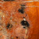
Auch Früchte können befallen werden, wobei der Pilz zumeist an der Narbe der Blütenansatzstelle, der Stelle, wo die Reste der Blüte an der Frucht verbleiben, eintritt. In fortgeschrittenem Stadium der Frucht sieht die Spitze zusammengezogen aus. Beim Aufschneiden von der Spitze her ist der Fruchtkern hell- bis dunkelbraun verfärbt. Später schrumpft die Spitze vollständig ein. Auch dort bilden sich sehr viele Pyknidien, was die Fruchtspitze schwarz aussehen lässt.[13] Auch nach der Ernte kann sich der Pilz noch weiterentwickeln und Früchte zerstören. Bei Melonen und Kürbissen bilden sich braune, je nach Sorte und Art teils bis schwarze, wässrige, eingesunken runde Stellen. In diesen können sich in fortgeschrittenem Stadium auch Fruchtkörper, die Pyknidien oder Pseudothecien bilden. Werden die Früchte länger unter feuchten Bedingungen gelagert, kann sich auf den eingesunkenen Stellen (Läsionen) auch weißlicher Mycelrasen bilden.[14] Der Pilz bildet teilweise konzentrisch eingesunkene verfärbte runde Flecke. Bei Früchten wird eine Infektion auch als Didymella-Fäule bezeichnet, schon bei beginnenden Symptomen gelten die Früchte als nicht gesund und verdorben.[15]
Der wirtschaftliche Schaden, der durch Didymella bryoniae verursacht wird, ist immens. Im Bundesstaat New York in den Vereinigten Staaten sind etwa 50 % der Wintersquash-Produktion betroffen, was Ernteausfälle von bis zu 75 % bei den betroffenen Kulturen verursacht (Zahlen 2001), obgleich New York nicht in den Subtropen liegt.[16] Im Lower Rio Grande Valley in Texas waren im Jahr 1997 68,4 % der Produktion von Cantaloupe-Melonen von Didymella bryoniae betroffen, was einen wirtschaftlichen Schaden von etwa 15 Millionen US-Dollar nur in diesem kleinen, begrenzten Gebiet verursachte.[17] Für Fidschi, wo ein tropisches Klima herrscht, wurden bei Wassermelonen (Citrullus lanatus) im Jahr 2000 Ernteausfälle von 30 % geschätzt.[18]
In der Landwirtschaft wird Didymella bryoniae mit Fungiziden chemisch bekämpft. Vor allem die Wirkstoffe Trifloxystrobin – ein Strobilurin – und Iprodion kommen hierbei zum Einsatz. Daraus hat sich ein erheblicher Markt entwickelt. Allein Bayer CropScience schätzt einen jährlichen Umsatz von 300 Millionen Euro mit Trifloxystrobin-Präparaten.[19]
Didymella bryoniae gehört der Gattung Didymella an. Die Gattung ist bislang noch nicht monographisch behandelt worden und sehr unübersichtlich. MycoBank listet über 400 Arten in der Gattung, von denen aber viele umstritten sind.[20]
Die Position der Gattung Didymella innerhalb des taxonomischen Systems ist bewegt. Ursprünglich bildeten die Gattungen Didymella und Mycosphaerella die Familie der Mycosphaerellaceae. Danach wurde die Gattung zunächst zu den Pleosporaceae, dann den Phaeosphaeriaceae oder den Venturiaceae gerechnet. Im Jahr 2007 stellten Forscher die Gattung gar incertae sedis. Erst im Jahr 2009 ergaben molekulargenetische Studien, dass eine neue Familie, die Didymellaceae, die taxonomische Position der Gattung am besten beschreibt.[21] Unumstritten ist jedoch, dass die Art innerhalb der Ordnung der Pleosporales liegt.
Corlett legte 1981 eine Arbeit vor, die verschiedene Didymella nach morphologischen Gesichtspunkten ordnet. Demzufolge sieht Didymella castillejae – eine Art die vor allem Castilleja befällt – Didymella bryoniae am ähnlichsten.[22]
Eine Arbeit aus dem Jahr 2009 die eine DNA-Sequenzanalyse der ribosomalen RNA in zwei Regionen (LSU = 60S und SSU = 40S) auswertet kommt zu folgenden Verwandtschaftsverhältnissen:
Didymella bryoniae
Da Didymella bryoniae näher zu diesen Arten als zu anderen Arten der Gattung Didymella steht, scheint klar, dass Didymella paraphyletisch ist. Eine ältere Studie aus dem Jahr 1999 kommt zu einer nahen Verwandtschaft zu Leptosphaerulina chartarum und Leptosphaerulina crassiasca.[23] Diese nahe Verwandtschaft wird in der Studie von 2009 bestätigt.
Die Anamorphe der Art wurde lange vor der Teleomorphe bereits im Jahr 1823 durch Elias Magnus Fries in seiner Systema mycologicum als Sphaeria cucurbitacearum erstbeschrieben. Pier Andrea Saccardo stellte die Art im Jahr 1884 dann korrekt in das Formtaxon Phoma.
Die Erstbeschreibung der Teleomorphe geht auf Bernhard Auerswald im Jahr 1869 zurück. In seinem Werk Mycologia europaea. Abbildungen sämmtlicher Schwämme Europas, an dem er zusammen mit Ludwig Rabenhorst geschrieben hat und das mit sehr aufwändigen Lithographien von Wilhelm Gonnermann illustriert ist, beschrieb er die Art als Sphaerella bryoniae. Das Artepipheton bryoniae wählte er nach der Wirtspflanze einer Zaunrübe (Bryonia sp.) von der er den Typus isoliert hatte.
Nur ein Jahr später beschrieb Leopold Fuckel die gleiche Art als Sphaeria bryoniae in den Jahrbüchern des Nassauischen Vereins für Naturkunde. Im Jahr 1880 erkannte Heinrich Rehm jedoch richtig, dass die Art der Gattung Didymella zuzuschlagen ist.[24]
Die Informationen dieses Artikels entstammen, wenn nicht anders angegeben, den unter Literatur bezeichneten Quellen:
Didymella bryoniae ist ein mikroskopisch kleiner Schlauchpilz (Ascomycota) aus der Familie der Didymellaceae. Der pflanzenpathogene Pilz ruft bei verschiedenen Kulturpflanzen die sogenannte Gummistängelkrankheit hervor. Mitunter wird die Krankheit auch als Stängelbrand bezeichnet. Die heute weltweit verbreitete Art befällt vor allem Kürbisgewächse (Cucurbitaceae) wie Kürbisse, Gurken und Melonen und richtet dadurch bedeutenden ökonomischen Schaden an.
Didymella bryoniae, syn. Mycosphaerella melonis, is an ascomycete fungal plant pathogen that causes gummy stem blight on the family Cucurbitaceae (the family of gourds and melons), which includes cantaloupe, cucumber, muskmelon and watermelon plants.[1][2][3][4] The anamorph/asexual stage for this fungus is called Phoma cucurbitacearum.[2] When this pathogen infects the fruit of cucurbits it is called black rot.[2]
The first symptoms appear as grayish green, circular spots between the veins of the leaf lobes.[1] With age these spots darken to brown and black.[1][2] Lesions begin to develop on vines at the vine nodes and then elongate into water-soaked streaks, and these streaks are pale brown at first but turn gray with time.[1] The petioles and stems eventually become necrotic and often shrivel. Eventually all infected vines will become necrotic and occasionally the plant dies due to wilting and defoliation.[1][5] Another common sign following the stem lesions is a red to amber colored ooze.[6] Some regions report the presence of small pseudothecia as black specks inside the cankers.[7]
Gummy stem blight can be confused with anthracnose, which is caused by a fungal plant pathogen called Colletotrichum lagenarium.[1] To distinguish between anthracnose and gummy stem blight, gummy stem blight leaf lesions are darker, target-like and less deteriorated than anthracnose lesions.[1] Newly infected plants will begin to show symptoms within 7–12 days.
In vitro, the fungal growth on an agar plate looks rough and undulated.[5] When grown in vitro on agar, the fungus produces a white to olive-colored mycelium. In latter periods of growth, the mycelium is an olive to dark green or black color.[5]
Didymella bryoniae survives on deceased vines, crop debris and on seeds in between seasons and D. bryoniae can survive for 5 months on the soil surface in winter.[2][4] The fungus develops best under moist conditions, and cotyledons and young watermelon/melon leaves are especially susceptible to the fungus.[2] D. bryoniae produces ascospores (meiotic spores) in perithecia and conidia (mitotic spores) in pycnidia and both of these spores are dispersed by rain/rain-splash and UV light is needed in order for the fungus to sporulate.[3] Ideal ascospore dispersal occurs after nightly rainfall and dew periods.[2] In order to infect, ascospores must land on leaves that have free-standing water on them.[2] Next the ascospores penetrate through the leaf cuticle.[2] Stems may be infected by D. bryoniae ascospores through stem wounds or by the extension of leaf lesions.[2] Fruits are penetrated through wounds and pollination flower scars.[2] Conidia are produced on the lesion sites of leaves and stems. Certain Cucurbita species are resistant to D. bryoniae but become vulnerable once they mature.[2]
Didymella bryoniae is common in the Southern U.S. and other subtropical or tropical locations.[2] Most infections occur during rainy/wet seasons, in which the humid is greater than 90% and the temperature is roughly 20–24 °C.[8] Humidity seems to be a larger factor than temperature when it comes to infection success.[2] D. bryoniae can also be found in temperate regions, especially where winter squash and pumpkins are grown.[2] This pathogen is also common in greenhouses where cucumbers are grown.[2]
D. bryoniae can be spread by the transfer of conidia through a variety of fashions. The most common forms of transfer for these conidia are through the air and water splashing. The fungus is capable of surviving in dead plant tissue giving it the ability to infect the following crop planting.[9] The pathogen requires an entry site on the plant in order to infect so areas that also experience issues with pests are at higher risk.
In vitro, D. bryoniae does not form pycnidia without UV-light but if cultured in the presence of UV light and darkness, conidia/pycnidiospores produce mycelium rapidly.[10]
The standard management practice for D. bryoniae is to use pesticide treated/pathogen-free seeds and to rotate crops on a 2-year cycle to reduce inoculum prevalence.[2] There are no commercially acceptable resistant cucumbers, melons or watermelons available yet on the market, but some plant breeders have identified D. bryoniae resistant genes, such as the gene db in watermelon.[2][11] Regular benzimidazole fungicide applications can control this pathogen, but certain D. bryoniae isolates have been found to be resistant to benzimidazole fungicides in greenhouse settings and in the field.[2]
Along with fungicides, it is important to have proper ventilation and irrigation practices in greenhouse settings.[2] Proper irrigation and ventilation can be utilized to prevent water buildup on leaves.[2] Also to prevent disease onset in greenhouse settings, use UV-absorbing vinyl film, to prevent fungal sporulation.[12]
Currently cultural practices and fungicides work well in greenhouses and in the field only if D. bryoniae is diagnosed in the early stages of disease development.[4] Molecular tools such as polymerase chain reaction (PCR), PCR-enzyme-linked immunosorbent assay and magnetic-capture hybridization multiplex real-time PCR are used to diagnose D. bryoniae in the early stages disease development, although these molecular tools may only be useful for specific isolates of D. bryoniae.[4][13][14]
The United States consumed 15.69 pounds of watermelon per capita in the year 2018 after a rise in both total imports and locally produced watermelons.[15] Florida and Georgia characterized 35 isolates of Didymella and phoma spp. Associated with symptoms of gummy stem blight on watermelon. These two states produced 42% of the United States total watermelon value in 2013, and a combined 20,000 hectares in total farm area. Florida alone produced 907 million pounds of watermelon in 2019[16] meaning that this pathogen could have a direct effect on at least 25% of domestic watermelon crop in the United States.
Didymella bryoniae, syn. Mycosphaerella melonis, is an ascomycete fungal plant pathogen that causes gummy stem blight on the family Cucurbitaceae (the family of gourds and melons), which includes cantaloupe, cucumber, muskmelon and watermelon plants. The anamorph/asexual stage for this fungus is called Phoma cucurbitacearum. When this pathogen infects the fruit of cucurbits it is called black rot.
Didymella bryoniae je grzib[16], co go nojprzōd ôpisoł Karl Wilhelm Gottlieb Leopold Fuckel, a terŏźnõ nazwã doł mu Rehm 1881. Didymella bryoniae nŏleży do zorty Didymella, rzyndu Pleosporales, klasy Dothideomycetes, grōmady Ascomycota i krōlestwa grzibōw.[17][18] Żŏdne podgatōnki niy sōm wymianowane we Catalogue of Life.[17]


Didymella bryoniae je grzib, co go nojprzōd ôpisoł Karl Wilhelm Gottlieb Leopold Fuckel, a terŏźnõ nazwã doł mu Rehm 1881. Didymella bryoniae nŏleży do zorty Didymella, rzyndu Pleosporales, klasy Dothideomycetes, grōmady Ascomycota i krōlestwa grzibōw. Żŏdne podgatōnki niy sōm wymianowane we Catalogue of Life.
Phoma cucurbitacearum je porostyt[5], co go nojprzōd ôpisoł Elias Fries, a terŏźnõ nazwã doł mu Pier Andrea Saccardo 1884. Phoma cucurbitacearum nŏleży do zorty Phoma, rzyndu Pleosporales, klasy Dothideomycetes, grōmady Ascomycota i krōlestwa grzibōw.[6][7] Żŏdne podgatōnki niy sōm wymianowane we Catalogue of Life.[6]
Phoma cucurbitacearum je porostyt, co go nojprzōd ôpisoł Elias Fries, a terŏźnõ nazwã doł mu Pier Andrea Saccardo 1884. Phoma cucurbitacearum nŏleży do zorty Phoma, rzyndu Pleosporales, klasy Dothideomycetes, grōmady Ascomycota i krōlestwa grzibōw. Żŏdne podgatōnki niy sōm wymianowane we Catalogue of Life.


