-
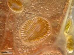
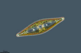
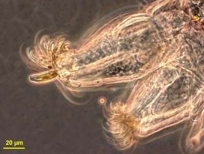
A habitat of Cocconeis scutellum on the red alga Polysiphonia. Collected from Bodden, the brackish waters lying between the isles of Hiddensee and Ruegen (German Baltic Sea). This image was taken using Zeiss Universal with Olympus C7070 CCD camera.
-
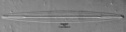
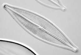
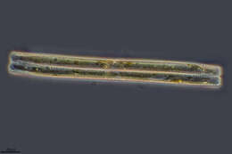
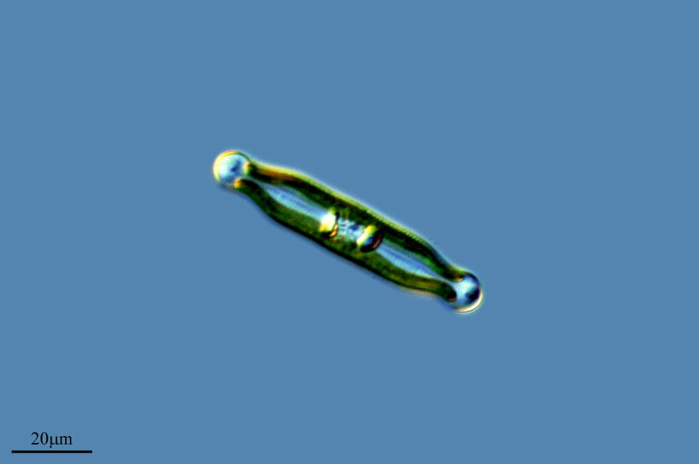
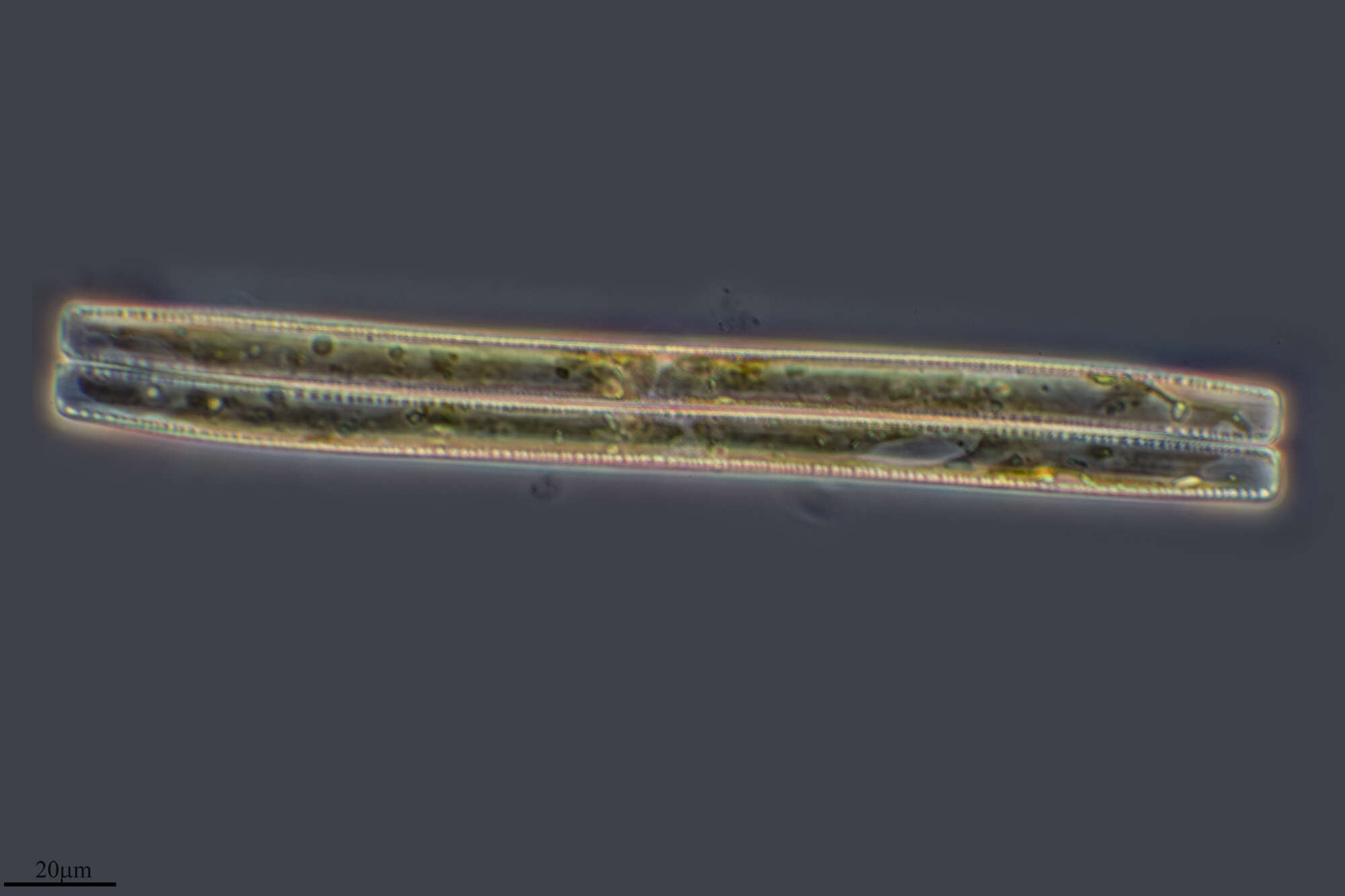
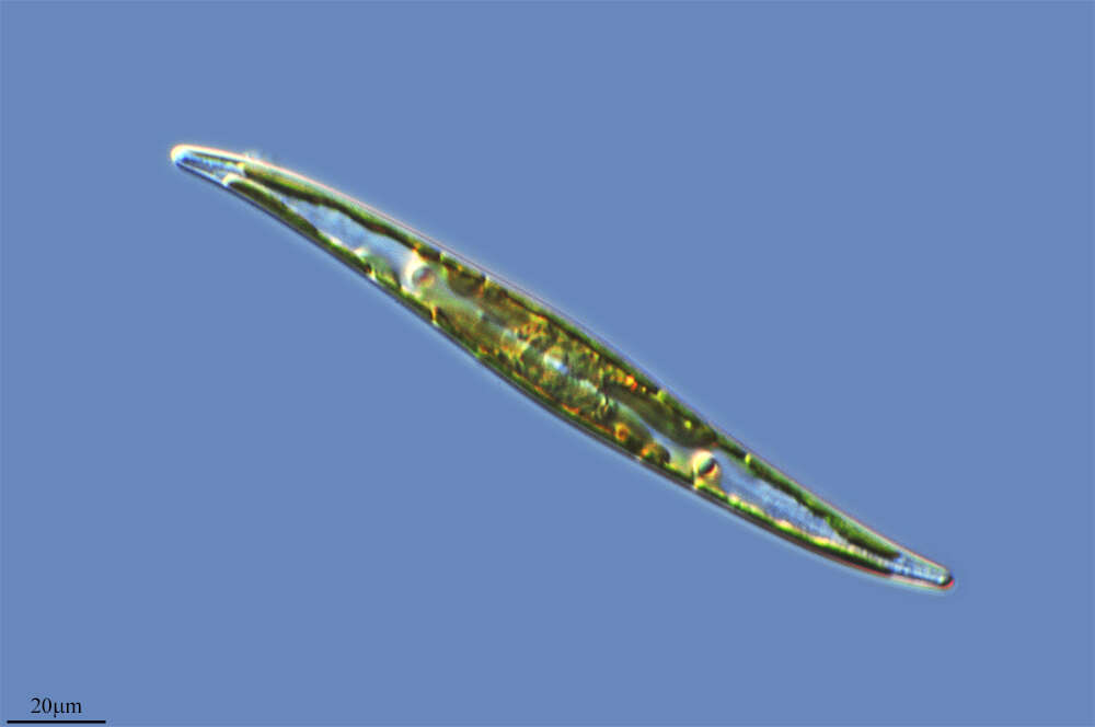
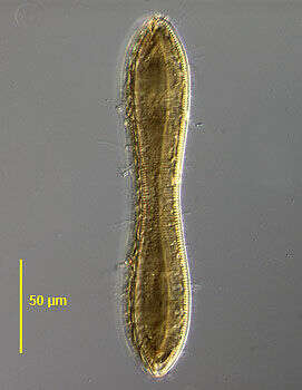
This image of Amphipleura pellucida was taken using an Olympus SPlanapo 100X/1.40, Zeiss 1.40 achro plan condenser, DIC, Wratten 47 deep blue filter, The Imaging Source 1024X768 digital camera, mosaic of 4 images (every image is an average of 32 frames in order to reduce noise). Software Panorama Maker 3.0 and Photoshop.
-
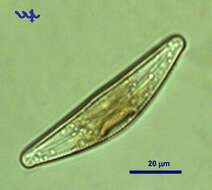
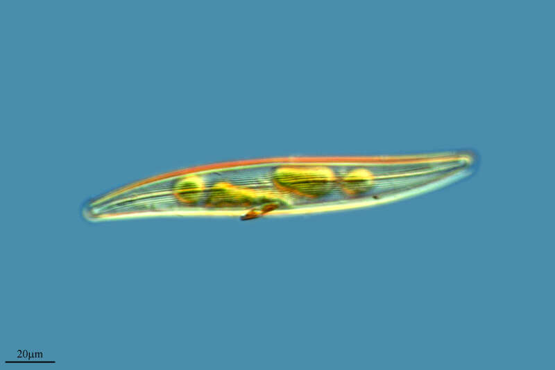
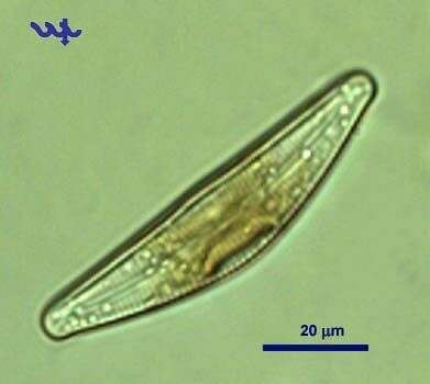
Cymbella (sim-bell-a) a pennate diatom with a slightly asymmetric body form, one face convex, other face flat. Differential interference contrast. This one stuck on the toe of a chironomid larva. Phase contrast.
-
Ajamil, La Rioja, Spain
-
Ribadelago, Castille and Leon, Spain
-
Luanco, Asturias, Spain
-
Villoslada de Cameros, La Rioja, Spain
-
Ribadelago de Franco, Castilla y Len, Espaa
-
Barcelona, Catalunya, Espaa
-
Luanco, Asturias, Spain
-
S Pedro, Galicia, Spain
-
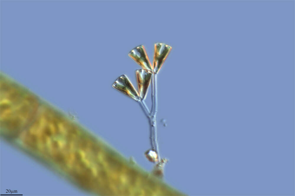
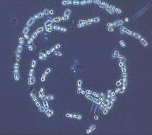
Fig 3: Part of a live colony showing some cells and a strand of several long setae (arrow) connecting different chains in the colony
-
Craticula cuspidata is regarded as the senior synonym of Navicula cuspidata, under which name this asset was submitted. The change of name has been made by the micro*scope team.
-
-
This pennate diatom usually occurs in the benthos as epilithic on stones of Lake Kinneret, infrequently it is found in the plankton.
-
Torrelles de Foix, Catalonia, Spain
-
Hoyo de Manzanares, Madrid, Spain
-
Ribadelago de Franco, Castille and Leon, Spain
-
Alcollarin, Extremadura, Spain
-
Madrid, Madrid, Spain
-
Campillos, Andaluca, Espaa
-
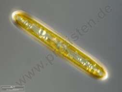
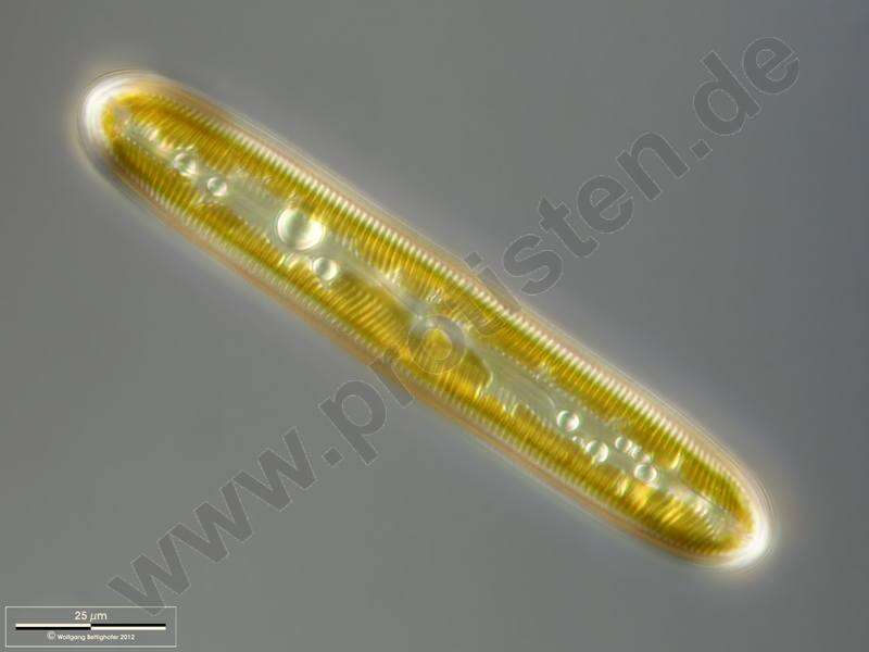
Pinnularia viridis.Valvar view. Scale bar indicates 25 m.Sample from a wetland at the Pillersee (Tyrol, Austria). The image was built up using several photomicrographic frames with manual stacking technique. Images were taken using Zeiss Universal with Olympus C7070 CCD camera.For more look at
www.protisten.de/english/gallery_main/gallery_main.htmlFor high-resolution images please ask postmaster@protisten.de.
-
Fig 6: Large colony of C. socialis cells under light microscope.
-
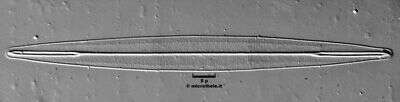
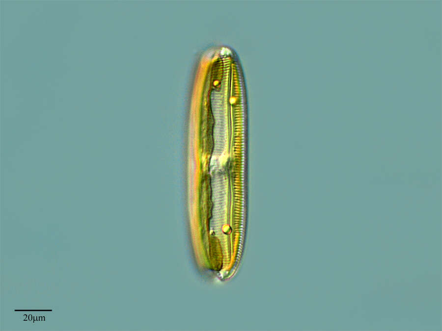
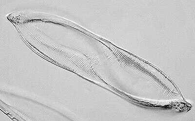
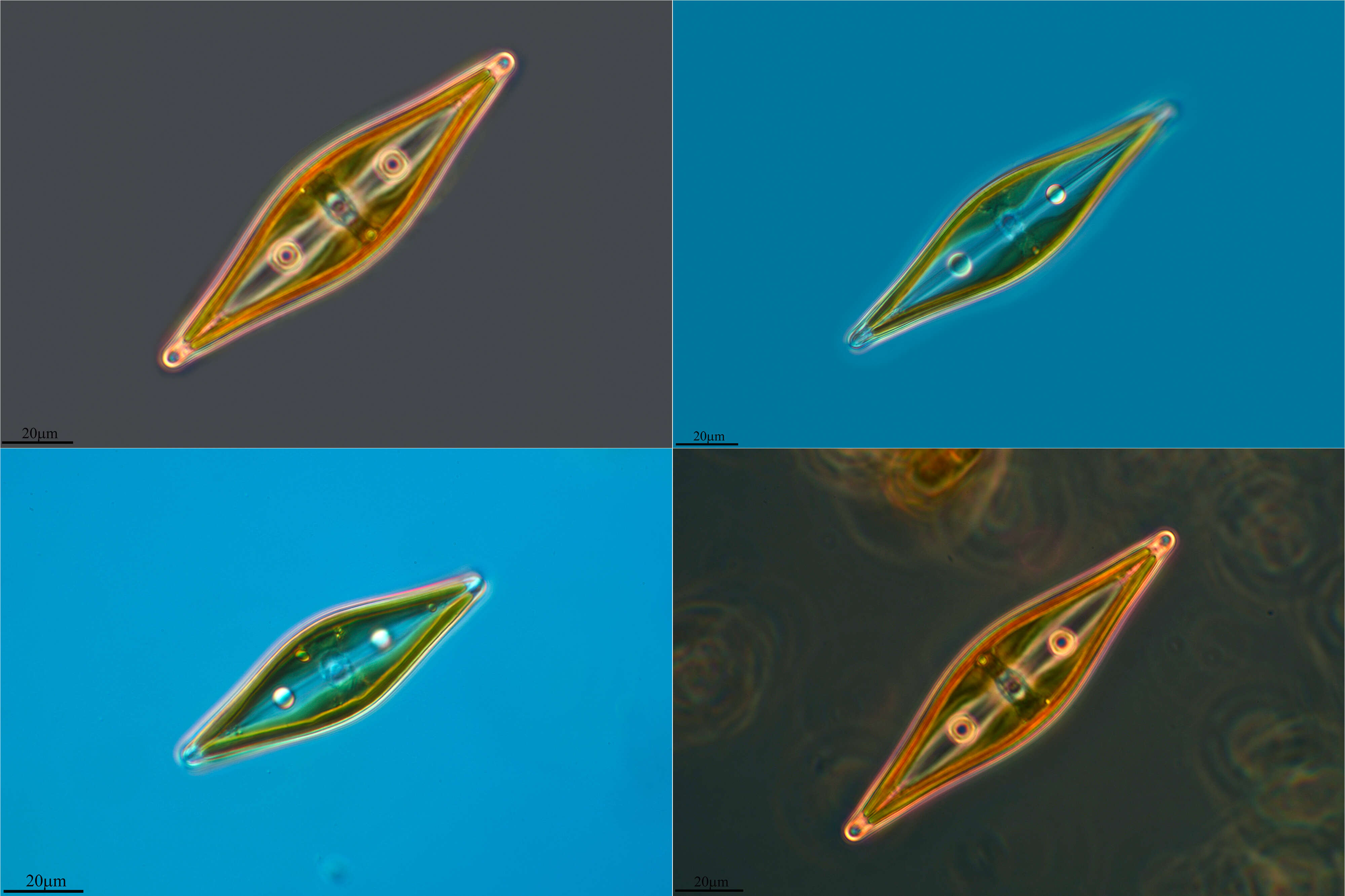
Valve view of the pennate diatom, Cymatopleura solea (Brébisson) W. Smith 1851. Valves are elliptical with a central waist and bluntly pointed apices. The valve surface is marked with several transverse undulations best seen in girdle view; striae and undulations are not interrupted in the median axis. Raphe are marginal. Very short costations appear as beading around the edge of the valve. There is a single lobulated plastid one half of which lies along the inner surface of the epivalve and the other along the inner surface of the hypovalve. Collected from the benthos of a freshwater pond near Boise, Idaho, January 2005. DIC.