Maria Cleide de Mendonça, Eduardo A. Abrantes, Ana Carolina R. Neves
Zookeys
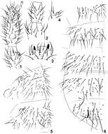
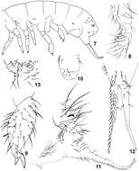
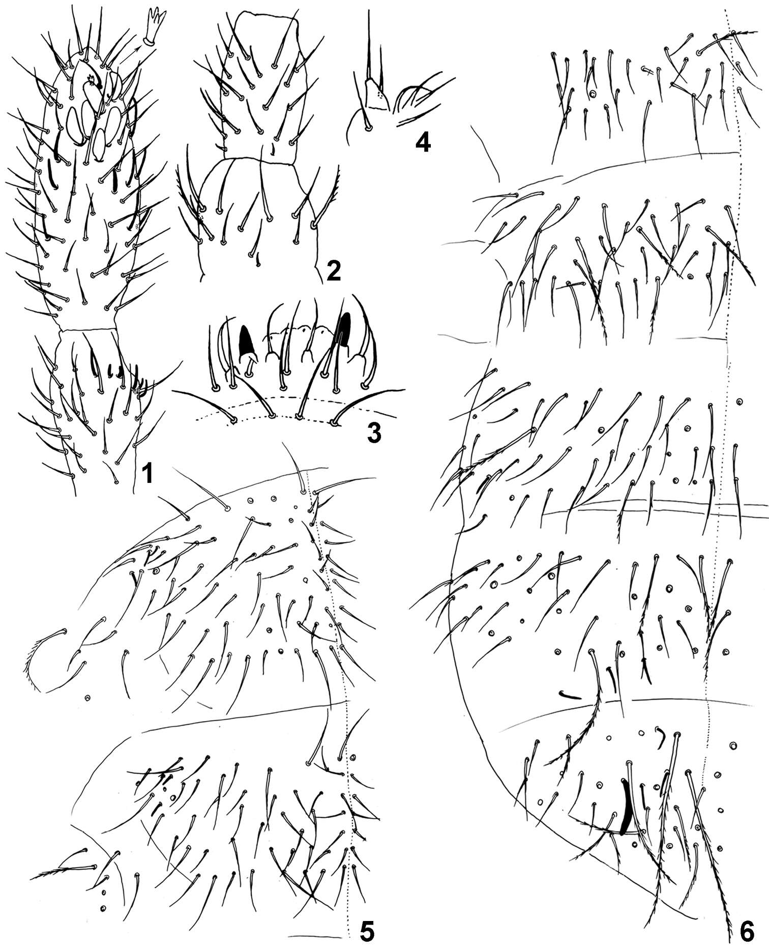
Figure 1–6.Isotomiella macedoi sp.n. 1 Ant III-IV Dorsal view, detail of the apical microsensillum 2 Ant I-II Dorsal view 3 Labral and prelabral chaetae 4 Outer lobe of maxilla 5 Dorsal chaetotaxy of Th II-III 6 Dorsal chaetotaxy of Abd I-VI.
Sopark Jantarit, Chutamas Satasook, Louis Deharveng
Zookeys
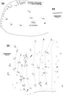
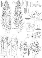
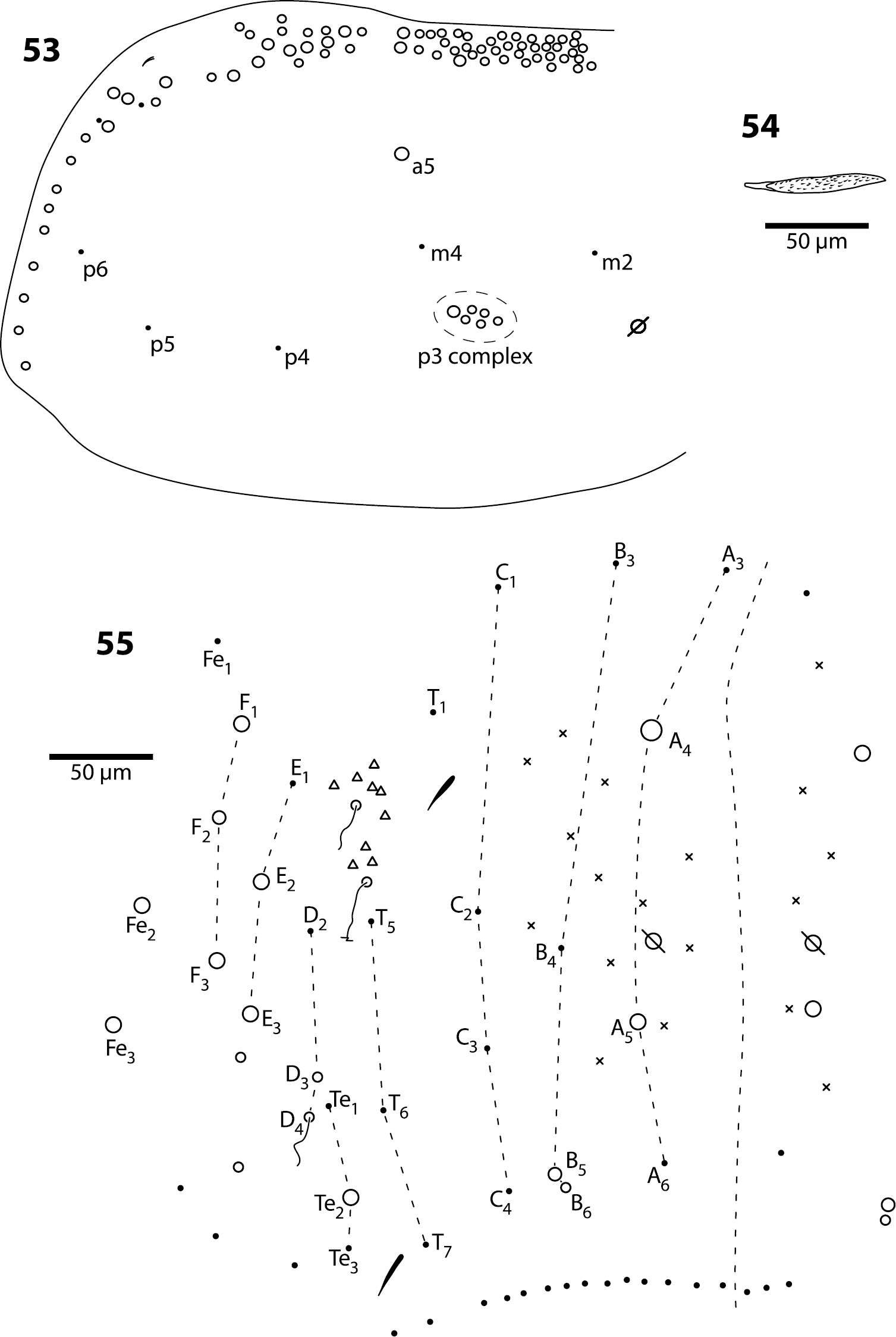
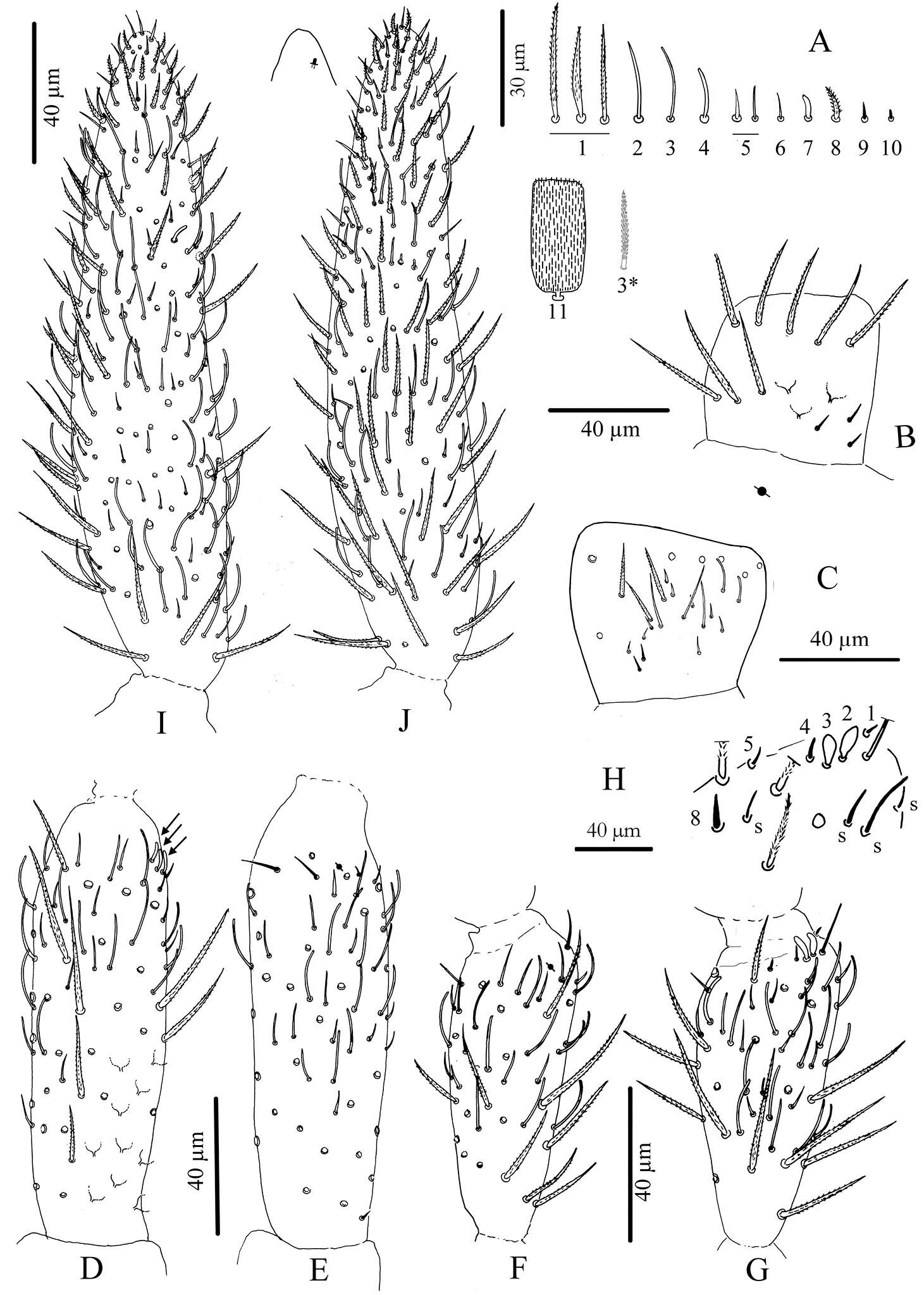
Figure 3.Cyphoderus songkhlaensis sp. n. continued A chaetae of antenna drawn from optical microscope, except 3* derived from SEM image B dorsal side of right Ant.I C ventral side of right Ant.I D dorsal side of right Ant.II; the apical swollen sens of type-7 are indicated by arrows E ventral side of right Ant.II with apical pseudopore F ventral side of right Ant.III with apical pseudopore G dorsal side of right Ant.III H distal organite of Ant.III I ventral side of Ant.IV J dorsal side of Ant.IV with separate view of the subapical organite (left).
Maria Cleide de Mendonça, Eduardo A. Abrantes, Ana Carolina R. Neves
Zookeys
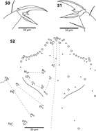
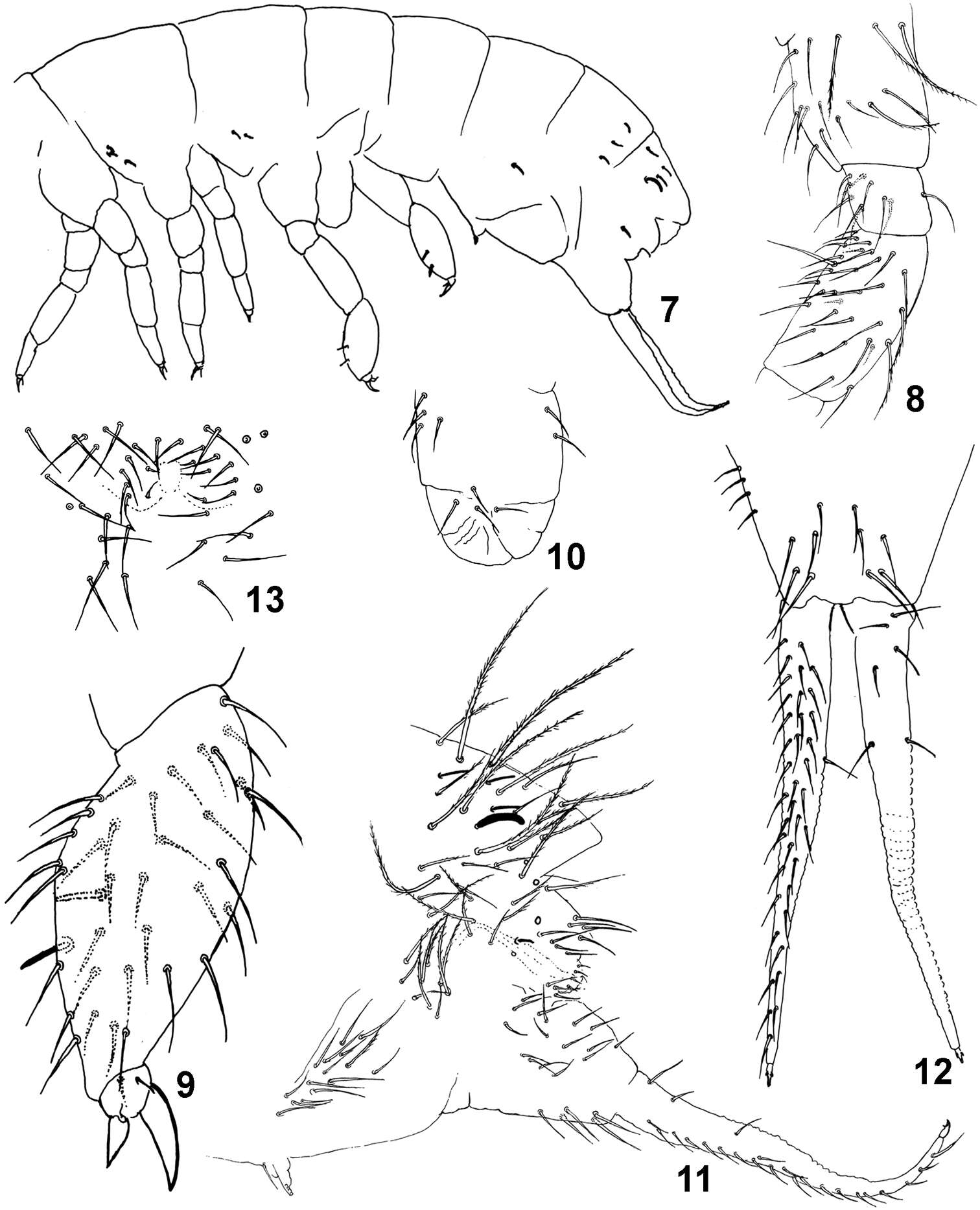
Figure 7–13.Isotomiella macedoi sp.n. 7 Sensillary pattern of the body 8 Subcoxa and femur of leg III 9 Tibiotarsus and unguis of leg III 10 Lateral view of ventral tube 11 Lateral view of abd. V-VI, subcoxa furcal, furca and tenaculum 12 Furca 13 Male genital plate.