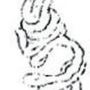
I have once obtained a very slender nemertean among sand and gravel on seashore in Enoura Bay, Shizuoka Prefecture. The worm was so soft and liable to cut that I could not obtain a complete specimen. Later, I found the same species among sand and gravel in several places along the coast of Tateyama Bay, Bohshuu [Chiba Prefecture], and finally collected a large number of complete specimens. When extended, the length reaches up to 200 mm, while the width does not reach 1 mm in its broadest portion. When fully extended, the worm may be much more slender. The head is dorsoventrally flattened, oval-shaped, and gradually leads to the oesophagus region. The oesophagus region is rather cylindrical. Both the regions are cream-white and translucent. The intestinal region is several times longer than the former two regions altogether, posteriorly tapered gradually. When the posterior part of the intestinal region is fully extended, it looks like an extremely thin, colorless fiber. When the worm is on sand, it is sometimes difficult to recognize its existence. Most part of the intestinal region may be dark yellow or dark brown, depending upon its contents. The posterior tip of the body is almost colorless for several centimeters. The mouth is a small pit, which is situated on the ventral surface of the posterior head region; the proboscis pore opens to the tip of the head.
The body wall consists of the epidermis, the dermis, the circular muscle layer, and the longitudinal layer. The interiors of them are the rhynchocoel and the alimentary canal, which are arranged dorsoventrally and run longitudinally. The central nervous system is situated between the dermis and the circular muscle layer. The brain is composed of a pair of nervous hemispheres, which consist of the proximal fiber cords and the neural cell layer that enclose the former. Each nervous hemisphere is further divided into dorsal and ventral ganglia. This division, however, is incomplete, i.e., the neural cell layer is continuous anywhere, and only the fibrous core is divided in its posterior region. The dorsal ganglion suddenly ends before it projects its fibrous cord to the cerebral sensory organ, whilst the ventral ganglion leads to the lateral nerve cord, which runs on each side of the body. In their anterior end, the nervous hemispheres are connected each other by the dorsal and ventral commissures, forming a cerebral ring. The rhynchocoel passes through this ring backwards. Both the commissures are situated between the dermis and the circular muscle layer.
The epidermis is thick, reaching up to half the thickness, or more, of the body wall. It is provided with ciliated cells and numerous unicellular gland cells. Beneath the epidermis is the thin dermis, which is composed of a connective tissue. The circular muscle layer is thin, the longitudinal muscle layer is several times thicker than the circular muscle layer. In the genus Hubrechtia, which resembles with the present species, the longitudinal muscle layer is present. The present species, however, completely lacks it.
Generally, nemerteans possess an excretory system. To the extent hitherto known, a pelagic form, Pelagonemertes, surely lacks the system. Until recently, the genus Cephalothrix had been thought to possess no excretory system. Some years ago, however, it was clarified by Wijnhoff’s study that this genus possesses an incomplete excretory system. Generally, the excretory system is situated on either side the of the oesophagus inside the body wall in the anterior body region, consisting of the collecting tubules and numerous branches that are derived from the collecting tubules. The collecting tubules are provided with some efferent ducts, which open to the exterior on either side of the body. In Cephalothrix, however, there are no collecting tubules, but only structures that correspond to the branches. The terminals of each structure open to the exterior. These excretory systems are extremely small, appearing only to be minute clumps of cells, thus seem not to have caught researcher’s attention. In the specimens that I collected, there found nothing that would represent an excretory organ, not only in living state but also in microscopic observations. Even cell clumps like in Cephalothrix could not have been found. Thus I believe in the present species there is surely no excretory organ, which, at least, resembles with those previously known.
The digestive tract is situated ventrally to the rhynchocoel, divided into the oesophagus and the intestine. The oesophageal epithelium contains numerous gland cells. The intestine has a general structure, in terms of being long and possessing numerous pairs of lateral diverticula. However, the morphology, in which the hind end of the intestine does not reach the posterior end of the body but ends a little in front of it, has never known except in this species. In a specimen whose length is 200 mm, the intestine is absent for about 10 mm of the posterior end of the body. It slightly resembles with the caudal cirri of Cerebratulus and Micrura in the order Heteronemertini. In both genera, however, the intestinal region and the caudal cirrus can be clearly distinguished, since the posterior end of the intestinal region suddenly narrows to become caudal cirrus. The distinction is apparent especially because their thickness is significantly different when the body contracts. In the present species, however, there is no distinction between the intestinal and non-intestinal regions on the external appearance, merely the intestine ends earlier than in the typical species, the situation is different from the caudal cirrus that is an appendage of the body. Except lacking the intestine, gonads and rhynchocoel, the structure of the non-intestinal region is almost the same as the intestinal region. It is also similar to the structure of caudal cirrus in terms of containing mid-dorsal and lateral blood vessels. Thus, the two structures are essentially not different, and should be considered to be the difference of degree.
The anus opens dorsally in the posterior tip of the intestine, like in Cerebratulus in which the anus opens dorsally in the basis of the caudal cirrus. In the
present species, however, its aperture is not distinct.
The blood vascular system, consisting of one mid-dorsal and two lateral vessels, possesses the identical structure as the general nemertean blood system. The mid-dorsal vessel extends between the rhynchocoel and the alimentary canal, the lateral vessels run laterally to the intestine. In the brain region they run along the rhynchocoel. In the non-intestinal region of the posterior tip of the body, the three vessels expand to fuse into a large lacuna, filling inside the body wall as in the caudal cirrus.
The rhynchocoel is short, only reaching to the anterior intestinal region.
The central nervous system is situated between the dermis and the circular muscle layer, as already written. Then a significant peripheral nervous layer, which is connected to the central nervous system, extends between the dermis and the circular muscle layer along all side of the body. This nerve layer, consisting of nervous cells and nervous fiber, is typical for the present, and related, species. Pre-cerebrally, it is well developed and becomes the same thickness as the body wall muscle layer. Posteriorly, however, it becomes thinner.
The cerebral sensory organs are well developed, forming a structure like those in heteronemerteans. It is retort-shaped, situated inside the body wall and
connected to the inner side of the posterior region of the brain. Its spherically expanded region is situated posteriorly to the brain, entering the swelled lateral blood vessel. The narrower neck region [of cerebral organ] extends between the dorsal and ventral ganglia, then leads to a small pore, which is situated on either side of the posterior head region, and makes a ciliated canal, which drills inside the organ, open to the exterior.
The eyes are absent.
Considering the above described structures, the present species apparently belongs to the order Protonemertini, since the central nervous system is situated between the dermis and the circular muscle layer, and the body wall is composed of the epidermis, dermis, circular and longitudinal muscle layers. In the present species, the cerebral sensory organs are well developed, spherical in shape, situated inside the body wall, and enter the lateral blood vessels; the mid-dorsal vessel is present, besides the lateral vessels. These are the points that differ from other families within this order, but correspond with the diagnosis of the family Hubrechtidae, which was proposed by Mr. Bürger in 1892. Thus the present species clearly belongs to this family. This family is originally established by him on the basis of the observation of the new genus and species, Hubrechtia desiderata Bürger, and has been a monotypic family. The species herein described significantly differs in its points from H. desiderata. So, a new genus is added in this family. That is, I believe that it would be inconvenient to include the present species to the genus Hubrechtia, because in the present form an excretory system nor an inner circular muscle are absent, the intestine does not reach to the posterior end of the body, the eyes are absent, the lateral diverticula are very shallow. Accordingly, I think it would be nice to establish a new genus Coeia in the family Hubrechtidae, and use a specific name Coeia ijimai for this form.