-
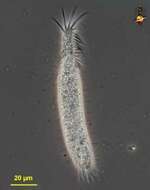
Originally described by Ehrenberg under the name Oxytricha gibba,
-
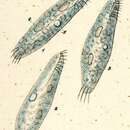
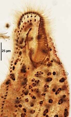
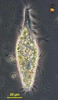
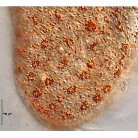
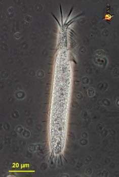
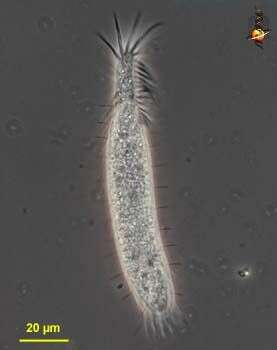
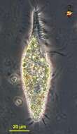
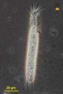
Portrait of Halteria oblonga (Kellicott, 1885; Kahl,1932), a spirotrich ciliate. The body is slightly elongate, rounded posteriorly and truncate anteriorly. There is a prominent anterior wreath of adoral membranelles that winds clockwise into the funnel-shaped peristome (seen in this image). The somatic ciliature is reduced to widely spaced longitudinal files of stout double cilia. The posterior cilia are longer with a characteristic J-shape. These trail behind the organism during swimming, which is accomplished by the rapid beating of the AZM. The single peripheral contractile vacuole is located in the anterior 1/3 adjacent to the oral aperture. The single spheroid granular macronucleus and single round micronucleus are seen posterior to the contractile vacuole. Although Kahl (I. Wimpertiere oder Ciliata. 3. Spirotricha pp. 505-506, Gustav Fischer Verlag, 1932) describes endosymbiotic zoochlorellae, at least some of the algae in these individuals appear to have been ingested. Collected from organically enriched freshwater pond near Boise, Idaho June 2003. DIC optics.
-
Ventral infraciliature of the marine urostylid ciliate, Pseudokeronopsis carnea (Cohn, 1866) Wirnsberger, Larsen & Uhlig, 1987. Collected from a salt water aquarium on the campus of Boise State University, Boise, Idaho. April 2009. Protargol. Brightfield.
-
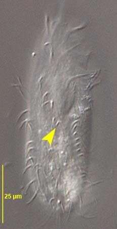
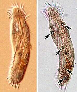
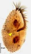
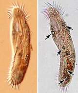
Ventral infraciliature of the oxytrichid,Gonostomum strenuum (ENGELMANN, 1862) STERKI, 1878. G. strenuum differs from G. affine by the greater number of frontoterminal (4-6 vs 2) and frontoventral cirri. In G. strenuum the last frontoventral cirrus is just posterior to the peristome (yellow arrowhead).Collected from a non-flooded Petri dish culture of soil from a park lawn in Boise, Idaho. January 2007.Stained by the protargol technique [Wilbert modification] (see Foissner, W. Europ. J. Protistol., 27:313-330;1991).Brightfield.
-
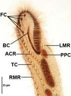
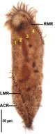
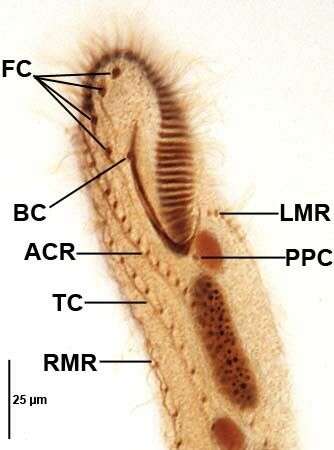
Pseudouroleptus caudatus Hemberger,1985. FC=frontal cirri.BC=buccal cirrus. PPC=postperistomial cirrus. RMR,LMR=right and left marginal cirral rows.ACR=amphisiellid median cirral row. TC=transverse cirral row.Specimen from rewetted soil sample from grass lawn of a public park in Boise,Idaho.January 2007.Protargol impregnation,Wilbert modification (see Foissner, W. Europ. J. Protistol., 27:313-330;1991).Brightfield.
-
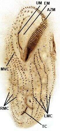
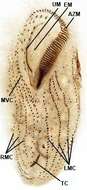
Ventral infraciliature of the large hypotrich ciliate, Urostyla grandis (EHRENBERG,1830). A file of buccal cirri parallels the undulating membrane (UM).Em=endoral membrane.AZM=adoral zone of membranelles.There is a zig-zag file of midventral cirri (MVC) between the right and left marginal cirral rows. There is an obliquely oriented row of about 12 transverse cirri (TC).Collected from a freshwater canal near Boise, Idaho.Stained by the protargol A technique (see Foissner, W. Europ. J. Protistol., 27:313-330;1991).Brightfield.
-
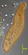
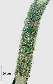
Chaetospira muelleri (LACHMAN,1856). This individual is completely retracted into the the flask-shaped lorica. Collected from tidal pools at Alki Beach, Seattle, Washington 47°35â41.25âN 122°23â19.60âW.January,2006. DIC.
-
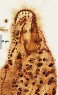
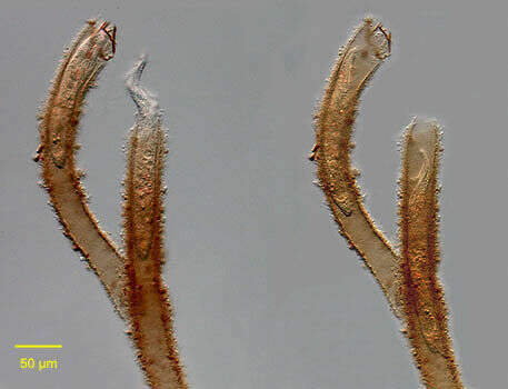
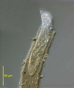
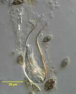
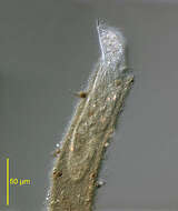
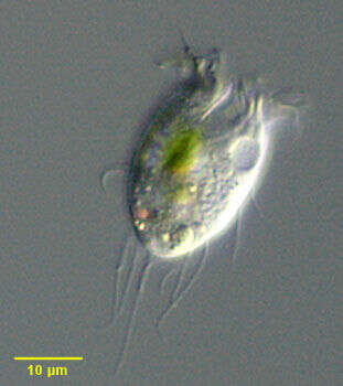
Portrait of the stichotrichine ciliate, Chaetospira remex (Hudson,1875; Kahl,1932). This species occupies a long, sometimes branched tubular lorica into which it intermittently retracts (as seen in this image). The lorica is attached to the substratum. C. muelleri has a flask-shaped lorica. The cell body is slender,elongate and very contractile. The corkscrew shaped anterior bears a prominent adoral zone of membranelles along the peristome. The somatic ciliature is reduced to right and left marginal and two ventral files of short cirri which spiral down the body. The macronucleus is bipartite. the contractile vacuole is in mid-body between the two macronuclei. Feeds mainly on bacteria, flagellates and diatoms. Collected from a freshwater pond near Boise, Idah May 2004. DIC optics.
-
Ventral infraciliature of the marine urostylid ciliate, Pseudokeronopsis carnea (Cohn, 1866) Wirnsberger, Larsen & Uhlig, 1987. Collected from a salt water aquarium on the campus of Boise State University, Boise, Idaho. April 2009. Protargol. Brightfield.
-
Ventral infraciliature of the oxytrichid,Gonostomum strenuum (ENGELMANN, 1862) STERKI, 1878. G. strenuum differs from G. affine by the greater number of frontoterminal and frontoventral cirri. In G. strenuum the last frontoventral cirrus is just posterior to the peristome (yellow arrowhead).Collected from a non-flooded Petri dish culture of soil from a park lawn in Boise, Idaho. January 2007.DIC.
-
Dorsal infraciliature of Pseudouroleptus caudatus (HEMBERGER,1985) .RMR,LMR right and left marginal cirral rows.ACR=amphisiellid median cirral row. 1-4+dorsal kineties.Specimen from rewetted soil sample from grass lawn of a public park in Boise,Idaho.January 2007.Protargol impregnation,Wilbert modification (see Foissner, W. Europ. J. Protistol., 27:313-330;1991).Brightfield.
-
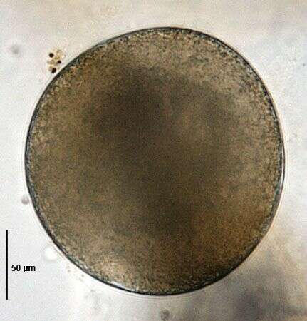
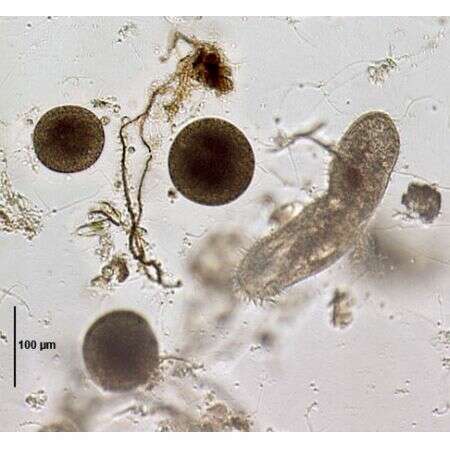
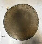
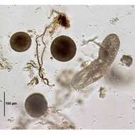
Resting cyst of Urostyla grandis (EHRENBERG,1830). Brightfield.
-
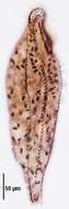
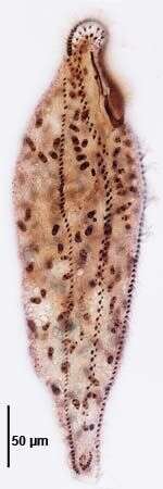
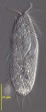
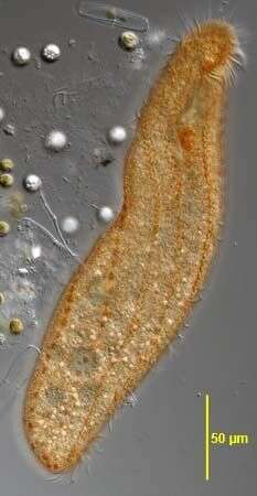
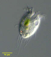
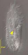
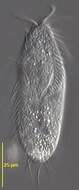
Trachelostyla (track-ell-owe-stike-a) (tentative identification) is one of a large number of stichotrichine hypotrich ciliates, and the genera can only be properly distinguished by careful mapping of the the distribution of the cirri - an exercise which requires special preparation of the cell. Trachelostyla is one of several genera which can have a narrowed front end bearing the adoral zone of membranelles. Phase contrast.
-
Portrait of the stichotrichine ciliate, Chaetospira remex (Hudson,1875; Kahl,1932). Slightly squashed. This species occupies a long, sometimes branched tubular lorica into which it intermittently retracts. The lorica is attached to the substratum. C. muelleri has a flask-shaped lorica. The cell body is slender,elongate and very contractile. The corkscrew shaped anterior bears a prominent adoral zone of membranelles along the peristome (seen well here). The somatic ciliature is reduced to right and left marginal and two ventral files of short cirri which spiral down the body. The left marginal and one of the ventral cirral files are seen here.The macronucleus is bipartite (not visible in this image). the contractile vacuole is in mid-body between the two macronuclei. Feeds mainly on bacteria, flagellates and diatoms. Collected from a freshwater pond near Boise, Idah May 2004. DIC optics.
-
Pigmentocysts (dorsal view) of the marine urostylid ciliate, Pseudokeronopsis carnea (Cohn, 1866) Wirnsberger, Larsen & Uhlig, 1987 clustered around bases of dorsal bristles. Paler, more numerous pigment vacuoles are also visible. Collected from a salt water aquarium on the campus of Boise State University, Boise, Idaho. April 2009. DIC.
-
Ventral view of the oxytrichid,Gonostomum strenuum (ENGELMANN, 1862) STERKI, 1878. G. strenuum differs from G. affine by the greater number of frontoterminal and frontoventral cirri. In G. strenuum the last frontoventral cirrus is just posterior to the peristome .Collected from a non-flooded Petri dish culture of soil from a park lawn in Boise, Idaho. January 2007.DIC.
-
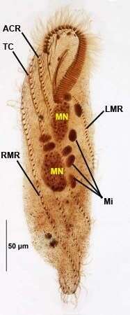
Ventral infraciliature of Pseudouroleptus caudatus (HEMBERGER,1985) .RMR,LMR right and left marginal cirral rows.TC=transverse cirral row.ACR=amphisiellid median cirral row.MN=two macronuclear nodules.Mi=multiple (up to eight) dense micronuclei.Specimen from rewetted soil sample from grass lawn of a public park in Boise,Idaho.January 2007.Protargol impregnation,Wilbert modification (see Foissner, W. Europ. J. Protistol., 27:313-330;1991).Brightfield.
-
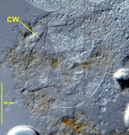
Ruptured resting cyst of Urostyla grandis (EHRENBERG,1830) showing very thin cyst wall (CW). DIC.
-
Trachelostyla (track-ell-owe-style-a), a hypotrich ciliate. With a narrowed anterior end to the body, with an adoral zone of membranelles. Phase contrast micrograph.
-
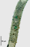
The stichotrichine ciliate, Chaetospira remex (Hudson,1875; Kahl,1932) stained with methyl green-pyronin to demonstrate the bipartite macronucleus. The animal is completely withdrawn into the tubular lorica. The two round macronuclei are stained green. The micronucleus is not seen in this image. The illustration in Kahl's compendium incorrectly depicts C. remex with a single macronucleus (Kahl,A.; Die Tierwelt Deutschlands und der angrenzenden Meeresteile. Teil 25 [Urtiere oder Protozoa I: Wimpertiere oder Ciliata (Infusoria) 3. Spirotricha. Germany:Verlag von Gustav Fischer. 1932, p. 542).Collected from a freshwater pond near Boise Idaho May 2004. Brightfield illumination with closed condenser.
-
In vivo view (right ventrolateral surface) of the marine urostylid ciliate, Pseudokeronopsis carnea(Cohn, 1866) Wirnsberger, Larsen & Uhlig, 1987. Collected from a salt water aquarium on the campus of Boise State University, Boise, Idaho. April 2009. DIC.
-
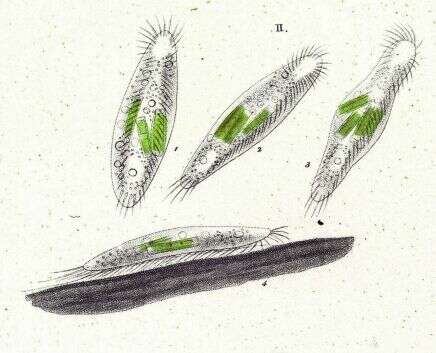
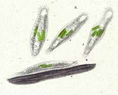
Image from Li et al., 2010. Acta Protozoologica, 49: 195-212.
-
Resting cysts of Urostyla grandis (EHRENBERG,1830). From starved culture.Brightfield.
-
Trachelostyla (track-ell-owe-style-a), a hypotrich ciliate. With a narrowed anterior end to the body, with an adoral zone of membranelles. Phase contrast micrograph.