-
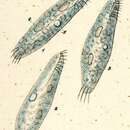
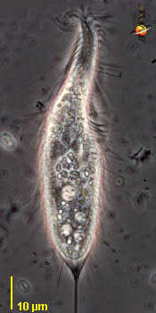
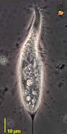
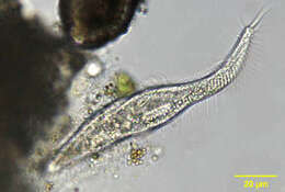
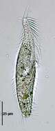
Stichotricha (stike-o-trike-a), un-named species. The body of the unidentified hypotrich ciliate has a broader posterior portion, and a narrowed anterior region. This is a detail of the cell surface showing the numerous long ectosymbiotic bacteria. Phase contrast.
-
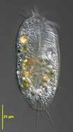
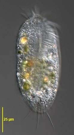
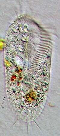
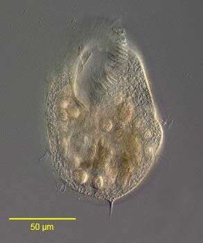
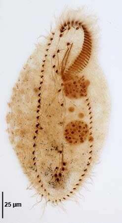
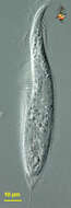
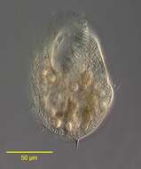
Ventral view of Stylonychia pustulata (MUELLER, 1786) EHRENBERG, 1835. Collected from a eutrophic freshwater pond in Boise, Idaho. June 2008. DIC.
-
Stichotricha (stike-o-trike-a), un-named species. The body of the unidentified hypotrich ciliate has a broader posterior portion, and a narrowed anterior region. The adoral zone of membranelles makes about one complete tour of the anterior part, winding down from the anterior to the cytostome located in the anterior part of the broader part of the cell. There are locomotor cirri over the body (not easily seen here), and also stiff projecting cilia / cirri. Some structures form a bracing structure to support the two very long cilia which extend 50 or 60 microns Phase contrast micrograph.
-
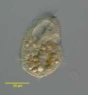
Ventral view of Stylonychia pustulata (MUELLER, 1786) EHRENBERG, 1835. Collected from a eutrophic freshwater pond in Boise, Idaho. January 2001. Oblique illumination.
-
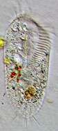
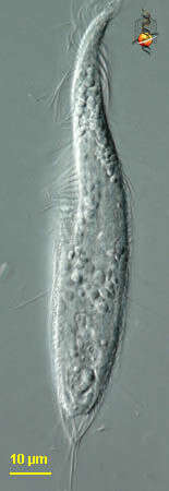
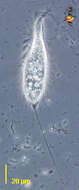
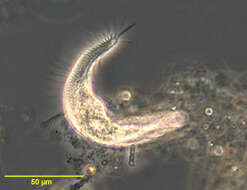
Stichotricha (stike-o-trike-a), un-named species. The body of the unidentified hypotrich ciliate has a broader posterior portion, and a narrowed anterior region. The adoral zone of membranelles can be seen to the right of the anterior-most part of the cell and then to the left near the cytostome located in the anterior part of the broader part of the cell - showing that it makes about one complete turn around the body. There is a sheath of material (ectosymbiotic bacteria?) which supports the caudal cilia. Differential interference contrast.
-
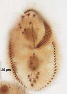
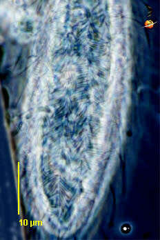
Dorsal infraciliature of Stylonychia pustulata (MUELLER, 1786) EHRENBERG, 1835.1-6+dorsal kineties. Collected from a eutrophic freshwater pond in Boise, Idaho. June 2008. Protargol A (see Foissner, W. Europ. J. Protistol., 27:313-330;1991).Brightfield.
-
Stichotricha (stike-o-trike-a), un-named species. The body of the unidentified hypotrich ciliate has a broader posterior portion, and a narrowed anterior region. The adoral zone of membranelles makes about one complete tour of the anterior part, winding down from the anterior to the cytostome located in the anterior part of the broader part of the cell. This image illustrates the two very long cilia which extend 50 or 60 microns Phase contrast micrograph.
-
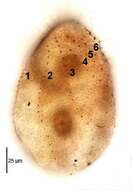
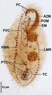
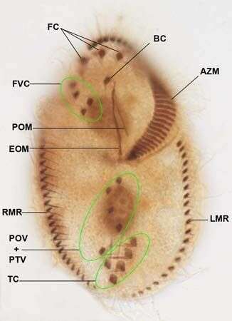
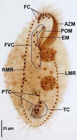
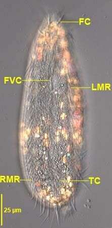
Ventral infraciliature of Stylonychia pustulata (MUELLER, 1786) EHRENBERG, 1835.FC=frontal cirri.FVC=frontoventral cirri.BC=buccal cirrus.AZM=adoral zone of membranelles.EOM=endoral membrane.POM=paroral membrane.RMR,LMR=right and left marginal cirral rows.POv+PTV=postoral and pretransverse ventral cirri.TC=transverse cirri. Collected from a eutrophic freshwater pond in Boise, Idaho. June 2008. Protargol A (see Foissner, W. Europ. J. Protistol., 27:313-330;1991).Brightfield.
-
-
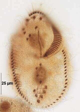
Ventral infraciliature of Stylonychia pustulata (MUELLER, 1786) EHRENBERG, 1835. Collected from a eutrophic freshwater pond in Boise, Idaho. June 2008. Protargol A (see Foissner, W. Europ. J. Protistol., 27:313-330;1991).Brightfield.
-
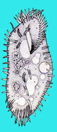
Stichotricha, hypotrich ciliate usually located in a flocculent mucoid mass adhering to submerged vegetation or other debris. The adoral zone of membranelles (AZM) is supported on a narrow and twisted anterior projection. Rows of cirri follow a curving path on the body.
-
-
Stichotricha, hypotrich ciliate usually located in a flocculent mucoid mass adhering to submerged vegetation or other debris. The adoral zone of membranelles (AZM) is supported on a narrow and twisted anterior projection. Rows of cirri follow a curving path on the body. Phase contrast optics.
-
-
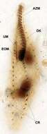
Stichotricha aculeata (WRZEÅNIOWSKI,1866).AZM=adoral zone of membranelles;DK=dorsal kinety;CR=2 of the 4 cirral rows;UM=undulating membrane;EOM=endoral membrane.Protargol protocol A (see Foissner, W. Europ. J. Protistol., 27:313-330;1991).Brightfield.
-
-
Stichotricha aculeata (WRZEÅNIOWSKI,1866).Brightfield,closed condenser.
-
Ventral infracilature of Oxytricha fallax (STEIN,1859).FC=frontal cirri,FVC=frontoventral cirri.PTC=pretransverse ventral cirri,TC=transverse cirri.RMR/LMR=right and left marginal cirral rows,AZM=adoral zone of membranelles,EM=endoral membrane,POM=paraoral membrane.Collected from a eutrophic artificial freshwater pond in Boise, Idaho.May 2008.Stained by the protargol technique (Wilbert modification). See Foissner, W. Europ. J. Protistol., 27:313-330;1991.Brightfield.
-
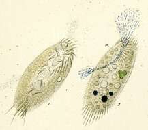
Portrait of the stichotrichine ciliate Hypotrichidium conicum (Ilowaisky, 1921). Collected from a freshwater pond near Boise, Idaho. July 2005. DIC.
-
Ventral infracilature of Oxytricha fallax (STEIN,1859).Collected from a eutrophic artificial freshwater pond in Boise, Idaho.May 2008.Stained by the protargol technique (Wilbert modification). See Foissner, W. Europ. J. Protistol., 27:313-330;1991.Brightfield.
-
in vivo portrait of the stichotrichine ciliate, Hypotrichidium conicum (Ilowaisky, 1921). Collected from a freshwater pond near Boise, Idaho. july 2005. DIC.
-
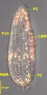
Oxytricha fallax (STEIN,1859).Ventral view.FC=frontal cirrus,RMR/LMR=right and left marginal cirral rows,FVC=frontoventral cirri,TC-transverse cirri.Collected from a eutrophic artificial freshwater pond in Boise, Idaho.May 2008.DIC.
-
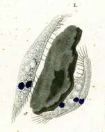
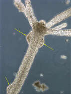
Kerona pediculus (O. F. Muller, 1773) Blochmann, 1886) , a hypotrich ciliate that lives on the surface of freshwater Hydra, and can consume epithelial and other cells from its host. Phase contrast optics.
-
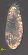
Oxytricha fallax (STEIN,1859).The cytoplasm contains many highly refractile crystals which appear yellow-orange in this image.Under brightfield illumination they appear dark.Collected from a eutrophic artificial freshwater pond in Boise, Idaho.May 2008.DIC.