-
-
-
-
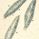
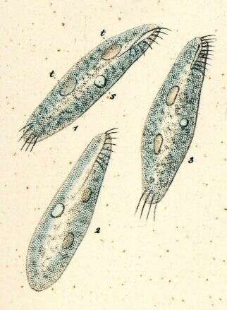
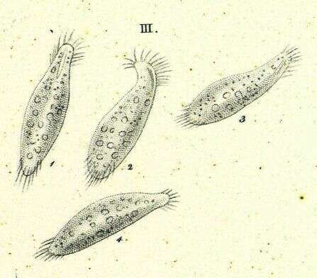
Originally described by Ehrenberg under the name Oxytricha pullaster.
-
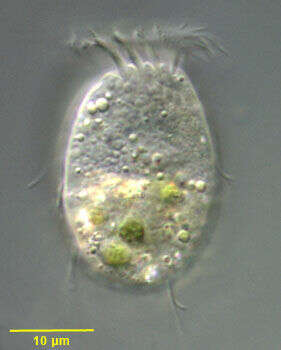
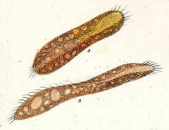
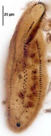
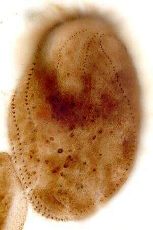
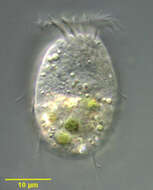
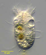
Dorsal view of Halteria oblonga (Kellicott, 1885;Kahl,1932), a spirotrich ciliate. The body is slightly elongate, rounded posteriorly and truncate anteriorly. There is a prominent anterior wreath of adoral membranelles that winds clockwise into the funnel-shaped peristome (seen in this image). The somatic ciliature is reduced to widely spaced longitudinal files of stout double cilia. The posterior cilia are longer with a characteristic J-shape. These trail behind the organism during swimming, which is accomplished by the rapid beating of the AZM. The single peripheral contractile vacuole is located in the anterior 1/3 adjacent to the oral aperture. The single spheroid granular macronucleus and single round micronucleus are seen posterior to the contractile vacuole. Although Kahl (I. Wimpertiere oder Ciliata. 3. Spirotricha pp. 505-506, Gustav Fischer Verlag, 1932) describes endosymbiotic zoochlorellae, at least some of the algae in these individuals appear to have been ingested. Collected from organically enriched freshwater pond near Boise, Idaho June 2003. DIC optics.
-
Originally described by Ehrenberg under the name Oxytricha rubra.
-
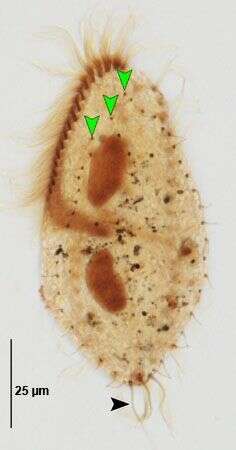
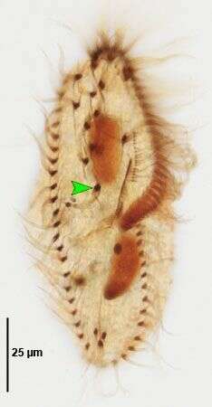
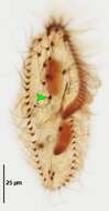
Gonostomum affine (Stein, 1859) Sterki, 1878. Dorsal kineties (green arrowheads) and caudal cirri (black arrowhead). Non-flooded Petri dish soil sample collected from flood-irrigated lawn in Boise, Idaho, June 2008. Protargol (Wilbert modification). Brightfield.
-
-
in vivo portrait (ventral view) of the large hypotrich ciliate, Urostyla grandis (EHRENBERG,1830). Collected from a freshwater canal near Boise, Idaho.Brightfield.
-
-
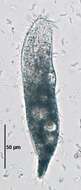
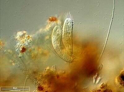
Conjugating couple. Scale bar indicates 50 µm. Collected from Bodden, the brackish waters lying between the isles of Hiddensee and Ruegen (German Baltic Sea). The image was built up using several photomicrographic frames with manual stacking technique. This image was taken using Zeiss Universal with Olympus C7070 CCD camera.
-
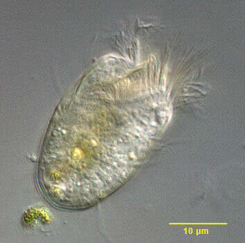
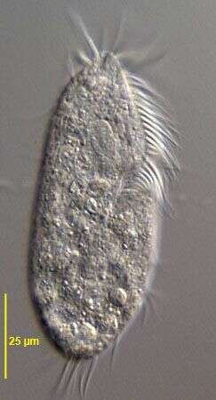
Ventral view of Halteria oblonga (Kellicott, 1885;Kahl,1932), a spirotrich ciliate. The body is slightly elongate, rounded posteriorly and truncate anteriorly. There is a prominent anterior wreath of adoral membranelles that winds clockwise into the funnel-shaped peristome (seen in this image). The somatic ciliature is reduced to widely spaced longitudinal files of stout double cilia. The posterior cilia are longer with a characteristic J-shape. These trail behind the organism during swimming, which is accomplished by the rapid beating of the AZM. The single peripheral contractile vacuole is located in the anterior 1/3 adjacent to the oral aperture. The single spheroid granular macronucleus and single round micronucleus are seen posterior to the contractile vacuole. Although Kahl (I. Wimpertiere oder Ciliata. 3. Spirotricha pp. 505-506, Gustav Fischer Verlag, 1932) describes endosymbiotic zoochlorellae, at least some of the algae in these individuals appear to have been ingested. Collected from organically enriched freshwater pond near Boise, Idaho June 2003. DIC optics.
-
Originally described by Ehrenberg under the name Oxytricha rubra.
-
Gonostomum affine (Stein, 1859) Sterki, 1878. Ventral infraciliature. Green arrowhead marks posterior-most "postoral" cirrus. Non-flooded Petri dish soil sample collected from flood-irrigated lawn in Boise, Idaho, June 2008. Protargol (Wilbert modification). Brightfield.
-
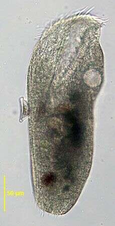
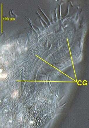
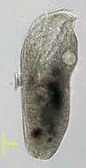
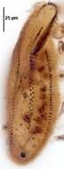
in vivo view of the amphisiellid hypotrich Pseudouroleptus caudatus (HEMBERGER,1985) showing colorless cortical granules (CG).Specimen from rewetted soil sample from grass lawn of a public park in Boise,Idaho.January 2007.DIC.
-
Ventral infraciliature of the large hypotrich ciliate, Urostyla grandis (EHRENBERG,1830). Collected from a freshwater canal near Boise, Idaho.Stained by the protargol A technique (see Foissner, W. Europ. J. Protistol., 27:313-330;1991).Brightfield.
-
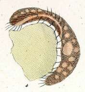
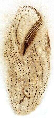
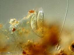
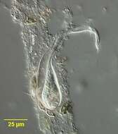
Portrait of Chaetospira, a loricate hypotrich ciliate. Elongate anterior end with prominent adoral zone of membranelles has a corkscrew configuration, distinguishing this genus from the similar Stichotricha. Usually only anterior portion protrudes and organism quickly retracts completely into lorica when disturbed. From freshwater pond near Boise, Idaho. Brightfield.
-
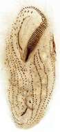
Ventral infraciliature of Anteholosticha monilata (KAHL, 1928)Berger2003. Collected from a freshwater irrigation canal. Boise,Idaho. December 2007.Protargol (see Foissner, W. Europ. J. Protistol., 27:313-330;1991).Brightfield.
-
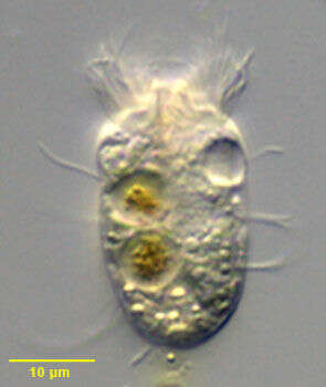
Portrait of Halteria oblonga (Kellicott, 1885;Kahl,1932), a spirotrich ciliate. The body is slightly elongate, rounded posteriorly and truncate anteriorly. There is a prominent anterior wreath of adoral membranelles that winds clockwise into the funnel-shaped peristome (seen in this image). The somatic ciliature is reduced to widely spaced longitudinal files of stout double cilia (seen well in this image). The posterior cilia are longer with a characteristic J-shape. These trail behind the organism during swimming, which is accomplished by the rapid beating of the AZM. The single peripheral contractile vacuole is located in the anterior 1/3 adjacent to the oral aperture. The single spheroid granular macronucleus and single round micronucleus are seen posterior to the contractile vacuole. Although Kahl (I. Wimpertiere oder Ciliata. 3. Spirotricha pp. 505-506, Gustav Fischer Verlag, 1932) describes endosymbiotic zoochlorellae, at least some of the algae in these individuals appear to have been ingested. Collected from organically enriched freshwater pond near Boise, Idaho June 2003. DIC optics.
-
Originally described by Ehrenberg under the name Oxytricha rubra.
-
Gonostomum affine (Stein, 1859) Sterki, 1878. Non-flooded Petri dish soil sample collected from flood-irrigated lawn in Boise, Idaho, June 2008. Protargol (Wilbert modification). Brightfield.
-
in vivo view of the amphisiellid hypotrich Pseudouroleptus caudatus (HEMBERGER,1985) .Specimen from rewetted soil sample from grass lawn of a public park in Boise,Idaho.January 2007.Brightfield.
-
Dorsal infraciliature of the large hypotrich ciliate, Urostyla grandis (EHRENBERG,1830). Collected from a freshwater canal near Boise, Idaho.Stained by the protargol A technique (see Foissner, W. Europ. J. Protistol., 27:313-330;1991).Brightfield.
-
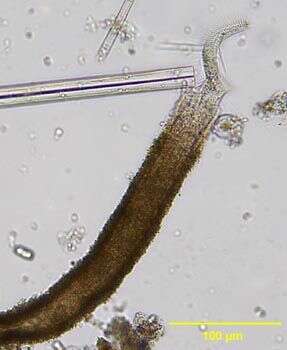
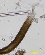
Chaetospira muelleri (LACHMAN,1856). The corkscrew shaped highly contractile anterior end of the cell is seen protriding through the opening of the flask-shaped lorica. Collected from tidal pools at Alki Beach, Seattle, Washington 47°35â41.25âN 122°23â19.60âW.January,2006. DIC.