-
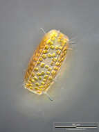
Thalassiosira punctigera The oblique view exhibits short silicous spines, the so called occluded processes. Some chitinous spines protruding from the fultoportulae (also called strutted processes) along the dotted valve margin are also visible. Scale bar indicates 50 µm. The image was built up using several photomicrographic frames with manual stacking technique. Sample from North Sea near Heligoland (spring diatom bloom). Images were taken using Zeiss Universal with Olympus C7070 CCD camera.Image under Creative Commons License V 3.0 (CC BY-NC-SA). Place name: North Sea around Heligoland Latitude: 54.186311 Longitude: 7.895034 Die schräge Sicht auf die Frusteln zeigt kurze Silikatfortsätze, die sogenannten occluded processes. Eine Reihe Schwebefortsätze aus Chitin, die aus den Fultoportulae (kleine Silikatröhren, auch strutted processes genannt) entlang der punktierten Kante der Frustel entspringen, sind sichtbar. Multiebenen-Abbildung, manuell gestapelt. Der Messbalken markiert eine Länge von 50 µm. Probe aus der Nordsee vor Helgoland in der Zeit der Frühjahrsblüte. Mikrotechnik: Zeiss Universal, Kamera: Olympus C7070.Creative Commons License V 3.0 (CC BY-NC-SA). For permission to use of (high-resolution) images please contact postmaster@protisten.de.
-
Thalassiosira punctigera The oblique view exhibits short silicous spines, the so called occluded processes. On the lower left, lower right and central above chitinous spines are visible. Scale bar indicates 50 µm. The image was built up using several photomicrographic frames with manual stacking technique. Sample from North Sea near Heligoland (spring diatom bloom). Images were taken using Zeiss Universal with Olympus C7070 CCD camera.Image under Creative Commons License V 3.0 (CC BY-NC-SA). Place name: North Sea around Heligoland Latitude: 54.186311 Longitude: 7.895034 Die schräge Sicht auf die Frusteln zeigt kurze Silikatfortsätze, die sogenannten occluded processes. Unten links, unten rechts und oberhalb der Mitte sieht man einzelne Schwebefortsätze aus Chitin. Multiebenen-Abbildung, manuell gestapelt. Der Messbalken markiert eine Länge von 50 µm. Probe aus der Nordsee vor Helgoland in der Zeit der Frühjahrsblüte. Mikrotechnik: Zeiss Universal, Kamera: Olympus C7070.Creative Commons License V 3.0 (CC BY-NC-SA). For permission to use of (high-resolution) images please contact postmaster@protisten.de.
-
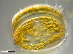
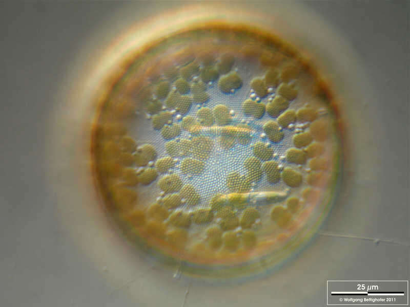
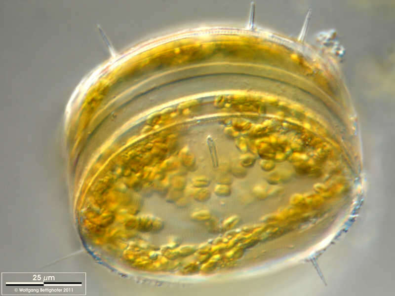
Thalassiosira punctigera Some specimen of this centric diatom carried naviculoid ones on the valve(s). Scale bar indicates 25 µm. The image was built up using several photomicrographic frames with manual stacking technique. Sample from North Sea near Heligoland (spring diatom bloom). Images were taken using Zeiss Universal with Olympus C7070 CCD camera.Image under Creative Commons License V 3.0 (CC BY-NC-SA). Place name: North Sea around Heligoland Latitude: 54.186311 Longitude: 7.895034 Einige Exemplare dieser Diatomeenart in der Probe trugen Navicula-ähnliche Diatomeen auf den Schalen. Multiebenen-Abbildung, manuell gestapelt. Der Messbalken markiert eine Länge von 25 µm. Probe aus der Nordsee vor Helgoland in der Zeit der Frühjahrsblüte. Mikrotechnik: Zeiss Universal, Kamera: Olympus C7070.Creative Commons License V 3.0 (CC BY-NC-SA). For permission to use of (high-resolution) images please contact postmaster@protisten.de.
-
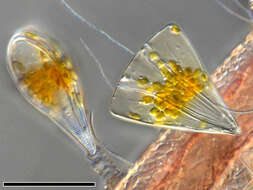
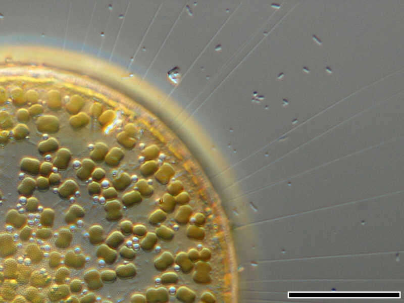
Thalassiosira punctigera The image shows numerous chitinous spines which minimize their sedimentation speed. Scale bar indicates 25 µm. The image was built up using several photomicrographic frames with manual stacking technique. Sample from North Sea near Heligoland (spring diatom bloom). Images were taken using Zeiss Universal with Olympus C7070 CCD camera.Image under Creative Commons License V 3.0 (CC BY-NC-SA). Place name: North Sea around Heligoland Latitude: 54.186311 Longitude: 7.895034 Darstellung der feinen Schwebefortsätze aus Chitin. Multiebenen-Abbildung, manuell gestapelt. Der Messbalken markiert eine Länge von 25 µm. Probe aus der Nordsee vor Helgoland in der Zeit der Frühjahrsblüte. Mikrotechnik: Zeiss Universal, Kamera: Olympus C7070.Creative Commons License V 3.0 (CC BY-NC-SA). For permission to use of (high-resolution) images please contact postmaster@protisten.de.
-
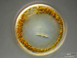
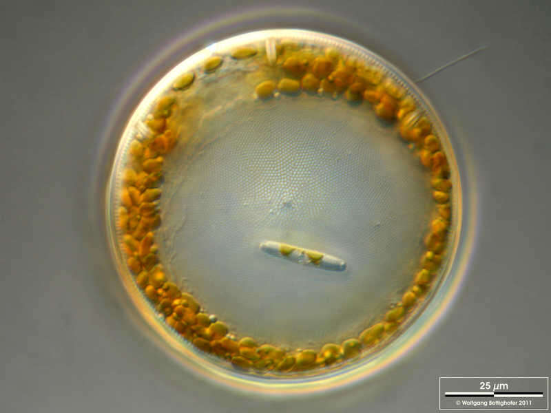
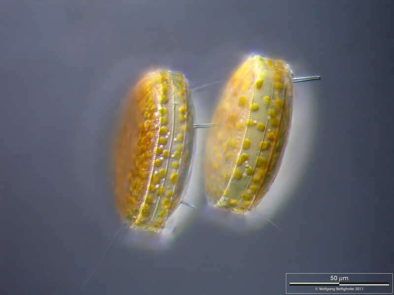
Thalassiosira punctigera The oblique view exhibits short silicous spines, the so called occluded processes. On the lower left near the scale bar a chitinous spine is visible. Scale bar indicates 25 µm. The image was built up using several photomicrographic frames with manual stacking technique. Sample from North Sea near Heligoland (spring diatom bloom). Images were taken using Zeiss Universal with Olympus C7070 CCD camera.Image under Creative Commons License V 3.0 (CC BY-NC-SA). Place name: North Sea around Heligoland Latitude: 54.186311 Longitude: 7.895034 Die schräge Sicht auf die Frusteln zeigt kurze Silikatfortsätze, die sogenannten occluded processes. Unten links nahe beim Maßbalken sieht man einen Schwebefortsatz aus Chitin. Multiebenen-Abbildung, manuell gestapelt. Der Messbalken markiert eine Länge von 25 µm. Probe aus der Nordsee vor Helgoland in der Zeit der Frühjahrsblüte. Mikrotechnik: Zeiss Universal, Kamera: Olympus C7070.Creative Commons License V 3.0 (CC BY-NC-SA). For permission to use of (high-resolution) images please contact postmaster@protisten.de.
-
Thalassiosira punctigera Some specimen of this centric diatom carried naviculoid ones on the valve(s). Scale bar indicates 25 µm. The image was built up using several photomicrographic frames with manual stacking technique. Sample from North Sea near Heligoland (spring diatom bloom). Images were taken using Zeiss Universal with Olympus C7070 CCD camera.Image under Creative Commons License V 3.0 (CC BY-NC-SA). Place name: North Sea around Heligoland Latitude: 54.186311 Longitude: 7.895034 Einige Exemplare dieser Diatomeenart in der Probe trugen Navicula-ähnliche Diatomeen auf den Schalen. Multiebenen-Abbildung, manuell gestapelt. Der Messbalken markiert eine Länge von 25 µm. Probe aus der Nordsee vor Helgoland in der Zeit der Frühjahrsblüte. Mikrotechnik: Zeiss Universal, Kamera: Olympus C7070.Creative Commons License V 3.0 (CC BY-NC-SA). For permission to use of (high-resolution) images please contact postmaster@protisten.de.
-
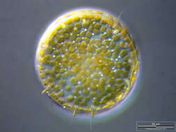
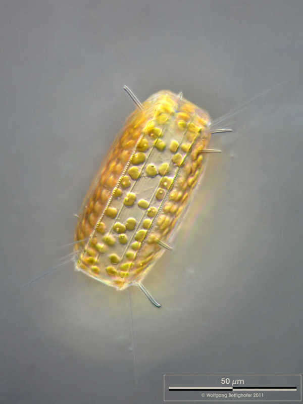
Thalassiosira punctigera Silicious processes (the labiate and the occluded ones) are visible. Scale bar indicates 25 µm. The image was built up using several photomicrographic frames with manual stacking technique. Sample from North Sea near Heligoland (spring diatom bloom). Images were taken using Zeiss Universal with Olympus C7070 CCD camera.Image under Creative Commons License V 3.0 (CC BY-NC-SA). Place name: North Sea around Heligoland Latitude: 54.186311 Longitude: 7.895034 Silikatfortsätze der Typen labiate und occluded sind sichtbar. Multiebenen-Abbildung, manuell gestapelt. Der Messbalken markiert eine Länge von 25 µm. Probe aus der Nordsee vor Helgoland in der Zeit der Frühjahrsblüte. Mikrotechnik: Zeiss Universal, Kamera: Olympus C7070.Creative Commons License V 3.0 (CC BY-NC-SA). For permission to use of (high-resolution) images please contact postmaster@protisten.de.
-
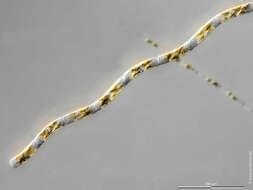
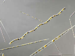
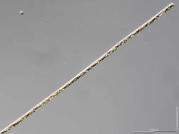
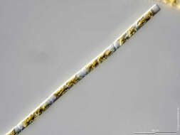
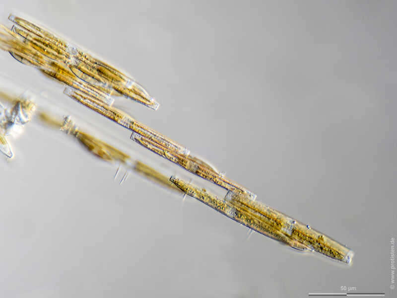
Bacillaria paxillifer Scale bar indicates 50 µm. Collected from Bodden, the brackish waters lying between the isles of Hiddensee and Ruegen (German Baltic Sea). Sampling date 9/2022. The image was built up using several photomicrographic frames with manual stacking technique. Images were taken using Zeiss Standard with Olympus OM-D M5 MKII. Image under Creative Commons License V 3.0 (CC BY-NC-ND). Place name: Hiddensee Bodden (Germany) Latitude: 54.582633 Longitude: 13.115051 Multiebenen-Abbildung, manuell gestapelt. Der Messbalken markiert eine Länge von 50 µm. Probe aus dem Hiddenseer Bodden. Datum der Aufsammlung: 9/2022. Mikrotechnik: Zeiss Standard, Kamera: Olympus OM-D M5 MKII. Creative Commons License V 3.0 (CC BY-NC-ND). For permission to use of (high-resolution) images please contact postmaster@protisten.de.
-
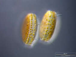
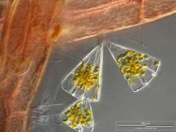
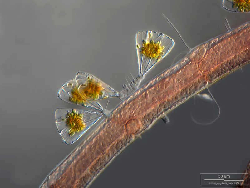
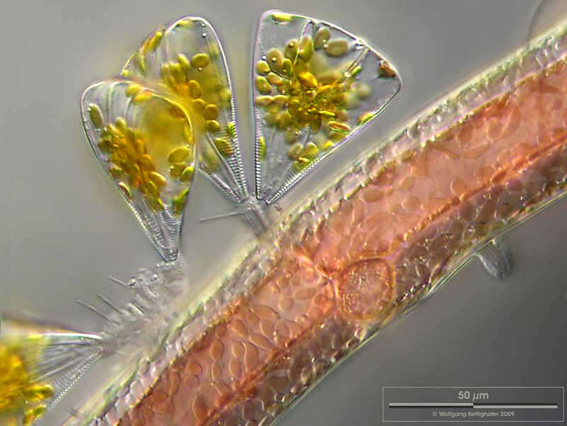
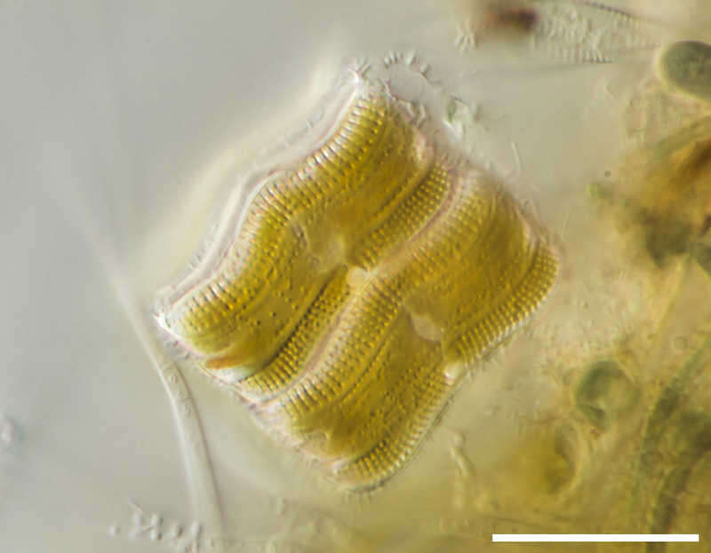
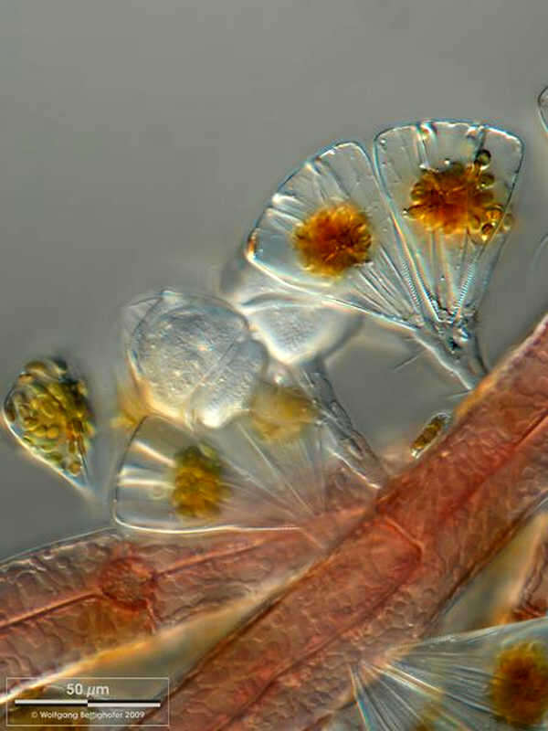
Licmophora juergensii On the left a Licmophora cell is visible in lateral (valvar) view, on the right we look at the girdle band (cingulim) of the cell, so this is called cingular view. Scale bar indicates 50 µm. Collected from Bodden, the brackish waters lying between the isles of Hiddensee and Ruegen (German Baltic Sea). This image was taken using Zeiss Universal with Olympus C7070 CCD camera.Image under Creative Commons License V 3.0 (CC BY-NC-SA). Place name: Hiddensee Bodden (Germany) Latitude: 54.582633 Longitude: 13.115051 Die Zelle links zeigt die Schalenansicht, rechts ist eine weitere in der Gürtelbandansicht zu sehen. Multiebenen-Abbildung, manuell gestapelt. Der Messbalken markiert eine Länge von 50 µm. Probe aus dem Hiddenseer Bodden. Mikrotechnik: Zeiss Universal, Kamera: Olympus C7070.Creative Commons License V 3.0 (CC BY-NC-SA). For permission to use of (high-resolution) images please contact postmaster@protisten.de.
-
Licmophora juergensii Licmophora juergensii as epibionts together with Cocconeis and Pseudanabaena on the red alga Polysiphonia fibrillosa. Scale bar indicates 25 µm. Collected from Bodden, the brackish waters lying between the isles of Hiddensee and Ruegen (German Baltic Sea). This image was taken using Zeiss Universal with Olympus C7070 CCD camera.Image under Creative Commons License V 3.0 (CC BY-NC-SA). Place name: Hiddensee Bodden (Germany) Latitude: 54.582633 Longitude: 13.115051 Licmophora juergensii zusammen mit Cocconeis als Aufwuchs auf der Rotalge Polysiphonia fibrillosa. Multiebenen-Abbildung, manuell gestapelt. Der Messbalken markiert eine Länge von 25 µm. Probe aus dem Hiddenseer Bodden. Mikrotechnik: Zeiss Universal, Kamera: Olympus C7070.Creative Commons License V 3.0 (CC BY-NC-SA). For permission to use of (high-resolution) images please contact postmaster@protisten.de.
-

Licmophora juergensii Licmophora on red alga Polysiphonia. Down to the left we see a deserted stalk. Sone of the Licmophora cells were broken away from their stalks through preparation. On the right of the deserted stalk there is such a cell which is now fixed with little mucilage an sister cell. Scale bar indicates 50 µm. Collected from Bodden, the brackish waters lying between the isles of Hiddensee and Ruegen (German Baltic Sea). This image was taken using Zeiss Universal with Olympus C7070 CCD camera.Image under Creative Commons License V 3.0 (CC BY-NC-SA). Place name: Hiddensee Bodden (Germany) Latitude: 54.582633 Longitude: 13.115051 Licmophora auf der Rotalge Polysiphonia. Links unten ist ein verlassener Gallertstiel zu sehen. Einige Licmophora-Zellen waren bei der Präparation von ihren Stielen abgebrochen. Recht neben diesem leeren Stiel sieht man solch eine Zelle, die sich mit wenig Gallerte an einer Schwesterzelle angeheftet hat. Multiebenen-Abbildung, manuell gestapelt. Der Messbalken markiert eine Länge von 50 µm. Probe aus dem Hiddenseer Bodden. Mikrotechnik: Zeiss Universal, Kamera: Olympus C7070.Creative Commons License V 3.0 (CC BY-NC-SA). For permission to use of (high-resolution) images please contact postmaster@protisten.de.
-
Licmophora juergensii Licmophora juergensii as epibionts together with the peritrich ciliat Carchesium, the diatom Cocconeis and the filamentous cyanobacteria Pseudanabaena on the red alga Polysiphonia fibrillosa. Scale bar indicates 50 µm. Collected from Bodden, the brackish waters lying between the isles of Hiddensee and Ruegen (German Baltic Sea). This image was taken using Zeiss Universal with Olympus C7070 CCD camera.Image under Creative Commons License V 3.0 (CC BY-NC-SA). Place name: Hiddensee Bodden (Germany) Latitude: 54.582633 Longitude: 13.115051 Licmophora juergensii zusammen mit dem peritrichen Ciliiaten Carchesium spec., der Kieselalge Cocconeis und der fädigen Cyanobakterie Pseudanabaena als Aufwuchs auf der Rotalge Polysiphonia fibrillosa. Multiebenen-Abbildung, manuell gestapelt. Der Messbalken markiert eine Länge von 50 µm. Probe aus dem Hiddenseer Bodden. Mikrotechnik: Zeiss Universal, Kamera: Olympus C7070.Creative Commons License V 3.0 (CC BY-NC-SA). For permission to use of (high-resolution) images please contact postmaster@protisten.de.
-
Licmophora juergensii Licmophora juergensii as epibionts together with filamentous cyanobacterial colonies of Pseudanabaena on the red alga Polysiphonia fibrillosa. Scale bar indicates 50 µm. Collected from Bodden, the brackish waters lying between the isles of Hiddensee and Ruegen (German Baltic Sea). This image was taken using Zeiss Universal with Olympus C7070 CCD camera.Image under Creative Commons License V 3.0 (CC BY-NC-SA). Place name: Hiddensee Bodden (Germany) Latitude: 54.582633 Longitude: 13.115051 Licmophora juergensii zusammen mit Cocconeis als Aufwuchs auf der Rotalge Polysiphonia fibrillosa. Multiebenen-Abbildung, manuell gestapelt. Der Messbalken markiert eine Länge von 50 µm. Probe aus dem Hiddenseer Bodden. Mikrotechnik: Zeiss Universal, Kamera: Olympus C7070.Creative Commons License V 3.0 (CC BY-NC-SA). For permission to use of (high-resolution) images please contact postmaster@protisten.de.
-
Licmophora juergensii Licmophora juergensii as epibionts together with Cocconeis on the red alga Polysiphonia fibrillosa. Scale bar indicates 25 µm. Collected from Bodden, the brackish waters lying between the isles of Hiddensee and Ruegen (German Baltic Sea). This image was taken using Zeiss Universal with Olympus C7070 CCD camera.Image under Creative Commons License V 3.0 (CC BY-NC-SA). Place name: Hiddensee Bodden (Germany) Latitude: 54.582633 Longitude: 13.115051 Licmophora juergensii zusammen mit Cocconeis als Aufwuchs auf der Rotalge Polysiphonia fibrillosa. Multiebenen-Abbildung, manuell gestapelt. Der Messbalken markiert eine Länge von 25 µm. Probe aus dem Hiddenseer Bodden. Mikrotechnik: Zeiss Universal, Kamera: Olympus C7070.Creative Commons License V 3.0 (CC BY-NC-SA). For permission to use of (high-resolution) images please contact postmaster@protisten.de.
-
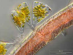
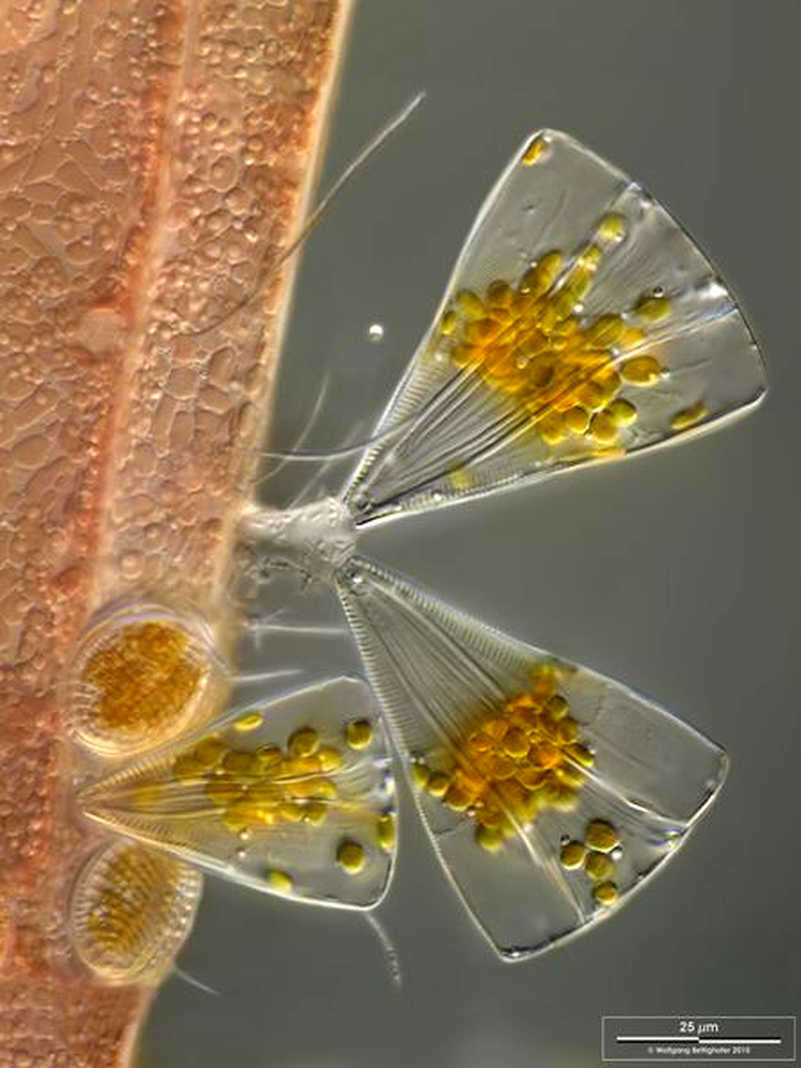
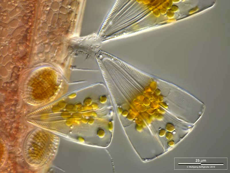
Licmophora juergensii Cells in valvar (left) and cingular (right) view. Scale bar indicates 50 µm. Collected from Bodden, the brackish waters lying between the isles of Hiddensee and Ruegen (German Baltic Sea). This image was taken using Zeiss Universal with Olympus C7070 CCD camera.Image under Creative Commons License V 3.0 (CC BY-NC-SA). Place name: Hiddensee Bodden (Germany) Latitude: 54.582633 Longitude: 13.115051 Zellen in Schalen- (links) und Gürtelbandansicht (rechts). Multiebenen-Abbildung, manuell gestapelt. Der Messbalken markiert eine Länge von 50 µm. Probe aus dem Hiddenseer Bodden. Mikrotechnik: Zeiss Universal, Kamera: Olympus C7070.Creative Commons License V 3.0 (CC BY-NC-SA). For permission to use of (high-resolution) images please contact postmaster@protisten.de.
-
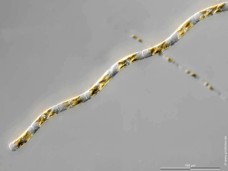
Guinardia delicatula Scale bar indicates 100 µm. The specimen was gathered in the Kieler Förde (German Baltic Sea). Sampling date 4/2018. The image was built up using several photomicrographic frames with manual stacking technique. Images were taken using Zeiss Axioplan with Olympus OM-D M5 MKII. Image under Creative Commons License V 3.0 (CC BY-NC-SA). Place name: Baltic Sea, Kieler Förde, Kiel Fjord (Germany) Latitude: 54.3894126 Longitude: 10.1749055 Multiebenen-Abbildung, manuell gestapelt. Der Messbalken markiert eine Länge von 100 µm. Probe aus der Kieler Förde. Datum der Aufsammlung: 4/2018. Mikrotechnik: Zeiss Axioplan, Kamera: Olympus OM-D M5 MKII. Creative Commons License V 3.0 (CC BY-NC-SA). For permission to use of (high-resolution) images please contact postmaster@protisten.de.
-
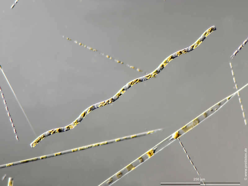
Guinardia delicatula Scale bar indicates 250 µm. The specimen was gathered in the Kieler Förde (German Baltic Sea). Sampling date 4/2018. The image was built up using several photomicrographic frames with manual stacking technique. Images were taken using Zeiss Axioplan with Olympus OM-D M5 MKII. Image under Creative Commons License V 3.0 (CC BY-NC-SA). Place name: Baltic Sea, Kieler Förde, Kiel Fjord (Germany) Latitude: 54.3894126 Longitude: 10.1749055 Multiebenen-Abbildung, manuell gestapelt. Der Messbalken markiert eine Länge von 250 µm. Probe aus der Kieler Förde. Datum der Aufsammlung: 4/2018. Mikrotechnik: Zeiss Axioplan, Kamera: Olympus OM-D M5 MKII. Creative Commons License V 3.0 (CC BY-NC-SA). For permission to use of (high-resolution) images please contact postmaster@protisten.de.
-
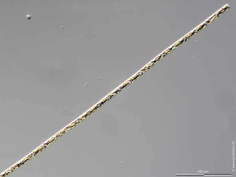
Guinardia delicatula Scale bar indicates 100 µm. The specimen was gathered in the Kieler Förde (Baltic Sea). Sampling date 2/2022. The image was built up using several photomicrographic frames with manual stacking technique. Images were taken using Zeiss Axioplan with Olympus OM-D M5 MKII. Image under Creative Commons License V 3.0 (CC BY-NC-SA). Place name: Baltic Sea, Kieler Förde, Kiel Fjord (Germany) Latitude: 54.3894126 Longitude: 10.1749055 Multiebenen-Abbildung, manuell gestapelt. Der Messbalken markiert eine Länge von 100 µm. Probe aus der Kieler Förde. Datum der Aufsammlung: 2/2022. Mikrotechnik: Zeiss Axioplan, Kamera: Olympus OM-D M5 MKII. Creative Commons License V 3.0 (CC BY-NC-SA). For permission to use of (high-resolution) images please contact postmaster@protisten.de.
-
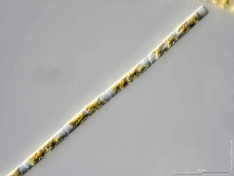
Guinardia delicatula Scale bar indicates 50 µm. The specimen was gathered in the Kieler Förde (Baltic Sea). Sampling date 2/2022. The image was built up using several photomicrographic frames with manual stacking technique. Images were taken using Zeiss Axioplan with Olympus OM-D M5 MKII. Image under Creative Commons License V 3.0 (CC BY-NC-SA). Place name: Baltic Sea, Kieler Förde, Kiel Fjord (Germany) Latitude: 54.3894126 Longitude: 10.1749055 Multiebenen-Abbildung, manuell gestapelt. Der Messbalken markiert eine Länge von 50 µm. Probe aus der Kieler Förde. Datum der Aufsammlung: 2/2022. Mikrotechnik: Zeiss Axioplan, Kamera: Olympus OM-D M5 MKII. Creative Commons License V 3.0 (CC BY-NC-SA). For permission to use of (high-resolution) images please contact postmaster@protisten.de.
-
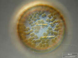
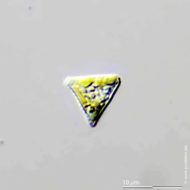
Goniochloris sculpta The scale bar indicates 10 µm. From a pond at Lemkendorf, isle of Fehmarn (Baltic Sea). This image was taken using Zeiss Axioplan with DSLR Canon 70D.Image under Creative Commons License V 3.0 (CC BY-NC-SA). Place name: Pond in Lemkendorf, isle Fehmarn (Baltic Sea, Germany) Latitude: 54.472296 Longitude: 11.095662 Der Messbalken markiert eine Länge von 10 µm. Probe aus einem Teich in Lemkendorf, Fehmarn. Mikrotechnik: Zeiss Axioplan, Kamera: Canon 70D. Creative Commons License V 3.0 (CC BY-NC-SA). For permission to use of (high-resolution) images please contact postmaster@protisten.de.
-
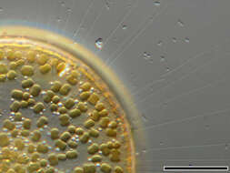
Cocconeis spec. Cocconeis spec. living on the green alga Cladophora. Sample from the North see near isle of Nordstrand. Images were taken using Zeiss Universal with Canon EOS 600D.Image under Creative Commons License V 3.0 (CC BY-NC-SA). Place name: North see near isle of Nordstrand (Schleswig-Holstein, Germany) Latitude: 54.49594138 Longitude: 8.80856752 Cocconeis auf der Grünalge Cladophora. Probe aus der Nordsee bei Nordstrand. Mikrotechnik: Zeiss Universal, Kamera: Canon EOS 600D.Creative Commons License V 3.0 (CC BY-NC-SA). For permission to use of (high-resolution) images please contact postmaster@protisten.de.
-
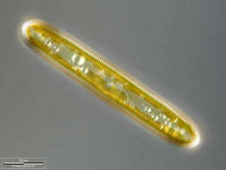
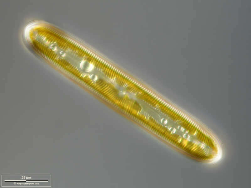
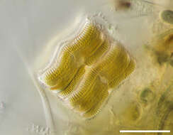
Pinnularia viridis Valvar view. Scale bar indicates 25 µm. Sample from a wetland at the Pillersee (Tyrol, Austria). The image was built up using several photomicrographic frames with manual stacking technique. Images were taken using Zeiss Universal with Olympus C7070 CCD camera.Image under Creative Commons License V 3.0 (CC BY-NC-SA). Place name: Wetland at the Pillersee (Tyrol, Austria) Latitude: 47.531785 Longitude: 12.573095 Schalenansicht. Multiebenen-Abbildung, manuell gestapelt. Der Messbalken markiert eine Länge von 25 µm. Probe aus dem Pillersee in Tirol. Mikrotechnik: Zeiss Universal, Kamera: Olympus C7070. Creative Commons License V 3.0 (CC BY-NC-SA). For permission to use of (high-resolution) images please contact postmaster@protisten.de.
-
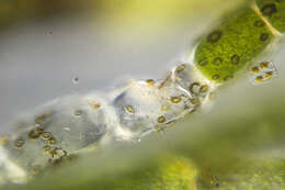
Achnanthes spec. Achnanthes spec. living on the plant Cryptocoryne wendtii. Scale bar indicates 25 µm. Sample from a tropical freshwater aquarium. Images were taken using Zeiss Universal with Canon EOS 600D.Image under Creative Commons License V 3.0 (CC BY-NC-SA). Place name: Tropical freshwater aquarium Latitude: 54.3018013 Longitude: 10.07120132 Achnanthes auf der Sumpfpflanze Cryptocoryne wendtii. Der Messbalken markiert eine Länge von 25 µm. Probe aus einem Süßwasseraquarium für tropische Fische. Mikrotechnik: Zeiss Universal, Kamera: Canon EOS 600D.Creative Commons License V 3.0 (CC BY-NC-SA). For permission to use of (high-resolution) images please contact postmaster@protisten.de.
-
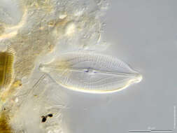
Caloneis amphisbaena Scale bar indicates 25 µm. Sample from the static fire tank of the village Kloster (Hiddensee Island). Sampling date 9/2022. Images were taken using Zeiss Universal with Olympus OM-D M5 MKII.Image under Creative Commons License V 3.0 (CC BY-NC-ND). Place name: Static fire tank Kloster, Hiddensee Latitude: 54.58447966 Longitude: 13.10767114 Der Messbalken markiert eine Länge von 25 µm. Probe aus dem Feuerlöschteich von Kloster/Hiddensee. Datum der Aufsammlung: 9/2022. Mikrotechnik: Zeiss Universal, Kamera: Olympus OM-D M5 MKII. Creative Commons License V 3.0 (CC BY-NC-ND). For permission to use of (high-resolution) images please contact postmaster@protisten.de.