-
-

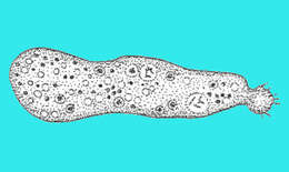
Multicilia marina Cienkowski, 1881. Cells are normally spherical in a culture, about 30-40 microns in diameter, however,some cells attain an oblong or irregular form. The majority of cells possess 20-30 long flagella, 1.5-2 times the cell diameter. The flagella differ from those of other heterotrophic flagellates by very weak movements, resulting in a similarity to heliozoan axopods. The locomotion of M ulticilia is slow, rotatory, without any definitive direction, therefore moving cells normally lack anterior and posterior ends. Cells glide in one direction for a very short distance and may change direction rapidly (including moving backwards). The cell rolls over when changing direction. The temporary anterior flagellum is then stretched and clasped along the substratum, only its apex continues to oscillate. The temporary posterior flagellum shows the same behaviour. Other flagella perform irregular movements without any visible coordination. After changing the salinity, cells become free floating and continue a weak oscillation of the flagella.
-
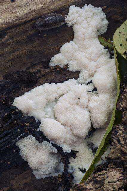
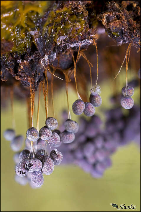
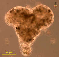
This is a image of a sorocarp. Notice the slime trail at the base of the fruiting body.
-
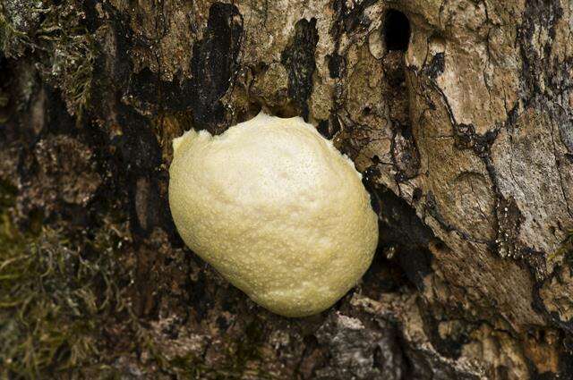
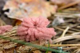
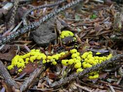
Badhamia panicea.
-
Pelomyxa (peal-o-mix-a), a large pelobiont which developed some reputation as possibly the most primitive eukaryote. This argument was based on the fact that it does not have mitochondria, conventional dictyosomes if any, flagella are aberrant, and nuclear division was also thought to be aberrant. The arguments for a primitive status now seem to be unsound. Cytoplasm with small particles of sand. eats algae and detritus. Moves with fountain-flow motion (cytoplasm moving forward up the centre of the cell and then spilling out near the front. Posterior end crumpled, to form a uroid. Phase contrast micrograph.
-
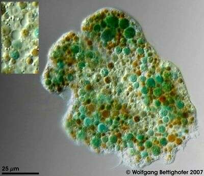
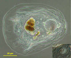
Enteromyxa paludosa cell was showing very colorful metabolites and numerous nuclei and contractile vacuoles. In order to deliver depth of focus 50 high resolution frames (Planapo 63/1.4) were processed. The picture inserted showes 4 nuclei and two contractile vacuoles in higher magnification. Sample from sphagnum pond situated in the northern alpine region of Austria near Salzburg. Images were taken using Zeiss Universal with Olympus C7070 CCD camera.
-
-

Description: Amoeba with a pellicle-like surface noticeable by a rather stiff appearance and, in this case, numerous wrinkles. Movement with a tractor-like rolling of the surface. Size of locomotive form in this specimen 90x70 µm. Hyaloplasm as an antero-lateral crescent. Contractile vacuole located in posterior end of the cell. Nucleus spherical to ovoid, nucleolus of granular texture (see picture in lower right angle), sometimes divided in 2 or 3 fragments. Phase contrast ; nucleus in modified phase contrast. Amoeba was cultured in filtered water from original habitat and fed with Oscillatoria (Cyanobacteria), rests of which are seen in a food-vacuole in the centre. It is one of the rugose species of the genus as described by Page and one of the typical representatives of Smirnovâs rugose morphotype. A future zip-file may contain tables showing the peculiar phagocytosis of oscillatoria by Thecamoeba sphaeronucleolus as well as the fusion of two cells followed by encystment and cell division.
-
Hårup Sande, Silkeborg, Jylland, Danmark
-
Suserup Skov, Danmark
-
Allindelille Fredskov, Denmark
-
Lille Øksø, Rold Skov, Jylland, Danmark
-
Øksedal, Sebbersund, Jylland, Danmark
-
Store Øksø, Rold Skov
-
Kridtstien, Nystrup Plantage, Thy, Danmark
-
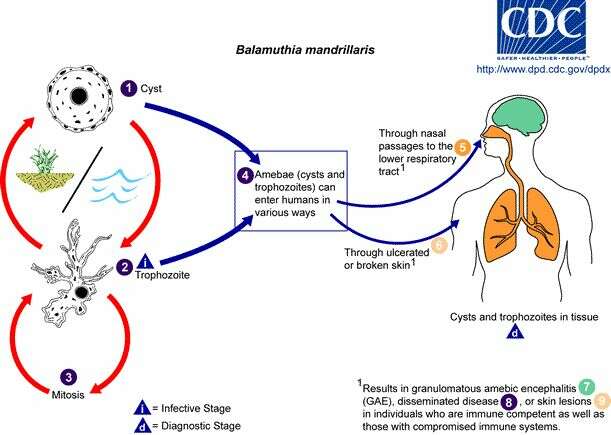
Centers for Disease Control/Division of Parasitic Diseases and Malaria
EOL staff
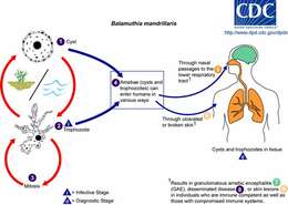
Life cycle of Balamuthia mandrillaris parasitizing humansBalamuthia mandrillaris has only recently been isolated from the environment and has also been isolated from autopsy specimens of infected humans and animals. The B. mandrillaris life cycle has only two stages, a dormant cyst stage (1) and an actively feeding and dividing trophozoite stage (2) (B. mandrillaris has no flagellated stage). The trophozoites replicate by mitosis (the nuclear membrane does not remain intact) (3). Although the trophozoites are the infective stage, both cysts and trophozoites gain entry into the body (4) through various means. Entry can occur through the nasal passages to the lower respiratory tract (5) or through ulcerated or broken skin (6). When B. mandrillaris enters the respiratory system or through the skin, it can invade the central nervous system by hematogenous dissemination causing
granulomatous amebic encephalitis (GAE) (7) or disseminated disease (8), or skin lesions (9) in individuals who are immune competent as well as those with compromised immune systems. Balamuthia mandrillaris cysts and trophozoites are found in tissue.From
Centers for Disease Control Parasites and Health website.
-
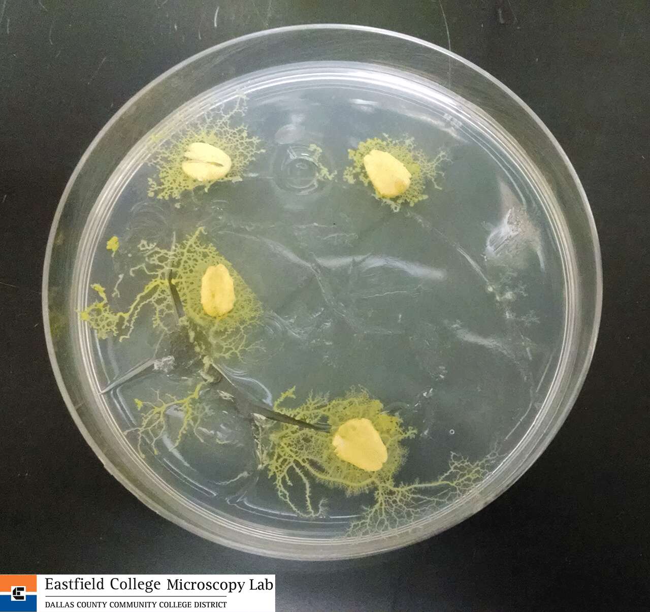
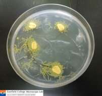
Eastfield College, Mesquite, TX
-
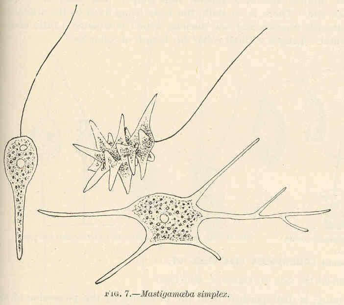
Mastigamoeba simplex.
-
Trichosphaerium sieboldi.
-
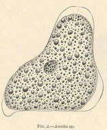
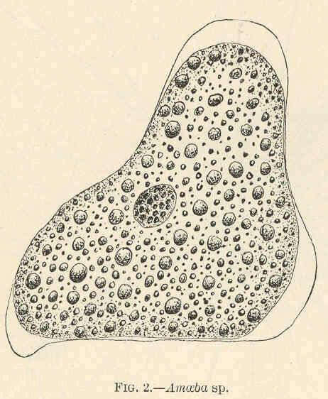
Amoeba sp..
-
-
taxonomy:binomial="Acanthamoeba castellanii"
-
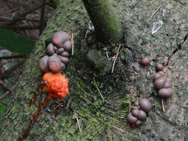
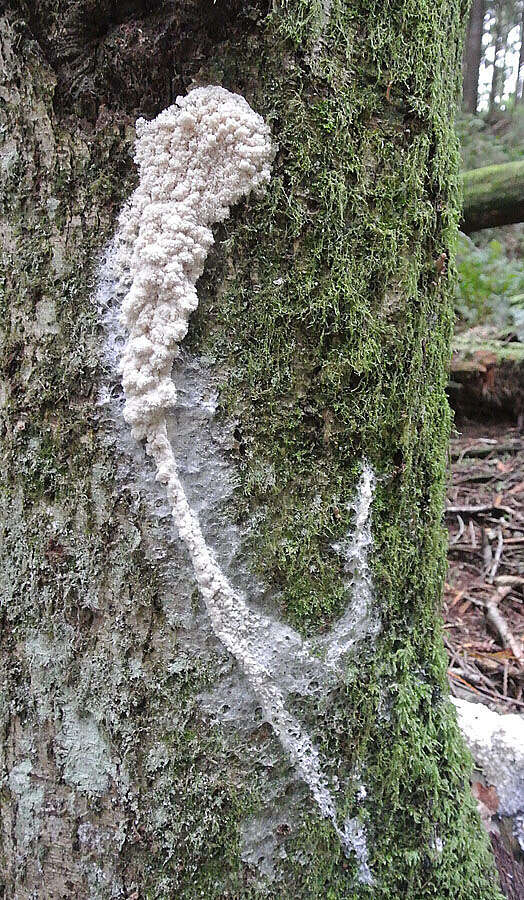
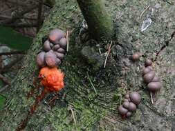
In Spring on Mt. Elphinstone, British Columbia. Stemonitidaceae Famly
-
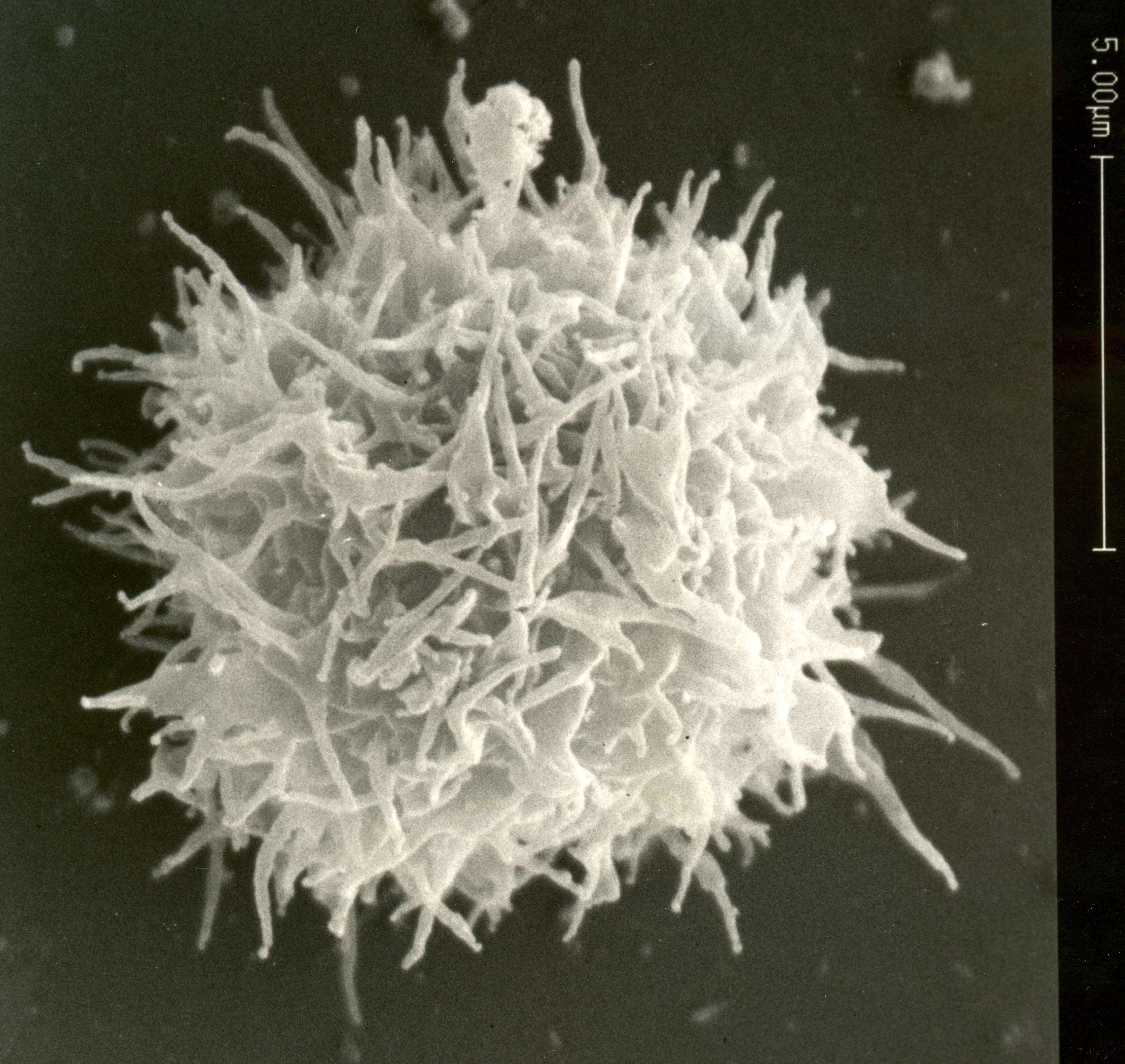
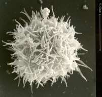
image courtesy of Dr. William F. Loomis


