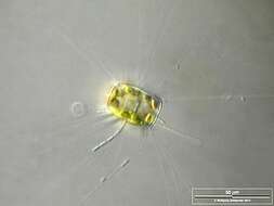
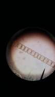
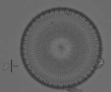
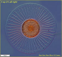
Optical tranversal section, showing the nucleus. The thin, barely visible floating extensions are made of chitin. Furthermore, filamentous bacteria colonies are attached. Scale bar indicates 50 µm. Sample from the Lake Constance (vicinity of Bodman). The image was built up using several photomicrographic frames with manual stacking technique. Images were taken using Zeiss Universal with Olympus C7070 CCD camera.Image under Creative Commons License V 3.0 (CC BY-NC-SA).
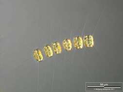
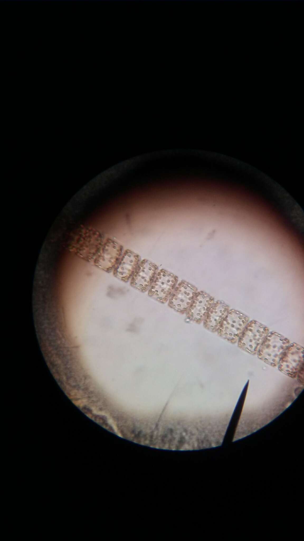
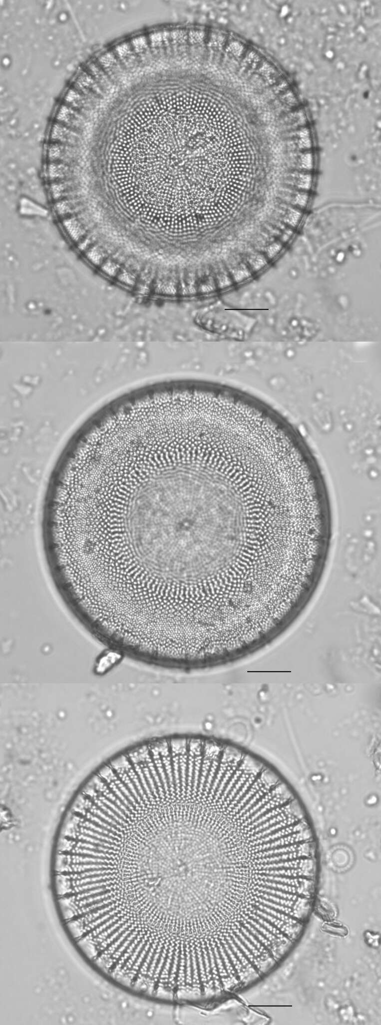
Long chain of Porosira glacialis. Note that the delicate spines are chitinous. Scale bar indicates 100 µm. The image was built up using several photomicrographic frames with manual stacking technique. Sample from North Sea near Heligoland (spring diatom bloom). Images were taken using Zeiss Universal with Olympus C7070 CCD camera.
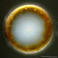
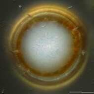
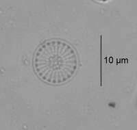
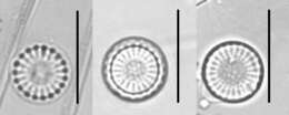
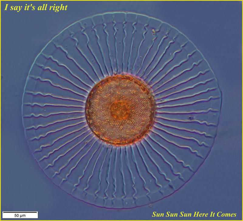
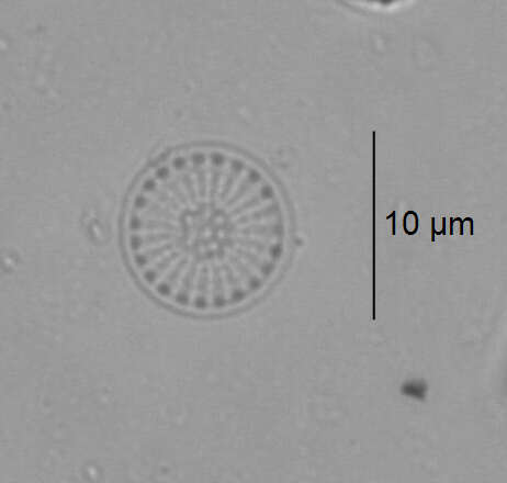
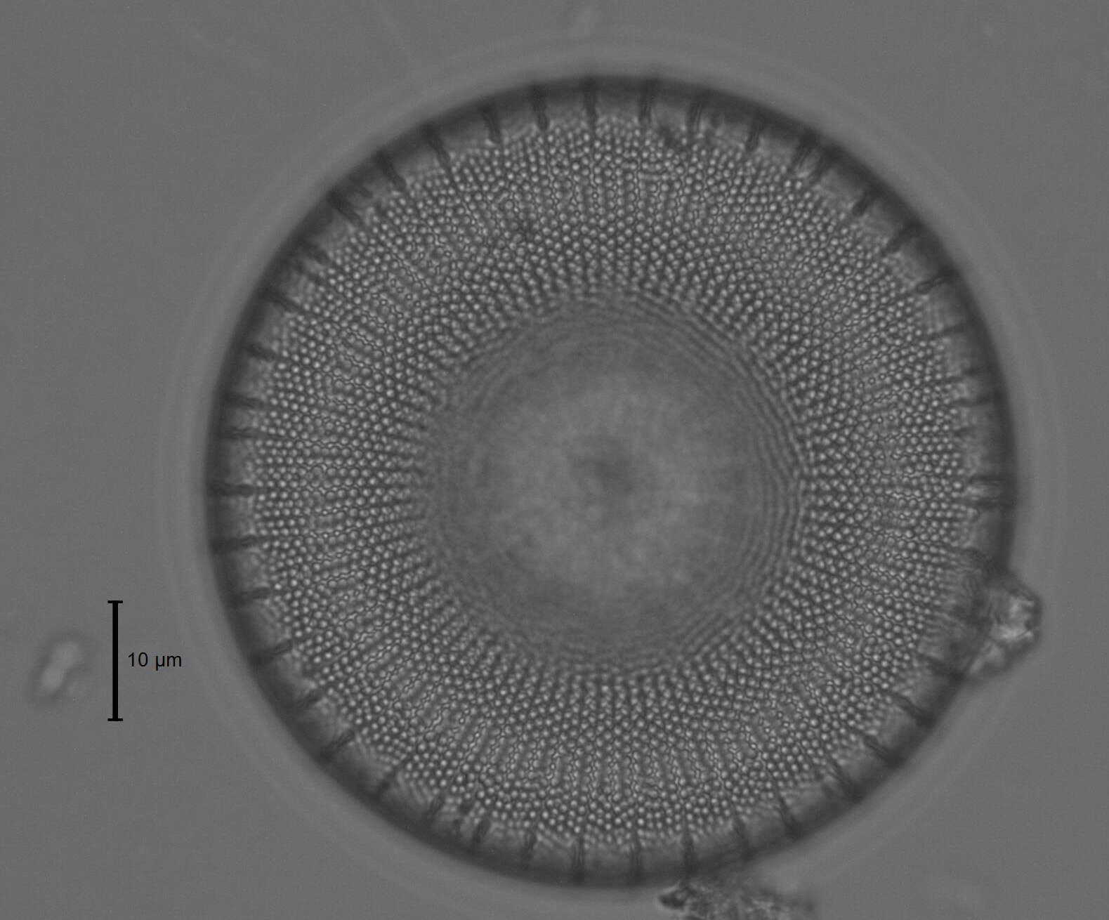
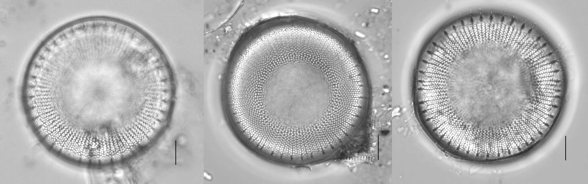
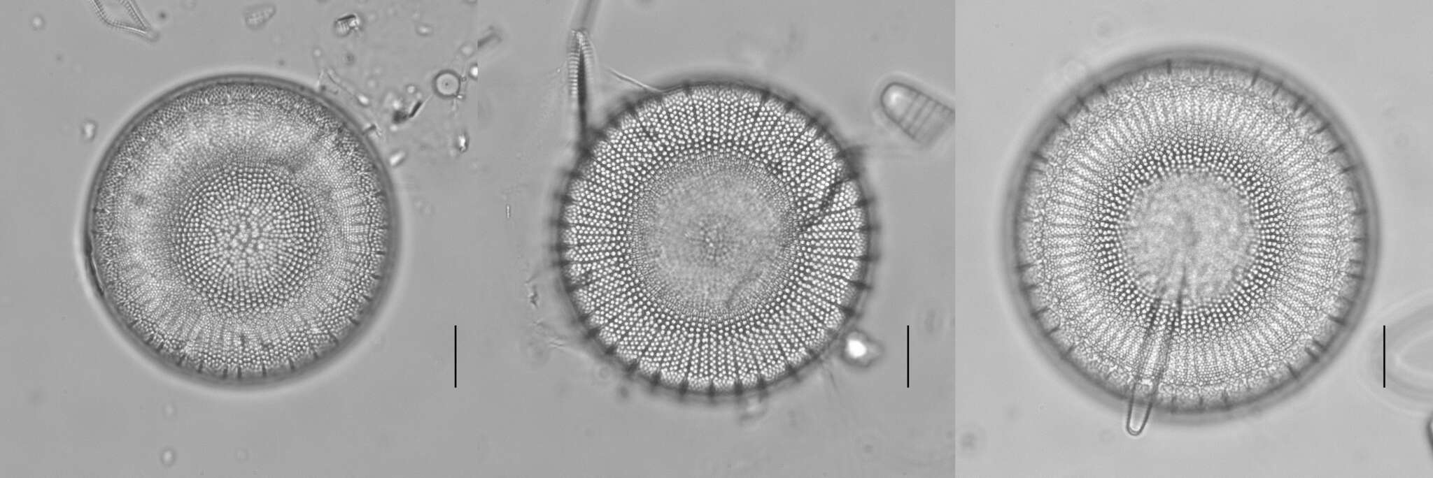
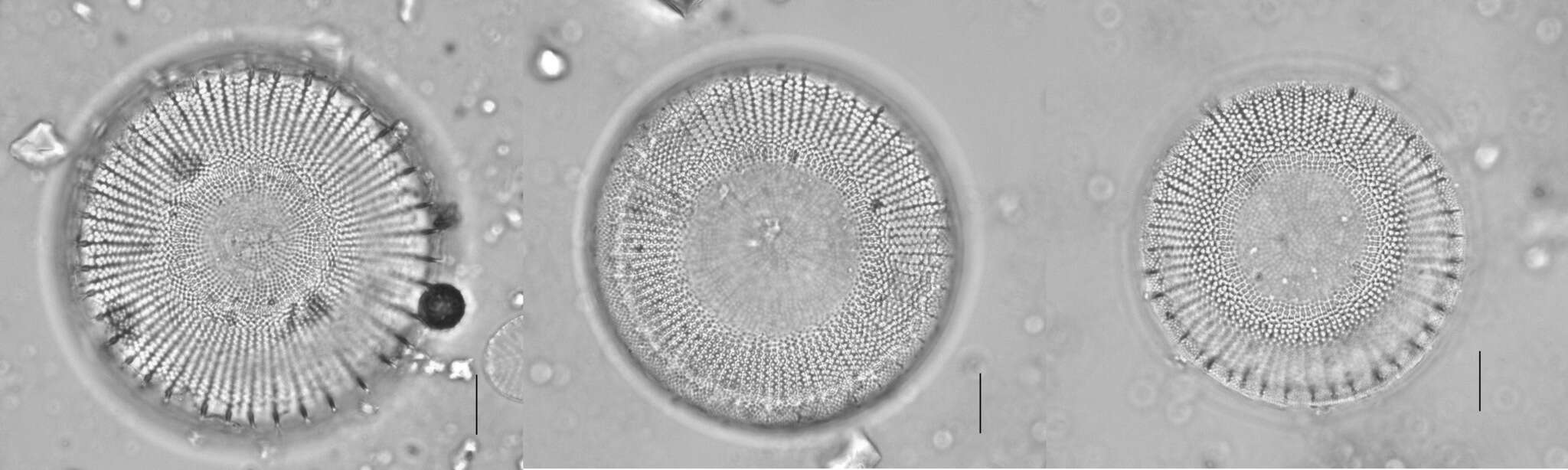
Silicious processes (the labiate and the occluded ones) are visible. Scale bar indicates 25 µm. The image was built up using several photomicrographic frames with manual stacking technique. Sample from North Sea near Heligoland (spring diatom bloom). Images were taken using Zeiss Universal with Olympus C7070 CCD camera.
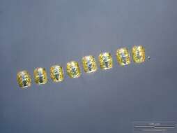
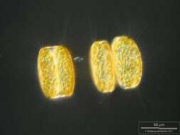
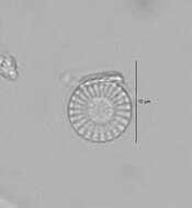
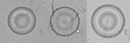
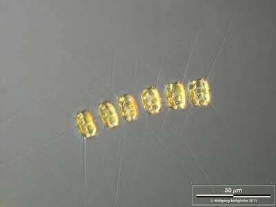
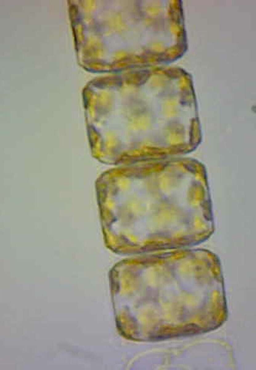
Short chain of Porosira glacialis. Note that the delicate spines are chitinous. Scale bar indicates 50 µm. The image was built up using several photomicrographic frames with manual stacking technique. Sample from North Sea near Heligoland (spring diatom bloom). Images were taken using Zeiss Universal with Olympus C7070 CCD camera.
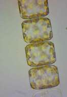
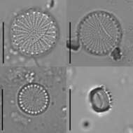
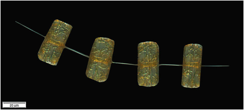
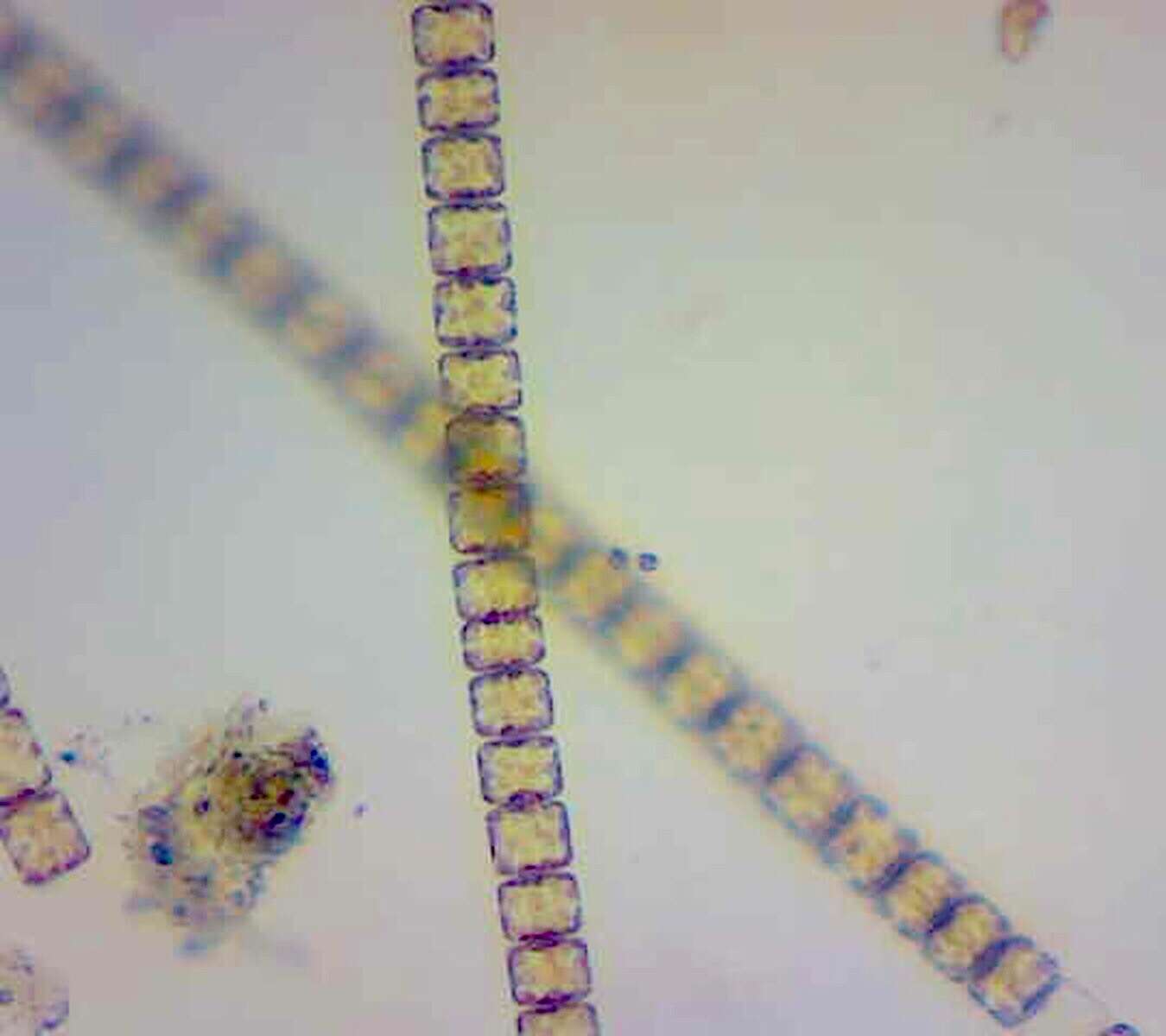
Labiate processes and chitinous spines are visible. Scale bar indicates 25 µm. The image was built up using several photomicrographic frames with manual stacking technique. Sample from North Sea near Heligoland (spring diatom bloom). Images were taken using Zeiss Universal with Olympus C7070 CCD camera.
Scale bar indicates 50 µm. The image was built up using several photomicrographic frames with manual stacking technique. Sample from North Sea near Heligoland (spring diatom bloom). Images were taken using Zeiss Universal with Olympus C7070 CCD camera.
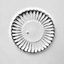
Public Domain, U.S. Government Work 2011 Barry H. Rosen Courtesy of life.nbii.gov
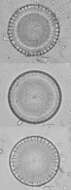
NBII images
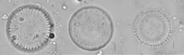
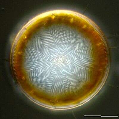
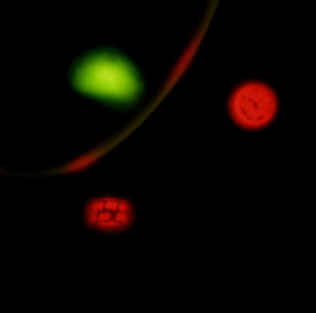
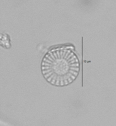
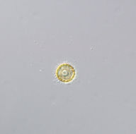
Category hierarchy: Microorganisms | AlgaeDescription: Discoid chromatophores and the nucleus easily observed with epifluorescent illumination. Sample was collected from Raccoon River, Iowa.Capture device: DP71Capture details: 400x with microscopeOriginal date: 20091002|||112306Locality: Latitude: 2.859009900000000e+001; Longitude: -8.119031699999999e+001
Public Domain, U.S. Government Work 2011 Barry H. Rosen Courtesy of life.nbii.gov
NBII images
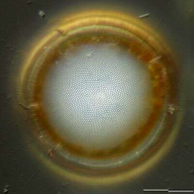
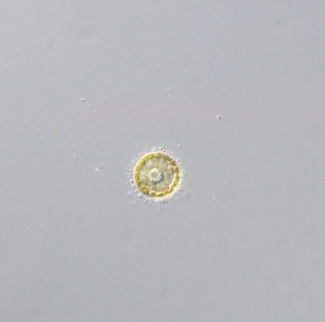
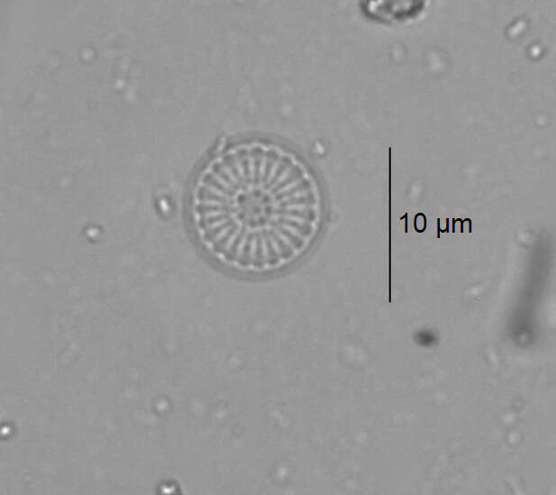
Category hierarchy: Microorganisms | MicrofloraDescription: Discoid chromatophores and the nucleus easily observed with normal illumination. Specimen was collected from Raccoon River, Iowa.Capture device: DP71Original date: 20091002|||113054Locality: Latitude: 2.859009900000000e+001; Longitude: -8.119031699999999e+001