-
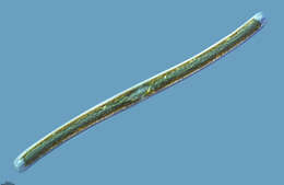
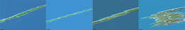
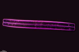
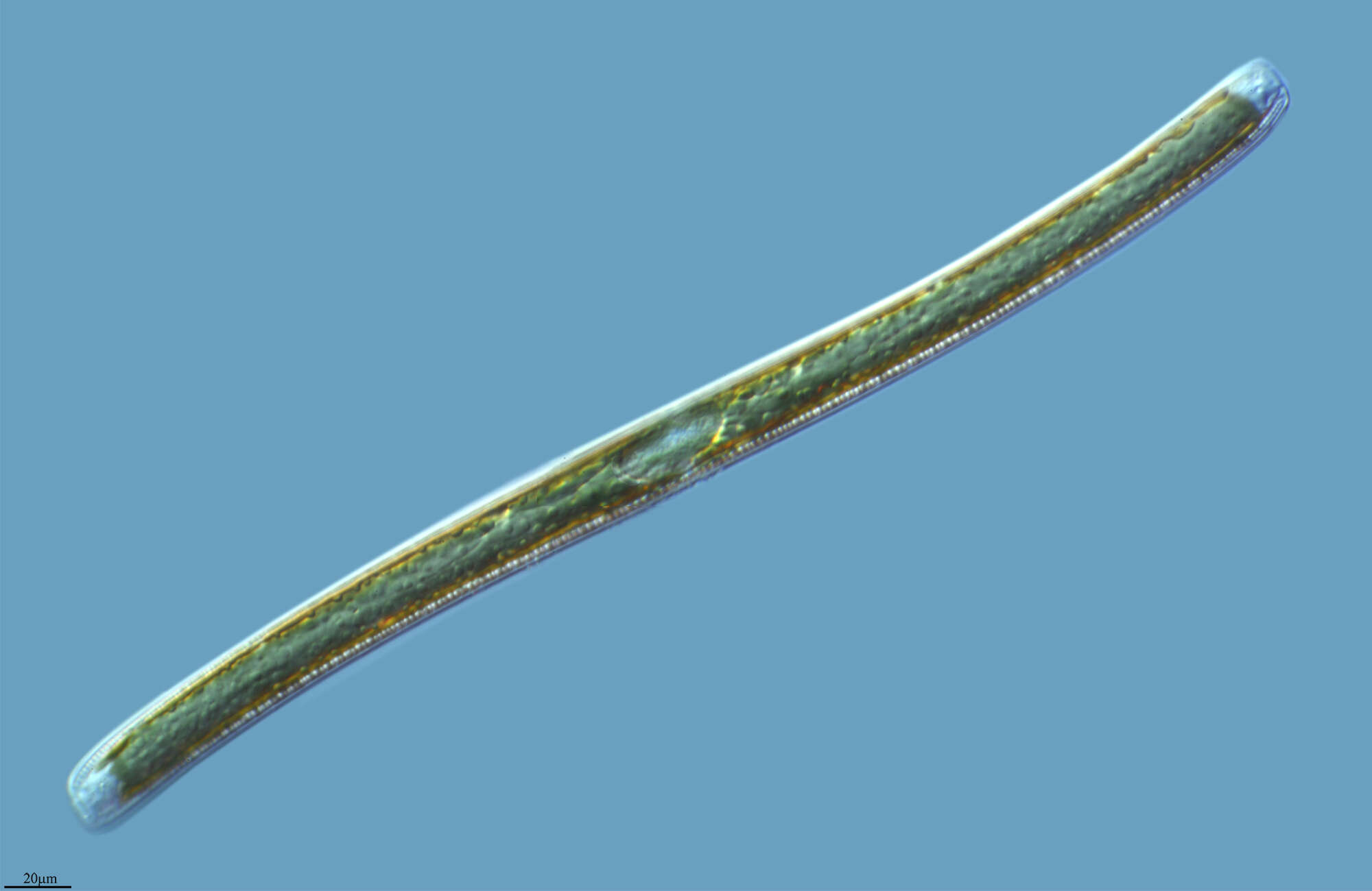
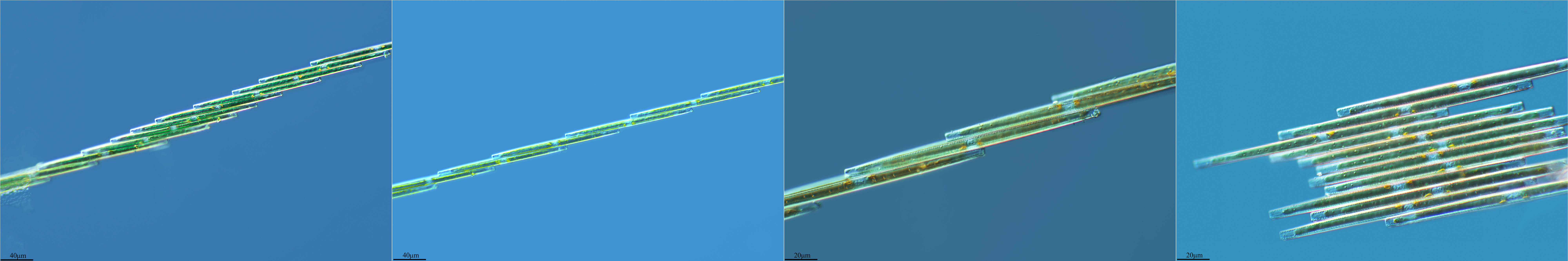
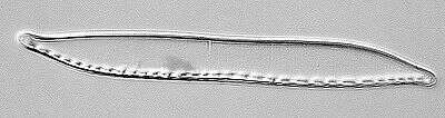
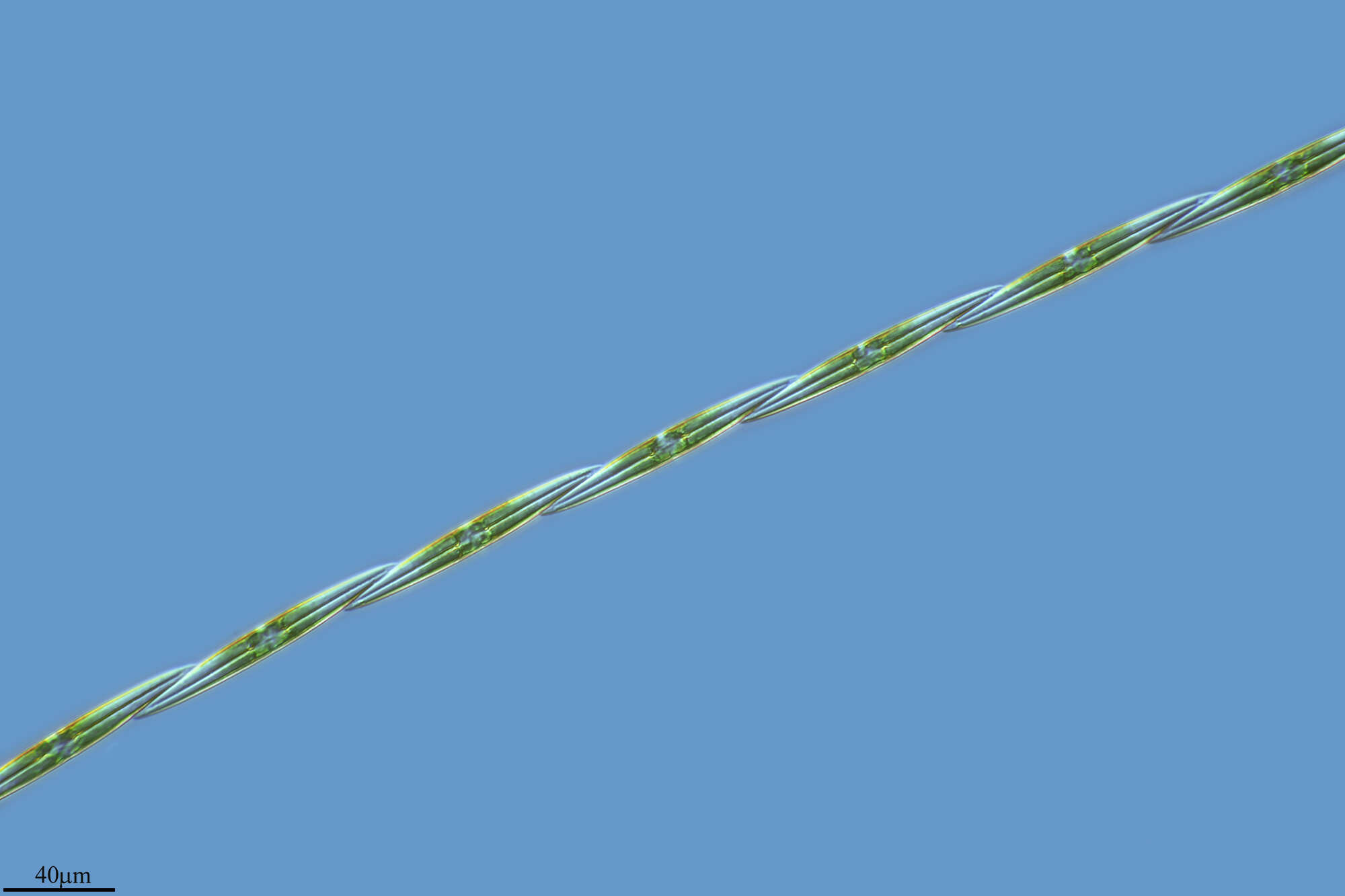
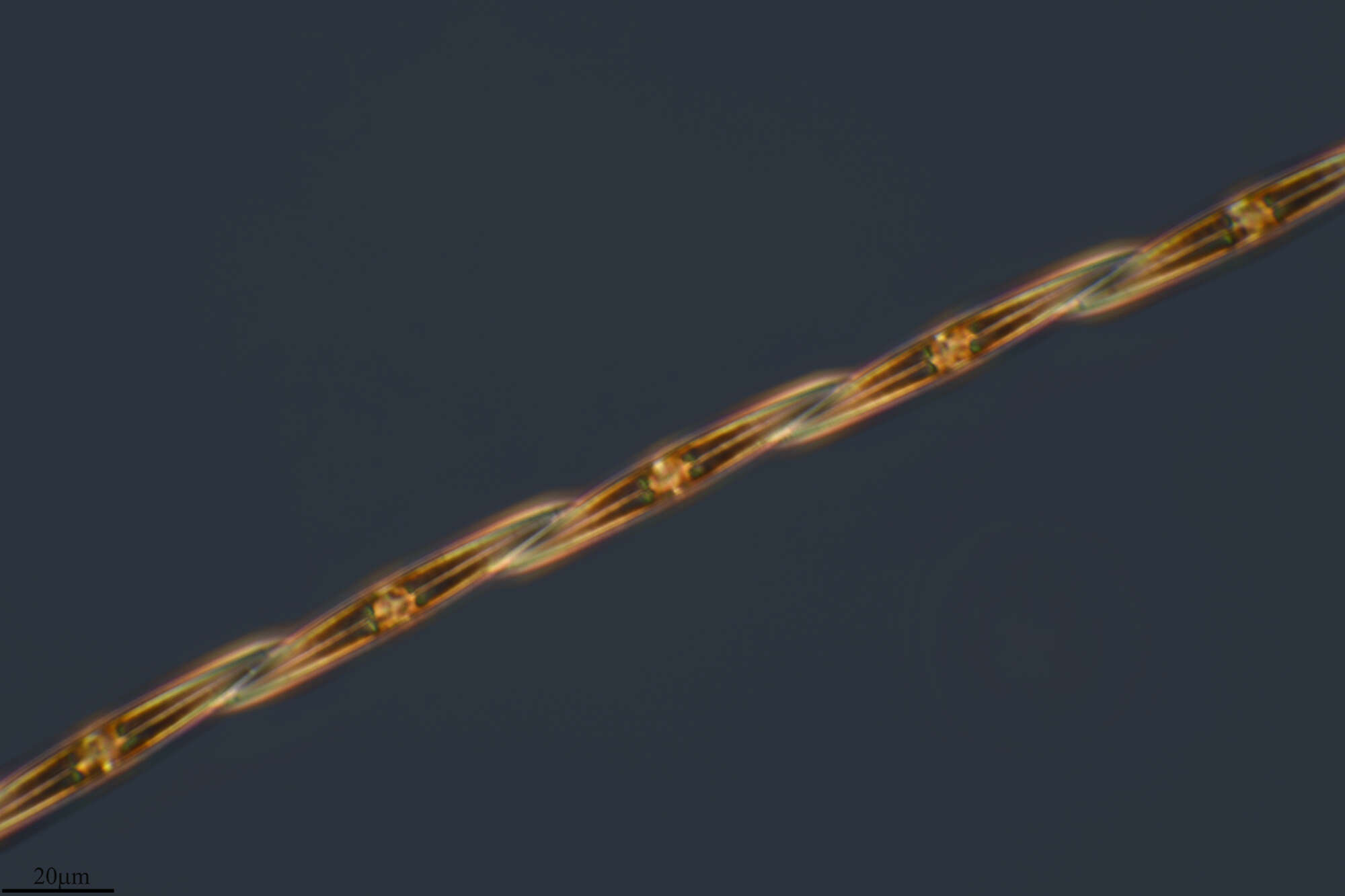
Cylindrotheca is a elongate pennate diatom is very common in sediments - the form apparently allowing the cell to penetrate through the pores of the sediments. Pennate diatoms are important in intertidal and illuminated subtidal sediments in marine ecosystems and primary producers. Pennate diatoms are capable of movement, relying on the raphe to produce thrust. Pennate diatoms are usually regarded as boat shaped, although some of the boats have very odd shapes. They can usually glide. Although enclosed in a siliceous shell, the shell of these rather delicate diatoms is flexible. The plastids contain chlorophylls a and c which gives the yellowy-brown colour. Phase contrast.
-
Miranda do Douro Municipality, Braganca, Portugal
-
Mahide, Castille and Leon, Spain
-
Mahide, Castilla y Len, Espaa
-
Corporales, La Rioja, Spain
-
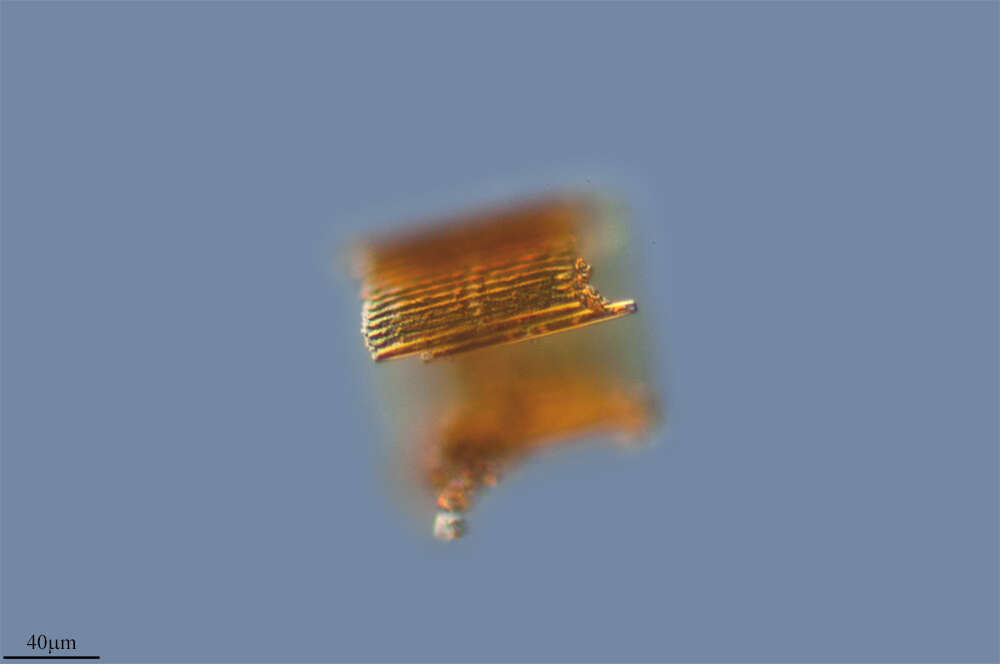
This EM isn't very good, but it's better than nothing.
-
Estella/Lizarra, Navarre, Spain
-
Grove, O, Galicia, Spain
-
Balea, Galicia, Spain
-
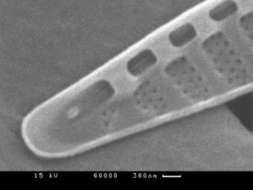
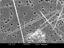
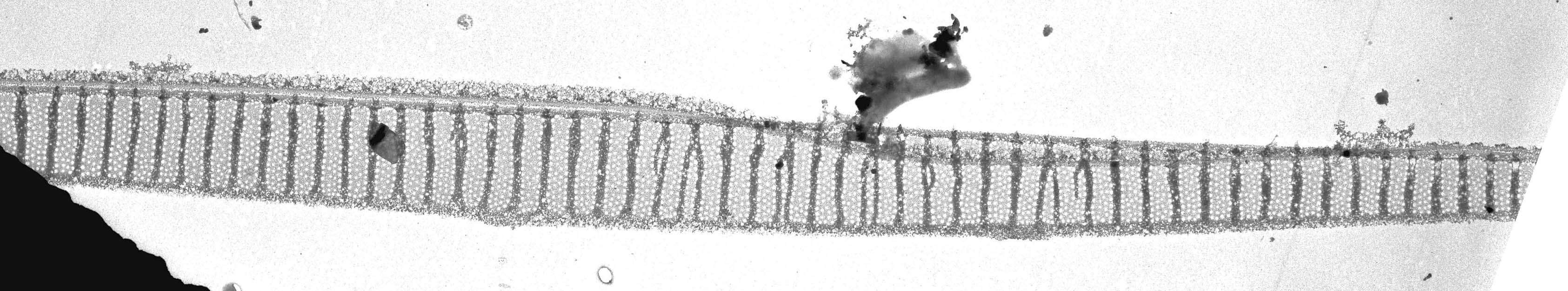
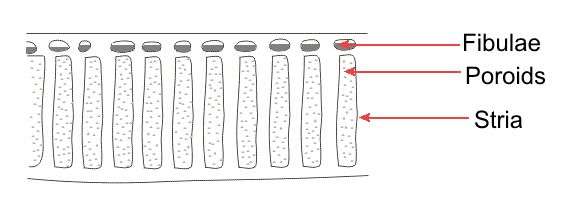
Fig 2: Pseudonitzschia multiseries Scanned electron micrograph showing detail of the stria, poroids and fibulae.
-
-
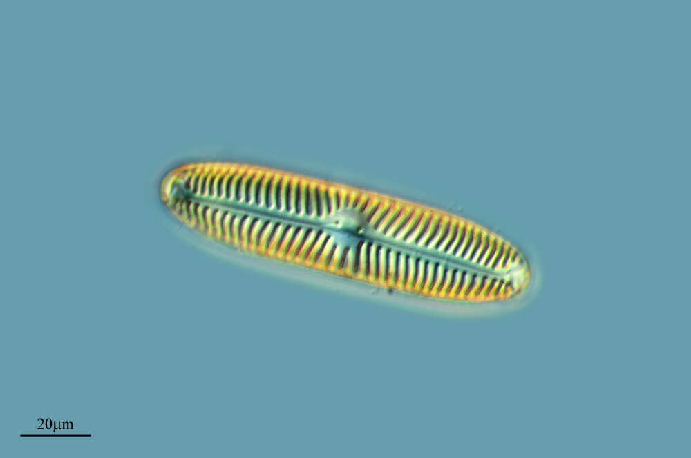
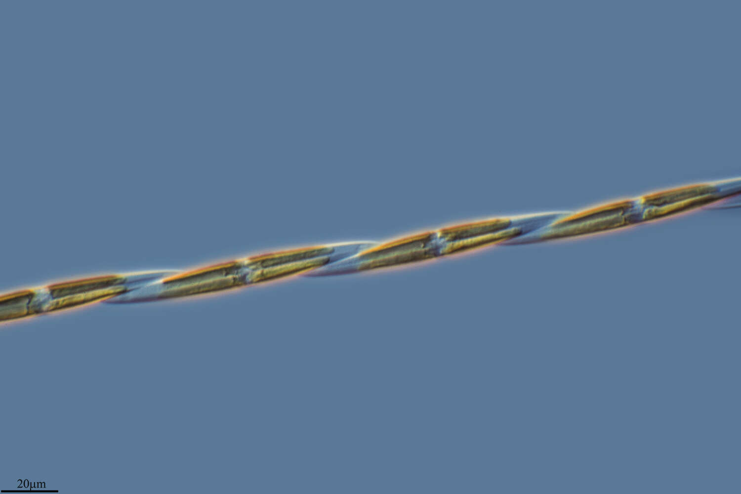
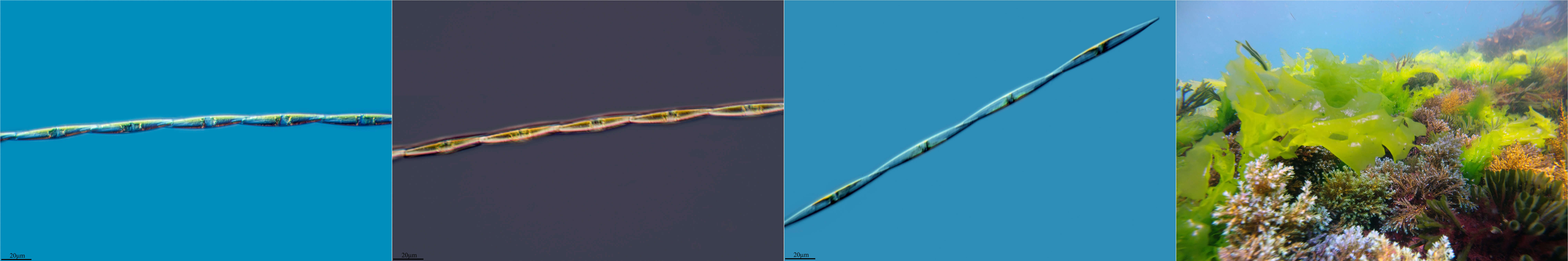
Cylindrotheca is a elongate pennate diatom usually found in sediments. Pennate diatoms are important in intertidal and illuminated subtidal sediments in marine ecosystems and primary producers. Pennate diatoms are capable of movement, relying on the raphe to produce thrust. Movement is needed so that diatoms can move towards the light, recover their location after disturbances by overlying water currents, wave actions, animal burial and so on. Several species illustrated to right. All have a siliceous shell (frustule) and chlorophyll a/c rich plastids. Phase contrast.
-
Hoyo de Manzanares, Madrid, Spain
-
Grove, O, Galicia, Spain
-
Fig 3: Pseudonitzschia multiseries Scanned electron micrograph image showing detailed stria, fibulae and poroids.
-
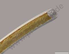
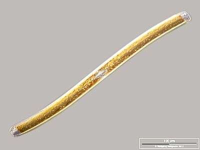
Cingular view. Scale bar indicates 100 µm. Sample from a wetland at the Pillersee (Tyrol, Austria). The image was built up using several photomicrographic frames with manual stacking technique. Images were taken using Zeiss Universal with Olympus C7070 CCD camera.Image under Creative Commons License V 3.0 (CC BY-NC-SA).
-
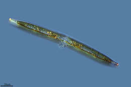
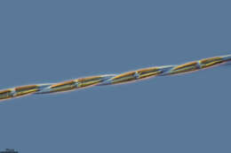
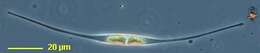
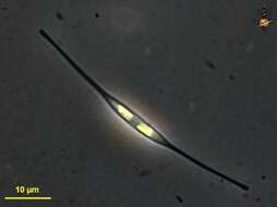
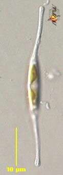
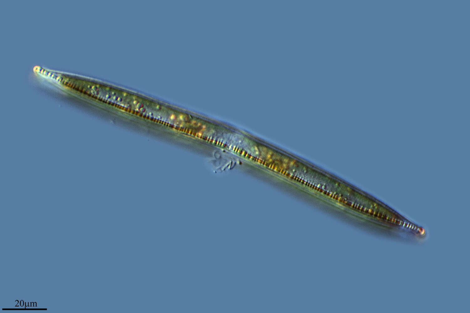
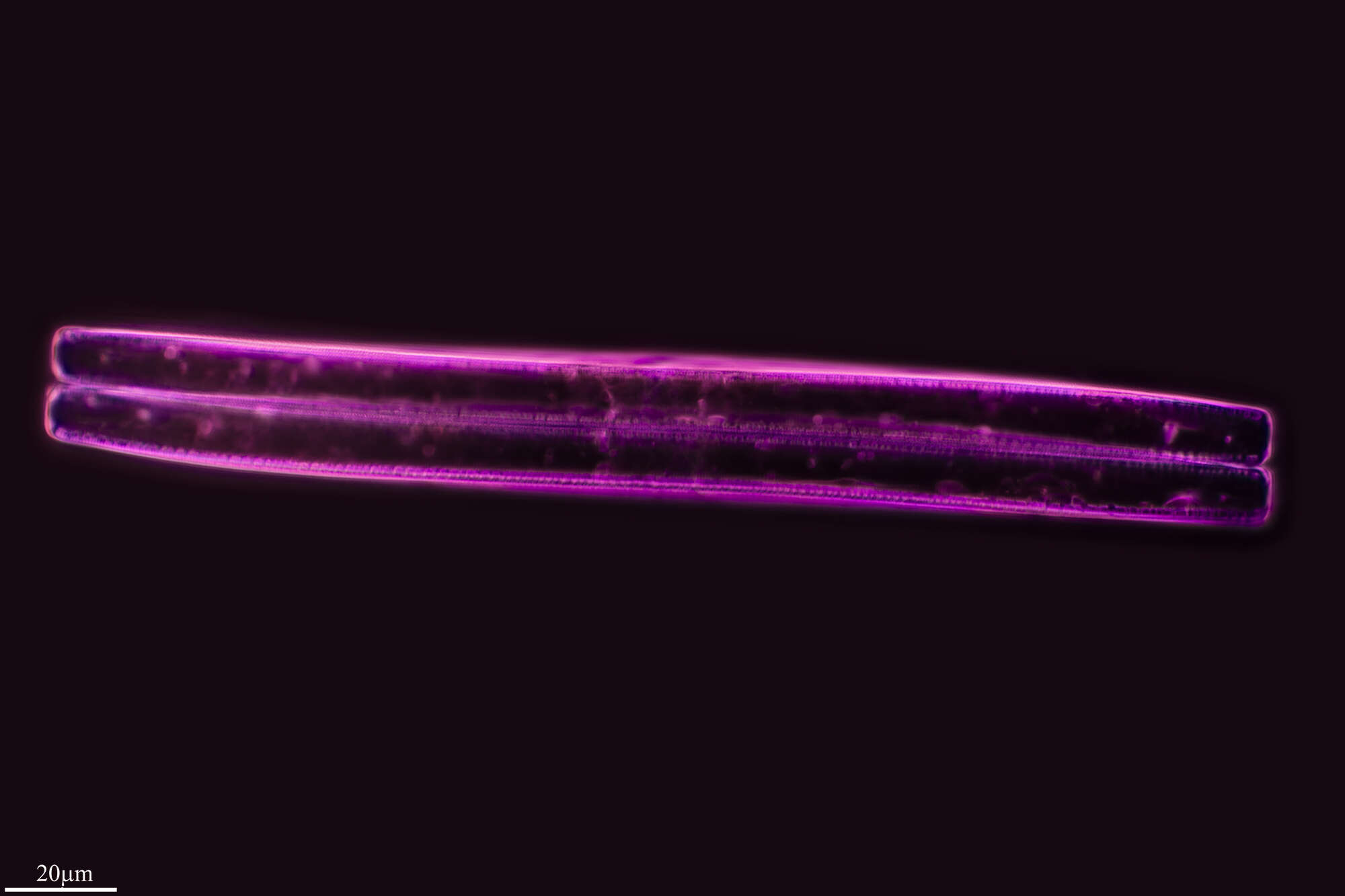
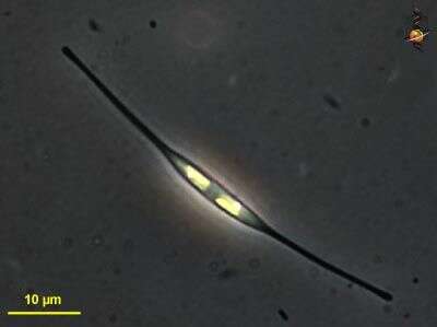
Cylindrotheca. Cell observed in sandy and muddy marine sediments in the vicinity of Broome, Western Australia in September 2003. This image was taken using phase contrast optics. This work was supported by the Australian Biological Resources Study.
-
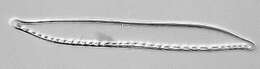
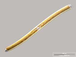
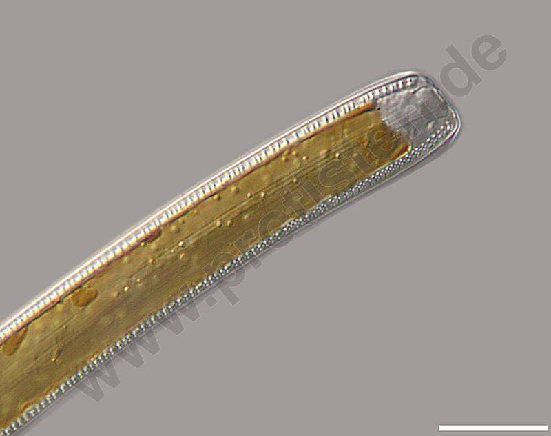
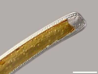
Nitzschia sigmoidea.Detail: Apex in cingular view, displaying cingulum (central strae and the two canal raphes on the edges. Scale bar indicates 25 m.Sample from a wetland at the Pillersee (Tyrol, Austria). The image was built up using several photomicrographic frames with manual stacking technique. Images were taken using Zeiss Universal with Olympus C7070 CCD camera.For more look at
www.protisten.de/english/gallery_main/gallery_main.htmlFor high-resolution images please ask postmaster@protisten.de.
-
Grove, O, Galicia, Spain
-
Fig 4: Pseudonitzschia multiseries Scanned electron micrograph image showing detail of the stria.
-
Detail: Apex in cingular view, displaying cingulum (central strae and the two canal raphes on the edges. Scale bar indicates 25 µm. Sample from a wetland at the Pillersee (Tyrol, Austria). The image was built up using several photomicrographic frames with manual stacking technique. Images were taken using Zeiss Universal with Olympus C7070 CCD camera.Image under Creative Commons License V 3.0 (CC BY-NC-SA).
-
Madrid, Madrid, Spain
-
Grove, O, Galicia, Spain
-
Fig 1: Schematic drawing of the cell in the valve view.