-
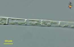
Mougeotia (moo-gee-oh-she-a) a filamentous green alga. With cellulosic cell walls and chloroplasts containing chlorophylls a and b. Differential interference contrast.
-
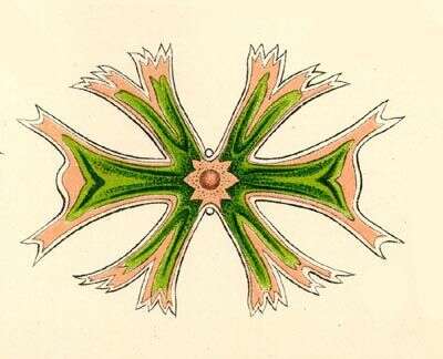
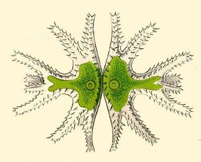
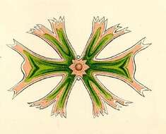
Micrasterias melitensis.
-
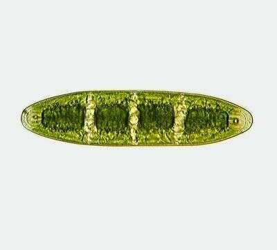
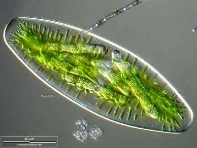
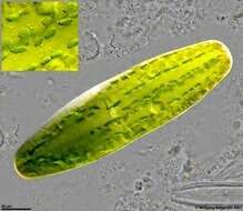
Netrium interruptum (BREB.) LÃTKEM. The cells are 4-5 times longer than wide. The central part is cylindrical and gradually converging towards the broadly rounded off ends. The terminating vacuoles are big with a spherical crystal each. The chromatophores are clearly partitioned in four segments. Dimension: Length 200 â 350 µm, width 30 â 70 µm Ecology: Sporadic in little acidic fens and bogs, in addition, in the plankton of oligotrophic lakes. Occurrence: Ubiquitous, apart from Australia
-
Scale bar indicates 50 µm. Sample from sphagnum pond situated in the northern alpine region of Austria near Salzburg. Images were taken using Zeiss Universal with Olympus C7070 CCD camera.
-
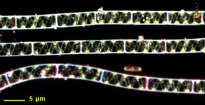
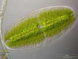
Zygotes after Conjugation. Scale bar indicates 100 µm.Sample from a wetland at the Pillersee (Tyrol, Austria). The image was built up using several photomicrographic frames with manual stacking technique. Images were taken using Zeiss Universal with Olympus C7070 CCD camera.Image under Creative Commons License V 3.0 (CC BY-NC-SA).
-
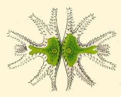
Micrasterias trigemina.
-
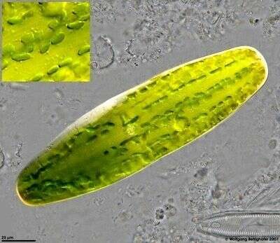
Netrium digitus has stelloid chloroplasts with pyrenoid grains at the center axes of the two chloroplasts. This high resolution depth of focus picture using Planapo 63/1.4 gives a synoptic view at the tongues of stelloid chloroplasts, the nucleus situated in center of the cell and (look at higher magnification inset) mitochondria on inner surface of cell wall (light circular dots). Another DOF taking in zip archive showes median layer with nucleus and pyrenoids. Scale bar indicates 25 µm. See zip archive for high resolution details. Sample from spagnum pond situated in the northern alpine region of Austria near Salzburg. Images were taken using Zeiss Universal with Olympus C7070 CCD camera.
-
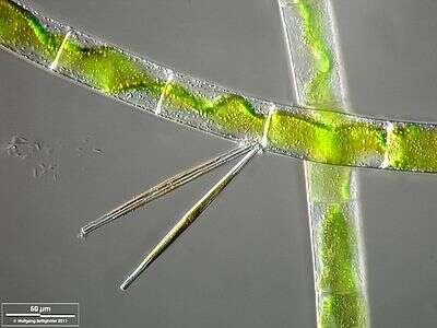
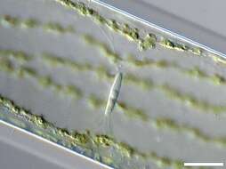
Mougeotia accompanied by the diatom Fragilaria ulna. The scale bar indicates 50 µm. The specimen was gathered in the wetlands of Oderbruch (Oder valley 100 km north east of Berlin). The image was built up using several photomicrographic frames with manual stacking technique. Images were taken using Zeiss Universal with Olympus C7070 CCD camera.Image under Creative Commons License V 3.0 (CC BY-NC-SA).
-
Scale bar indicates 50 µm. Sample from sphagnum pond situated in the northern alpine region of Austria near Salzburg. Images were taken using Zeiss Universal with Olympus C7070 CCD camera.
-
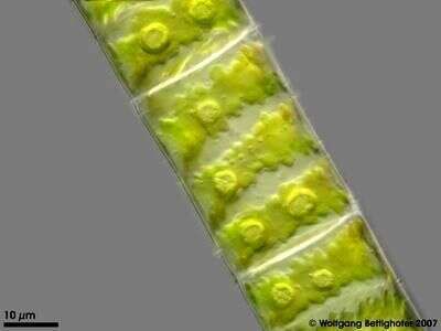
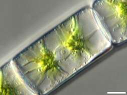
Zygnema, filamentous green algae with star shaped chloroplasts. Enclosed in cellulosic cell wall. Each plastid contains a pyrenoid. Nucleus lies between plastids. Differential interference contrast.
-
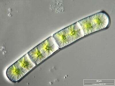
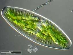
Multilayer image shows cell with lots of fungal sporangia (parasitism). Scale bar indicates 50 µm.Sample from ponds situated in the vicinity of Lake Constance (Bodensee, Southern Germany). The image was built up using several photomicrographic frames with manual stacking technique. Images were taken using Zeiss Universal with Olympus C7070 CCD camera.
-
The protective gelatinous layer around the cell is visible. Scale bar indicates 50 µm.Sample from the pond Hegne Moor situated in the vicinity of Lake Constance. The image was built up using several photomicrographic frames with manual stacking technique. Images were taken using Zeiss Universal with Olympus C7070 CCD camera.Image under Creative Commons License V 3.0 (CC BY-NC-SA).
-
Zoom-in. You can see the two stellate chloroplasts and amidst, the nucleus with its central nucleolus. Scale bar indicates 25 µm.Sample from the pond Hegne Moor situated in the vicinity of Lake Constance. The image was built up using several photomicrographic frames with manual stacking technique. Images were taken using Zeiss Universal with Olympus C7070 CCD camera.Image under Creative Commons License V 3.0 (CC BY-NC-SA).
-
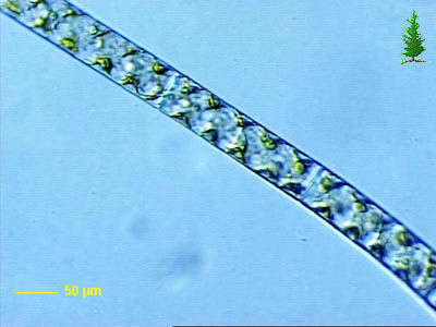
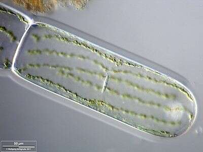
In Spyrogyra, the plastids (a type of chloroplast) are spiral around the interior of the cell. The nucleus is slightly to the right of the center. This alga was collected from the Gardner River near Sheepeater Cliffs.
-
Spirogyra observed in freshwater sediments in the vicinity of Broome, Western Australia in September 2003. This image was taken using phase contrast optics. This work was supported by the Australian Biological Resources Study.
-
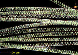
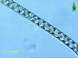
Dark ground illumination of this filamentous green alga. The chloroplasts are ribbon-like and spiral around the interior of the cell wall.
-
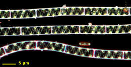
Small clump of filaments with spiral plastids viewed using dark-ground illumination.
-
Collected from Cumloden Swamp on October 7, 2002.
-
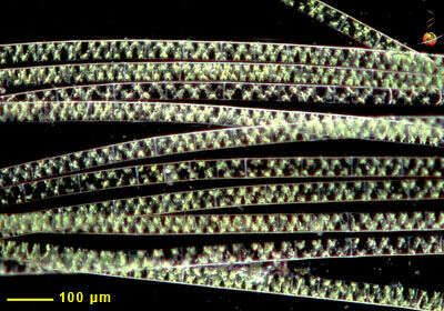
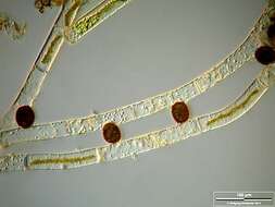
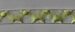
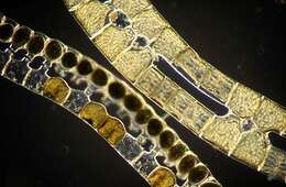
Dark ground image of preserved filaments showing various stages of the conjugation process. Paired filaments caught during conjugation are above, filaments with zygotes are below.
-
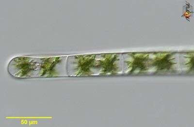
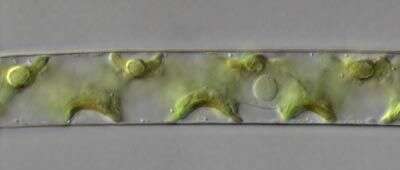
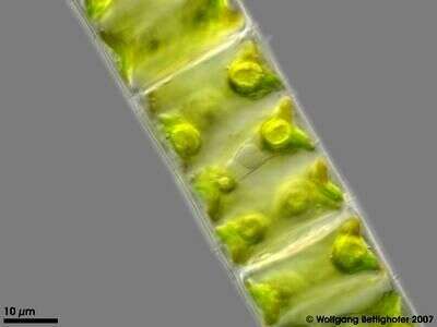
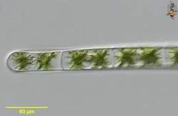
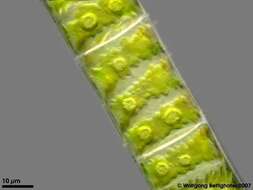
Part of a filament showing the shape of the ribbon like chloroplast with pyrenoids. Multi layer image (DOF) using 5 frames generating depth of focus, stacked manually using Corel Photopaint. Aufwuchs on roots dangling in a creek´s water. This image was taken using Zeiss Universal with Olympus C7070 CCD camera.
-
Part of a filament showing the nucleus and chloroplast´s pyrenoids. Multi layer image (DOF) using 3 frames generating depth of focus, stacked manually using Corel Photopaint. Aufwuchs on roots dangling in a creek´s water. This image was taken using Zeiss Universal with Olympus C7070 CCD camera.
-
Scale bar indicates 50 µm.Sample from the pond Hegne Moor situated in the vicinity of Lake Constance. The image was built up using several photomicrographic frames with manual stacking technique. Images were taken using Zeiss Universal with Olympus C7070 CCD camera.Image under Creative Commons License V 3.0 (CC BY-NC-SA).
-
Zoom-in. The nucleus with its central nucleolus is visible. The suspension of the nucleus with fine cytoplasmic filaments indicates that almost the whole cell is filled with a large water vacuole. The parietal chloroplast is also lying surrounded by cytoplasm. Scale bar indicates 25 µm.Sample from the pond Hegne Moor situated in the vicinity of Lake Constance. The image was built up using several photomicrographic frames with manual stacking technique. Images were taken using Zeiss Universal with Olympus C7070 CCD camera.Image under Creative Commons License V 3.0 (CC BY-NC-SA).
-
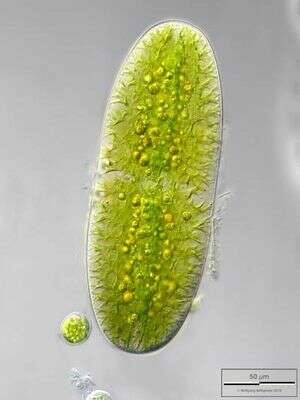
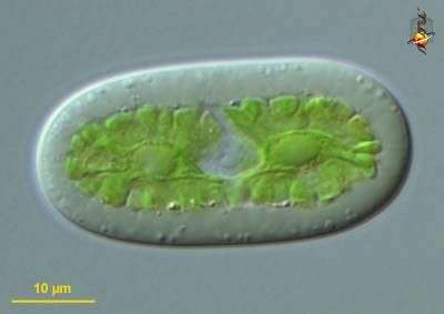
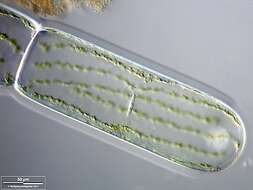
Netrium (knee-tree-um) a simple desmid cell - a type of green alga having, like other green algae, a cellulosic cell wall and chloroplasts with chlorophyll B. Although a desmid, without the more usual mirror-image form. Central nucleus and two flat crenulated plastids. The similar Cylindrocystis differs because it has star-shaped plastids. Differential interference contrast.