-
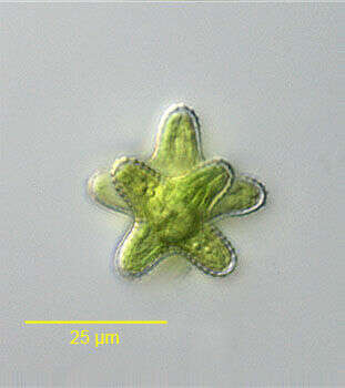
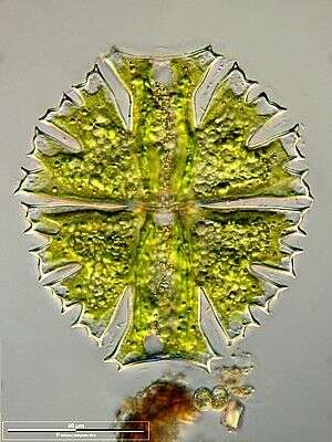
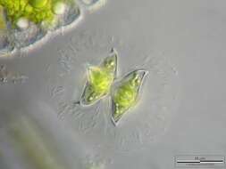
End view Staurastrum dilatatum (Ehrenberg,1838) Ralfs, 1848.The four-armed semi-cells are rotated 45 degrees with respect to each other.From freshwater aquaculture tub near Boise, Idaho December 2005. DIC.
-
Differential interference contrast.
-
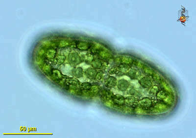
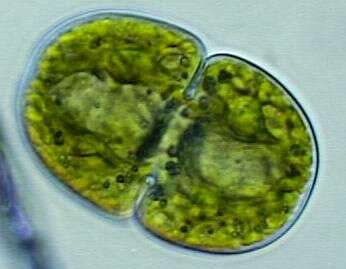
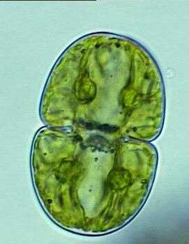
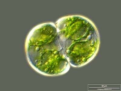
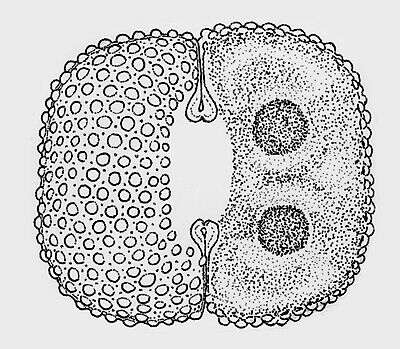
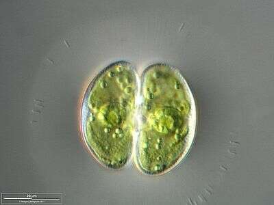
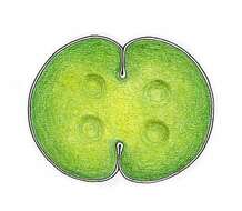
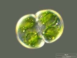
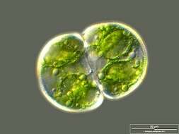
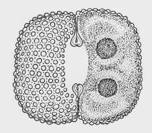
Cosmarium, one of the many desmids - most of which have the appearance of mirror imaged cells joined together, but typically with only one nucleus. With cellulose cell wall, bright green chloroplasts. Differential interference contrast.
-
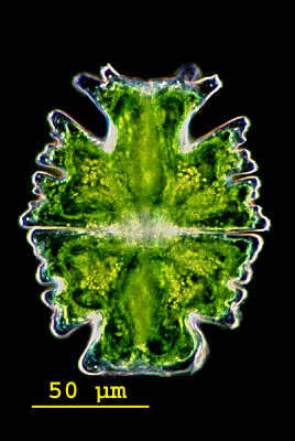
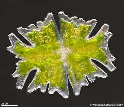
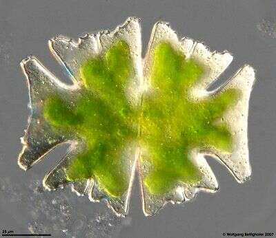
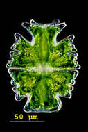
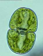
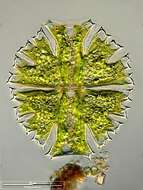
Micrasterias(mike-raz-tear-ee-ass) is a genus of unicellular algae in the family Desmidiaceae. The cells are flattened and disc-like. The cells of the genus Micrasterias are organized in two semi-cells that are mirror images of each other. The semicells have a distinctive shape with an intricate lobes and indentation. At the end of the lobes the cell wall may sometimes form notches or short spines. The nucleus is located in the centre between the semicells. Each semicell has a chloroplast with some pyrenoids. Usually found in oligotrophic, acid waters. This specimen was collected in a moor located in the Salzburger Land, Austria. Differential interference contrast.
-
Cosmarium pachydermum P. LUNDELL The cells are up to 1.4 times longer than wide and coarsely elliptical in shape. The cell halves are semicircular. The central cuts are narrowly rounded on the inside and extend strongly to the outside. The cell wall is thick. Some areas are covered closely, others are covered loosely with standing pores. The vertex view is broadly elliptical. Length 100 - 120 µm, width 70 - 90 µm. Occurrence: acidophilic alga, widely spread in sphagnum bogs
-
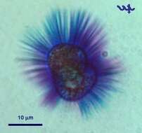
The image shows the jelly envelope with symbiontic bacteria. Scale bar indicates 25 µm. Sample from sphagnum pond situated in the northern alpine region of Austria near Salzburg. Images were taken using Zeiss Universal with Olympus C7070 CCD camera.
-
Differential interference contrast.
-
Differential interference contrast.
-
Micrasterias(mike-raz-tear-ee-ass) is a genus of unicellular algae in the family Desmidiaceae. The cells are flattened and disc-like. The cells of the genus Micrasterias are organized in two semi-cells that are mirror images of each other. The semicells have a distinctive shape with an intricate lobes and indentation. At the end of the lobes the cell wall may sometimes form notches or short spines. The nucleus is located in the centre between the semicells. Each semicell has a chloroplast with some pyrenoids. Usually found in oligotrophic, acid waters. This specimen was collected in a moor located in the Salzburger Land, Austria. Dark ground illumination.
-
Focus on cellwall surface. Scale bar indicates 50 µm.Sample from the pond Hegne Moor situated in the vicinity of Lake Constance. The image was built up using several photomicrographic frames with manual stacking technique. Images were taken using Zeiss Universal with Olympus C7070 CCD camera.Image under Creative Commons License V 3.0 (CC BY-NC-SA).
-
Collected from Cumloden Swamp on August 28, 2002.
-
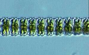
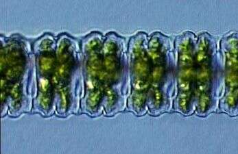
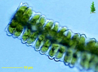
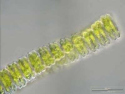
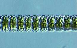
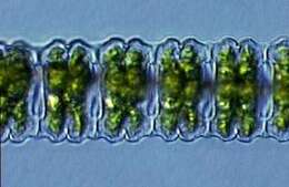
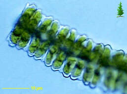
Colonial desmid in which adjacent cells link together to form elongate filaments. Differential interference contrast image.
-
Differential interference contrast.
-
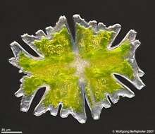
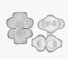
Micrasterias americana shows a very special morphological phenomenon. The four minor processes of apical lobes are optional. But if some of them are formed they show rotational symmetry according to direction of the appendices. This depth of focus picture assembling 26 high resolution shots showes a specimen with all four processes formed. See zip archive for details. Sample from sphagnum pond situated in the northern alpine region of Austria near Salzburg. Images were taken using Zeiss Universal with Olympus C7070 CCD camera.
-
Optical transversal section showing chloroplast. Scale bar indicates 50 µm.Sample from the pond Hegne Moor situated in the vicinity of Lake Constance. The image was built up using several photomicrographic frames with manual stacking technique. Images were taken using Zeiss Universal with Olympus C7070 CCD camera.Image under Creative Commons License V 3.0 (CC BY-NC-SA).
-
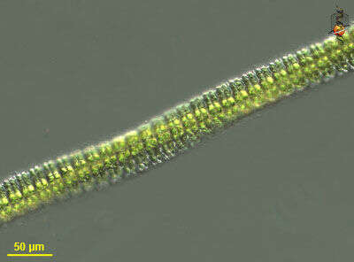
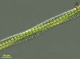
Multilayer image of the filamentous colony showing the triangular shape of the single cells. Scale bar indicates 50 µm. Sample from sphagnum pond situated in the northern alpine region of Austria near Salzburg. Images were taken using Zeiss Universal with Olympus C7070 CCD camera.
-
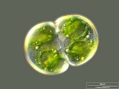
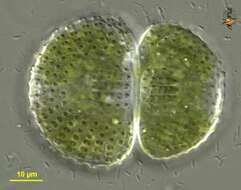
Cosmarium observed in freshwater sediments in the vicinity of Broome, Western Australia in September 2003. This image was taken using differential interference contrast optics. This work was supported by the Australian Biological Resources Study.
-
Micrasterias americana shows a very special morphological phenomenon. The four minor processes of apical lobes are optional. But if some of them are formed they show rotational symmetry according to direction of the appendices. This depth of focus picture assembling 14 high resolution shots showes a specimen with one appendix formed. See zip archive for details. Sample from spagnum pond situated in the northern alpine region of Austria near Salzburg. Images were taken using Zeiss Universal with Olympus C7070 CCD camera.
-
Cosmarium protractum (NÃG.) DE BARY var. procerum LENZENW. The cells are only little longer than wide. The cell halves form three lobes, the lateral lobes are unequally rounded off, whereby they appear gently rolling. The vertex lobes are slightly widened outwards, rounded off laterally and slightly concave in the center. The central cuts are deep and outward extended. At the basis of both cell halves there is a hump, which is covered with larger verrucae. The remaining cell wall is covered with smaller warts. The vertex view is oblong oval with one distinct surface bulg each at the sides. Length 95 - 100 µm, width 55 - 65 µm. Occurrence: New description, well-known only from Greenland so far.
-
Scale bar indicates 50 µm.Sample from a wetland at the Pillersee (Tyrol, Austria). The image was built up using several photomicrographic frames with manual stacking technique. Images were taken using Zeiss Universal with Olympus C7070 CCD camera.Image under Creative Commons License V 3.0 (CC BY-NC-SA).
-
A new desmid, (Cosmarium?) that recently appeared in the plankton of Lake Kinneret. Methylene blue staining shows a âhallowâ of thin mucilaginous strands extending out of the cell.
-
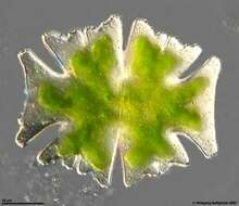
Micrasterias americana (EHR.) RALFS. Length 130 - 150 µm, width 100 - 120 µm. Not rare both in lowlands and in alpine waters, sometimes abundant.
-
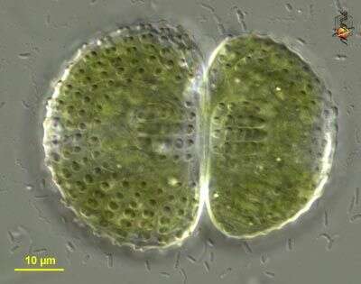
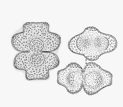
Cosmarium quadrum P. LUNDELL The cells are a little longer than wide, rounded off rectangular in shape. The sides are straight or flat convex. The central cuts are deep, linear, and extended outwards. The cell wall is covered with warts. They run in crossing rows (under an angle of appr. 45°). Around each of the warts are pores, which build symmetrical hexagon in their arrangement. Length 55 - 85 µm, width 50 - 80 µm. Occurrence: Common in littoral region and quaking bogs of moor ponds and in moderate acidic moorlands in Central Europe.
-
Scale bar indicates 25 µm.Sample from the pond Hegne Moor situated in the vicinity of Lake Constance. The image was built up using several photomicrographic frames with manual stacking technique. Images were taken using Zeiss Universal with Olympus C7070 CCD camera.Image under Creative Commons License V 3.0 (CC BY-NC-SA).