-
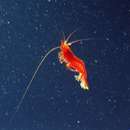
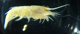
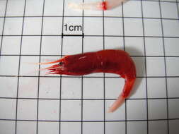
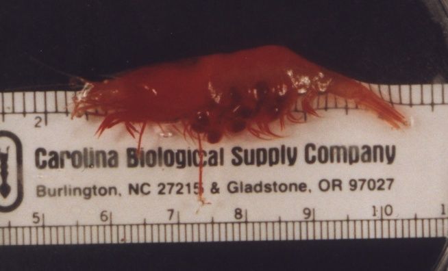
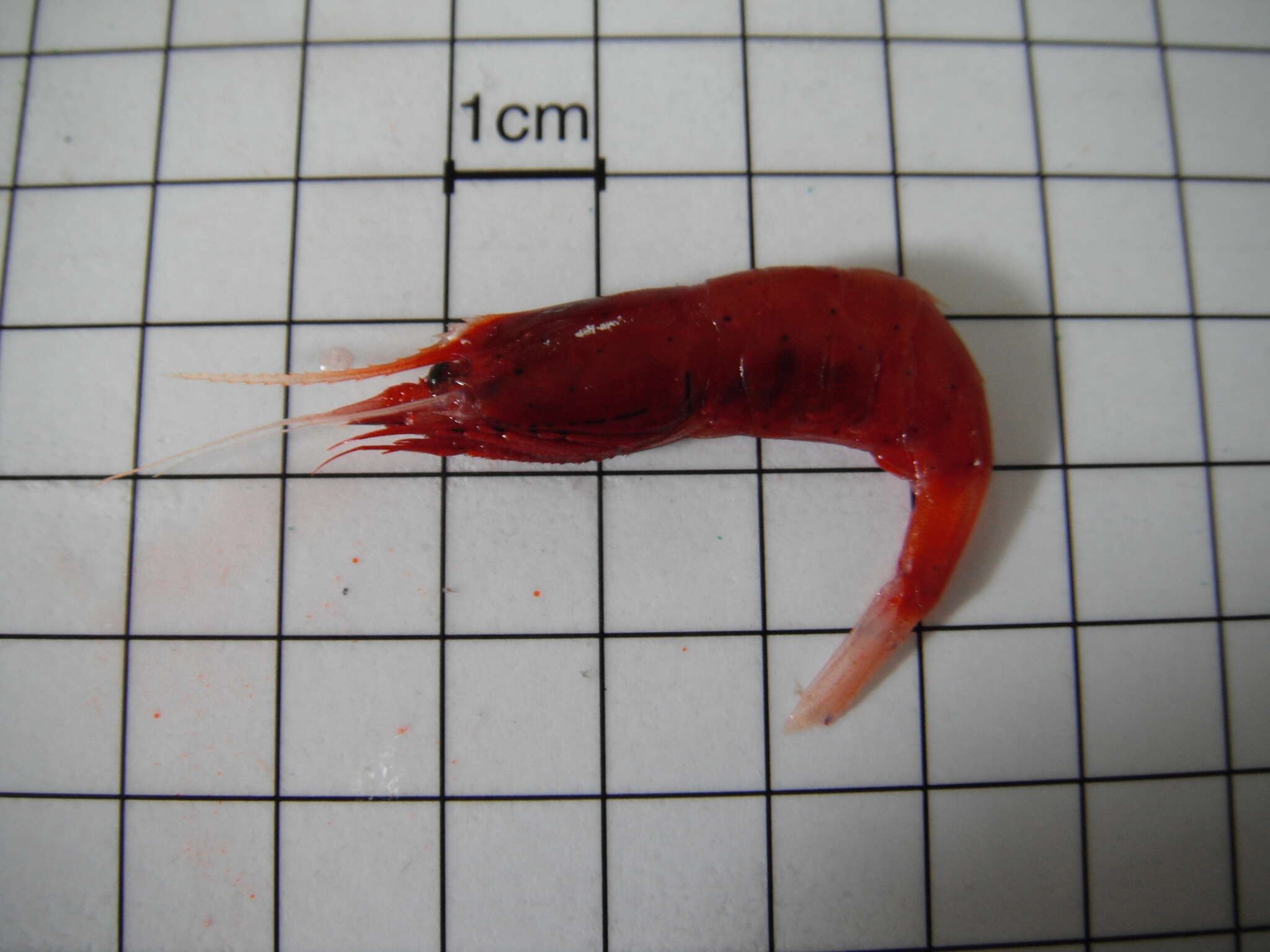
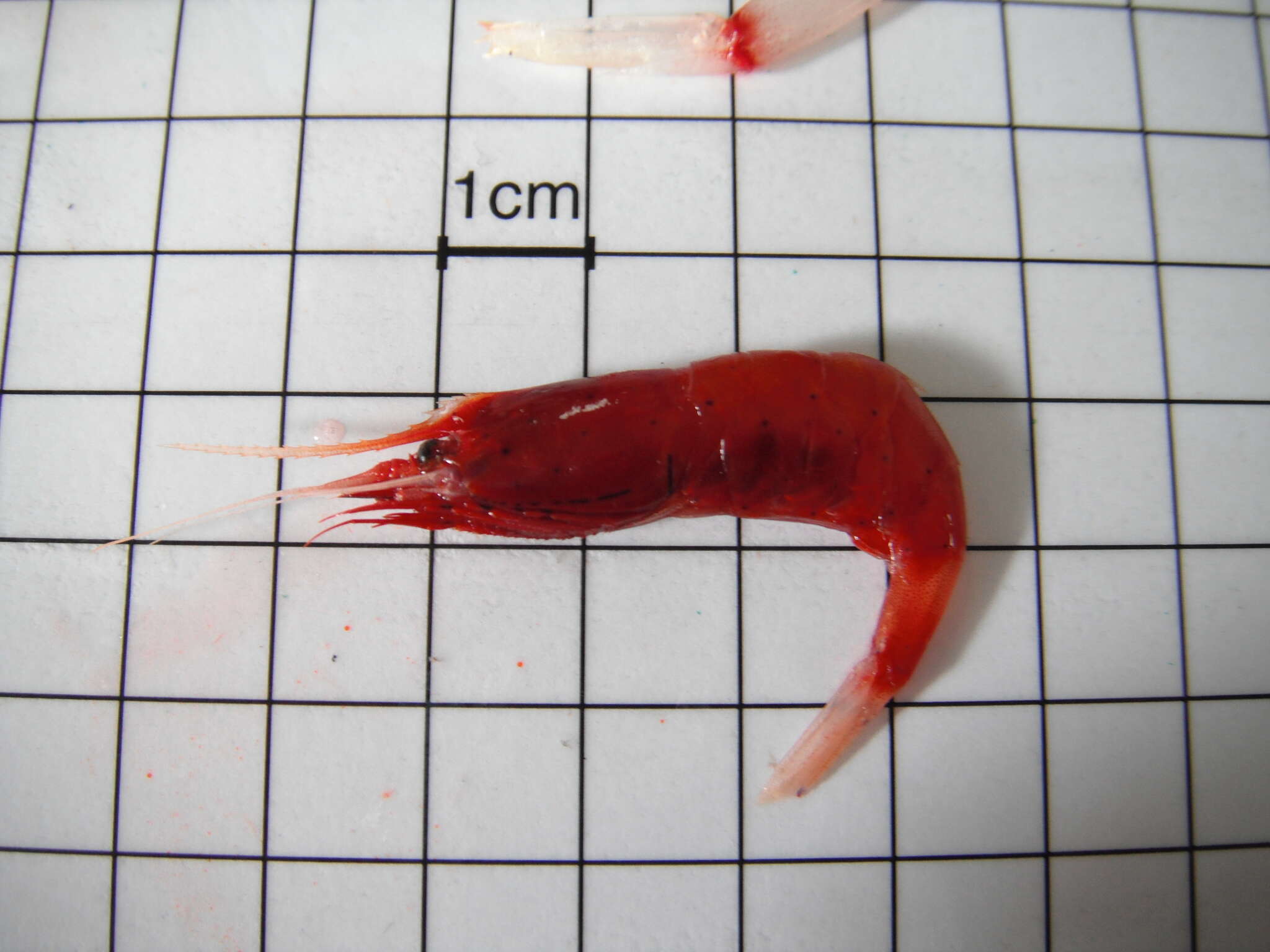
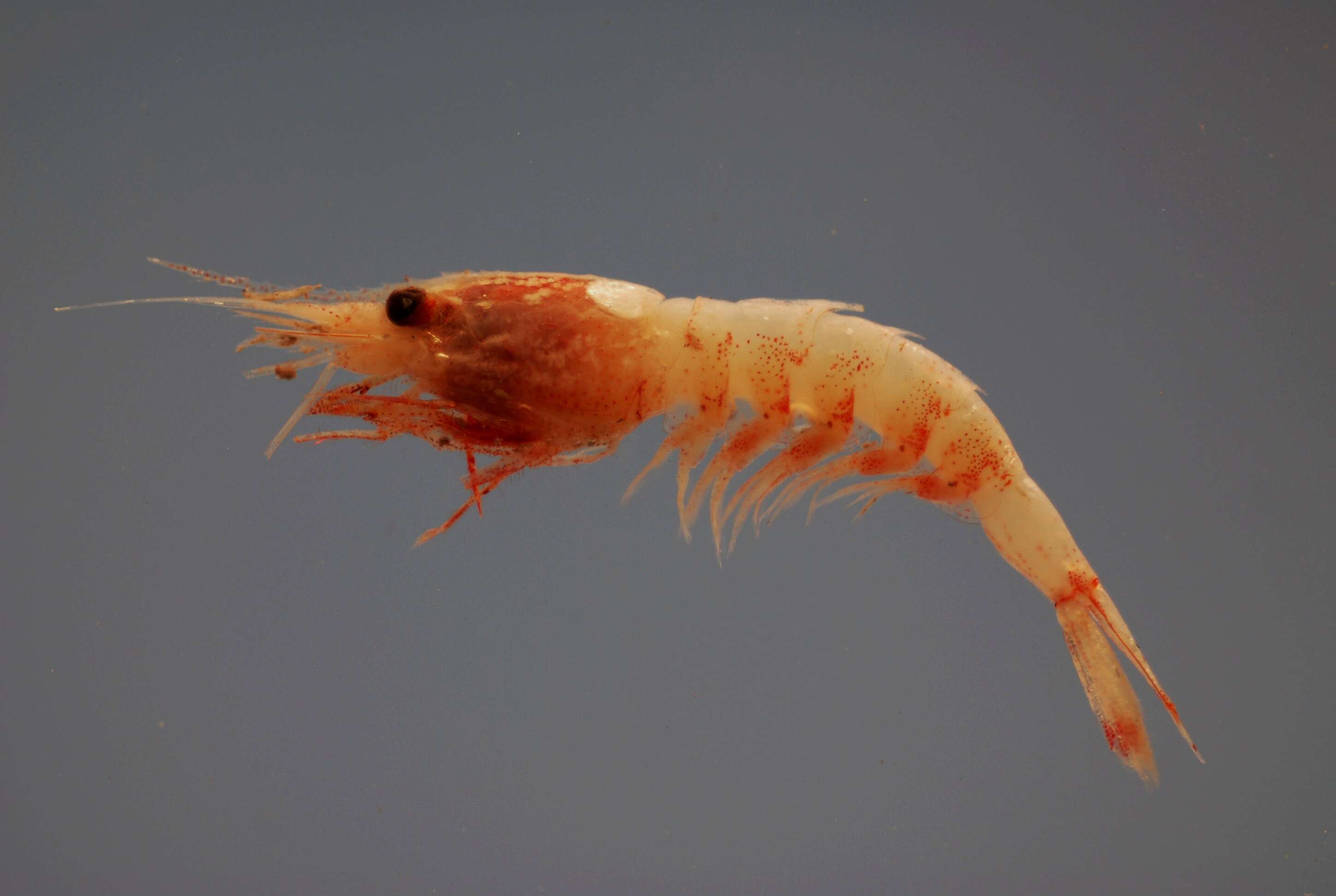
Acanthephyra curtirostris, caught at about 500 m depth off Point Conception, CA (Photo by: Dave Cowles, May 1996)
-
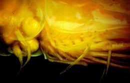
This side view of a preserved specimen shows the rostrum which extends well beyond the corneas of the eyes and even exceeds the peduncle of the first antenna. Note also the pereopods. Oplophorids, unlike most other families of true shrimp, has exopods (exopodites) on its pereopods. The exopodites of the pereopods are short, curved backward and used for swimming. The endopodites of the pereopods are longer, extended forward, and used for manipulating objects. The eye pigment in Hymenodora is always pale, even in living specimens.
-
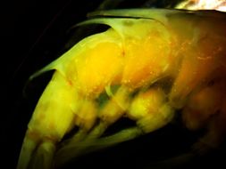
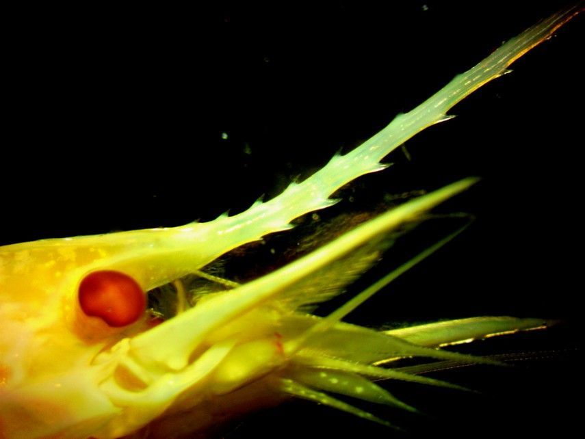
As with other Oplophorus species, O. spinosus has a long rostrum with many spines on both the dorsal and ventral surface. Note the well-developed eye as well.
-
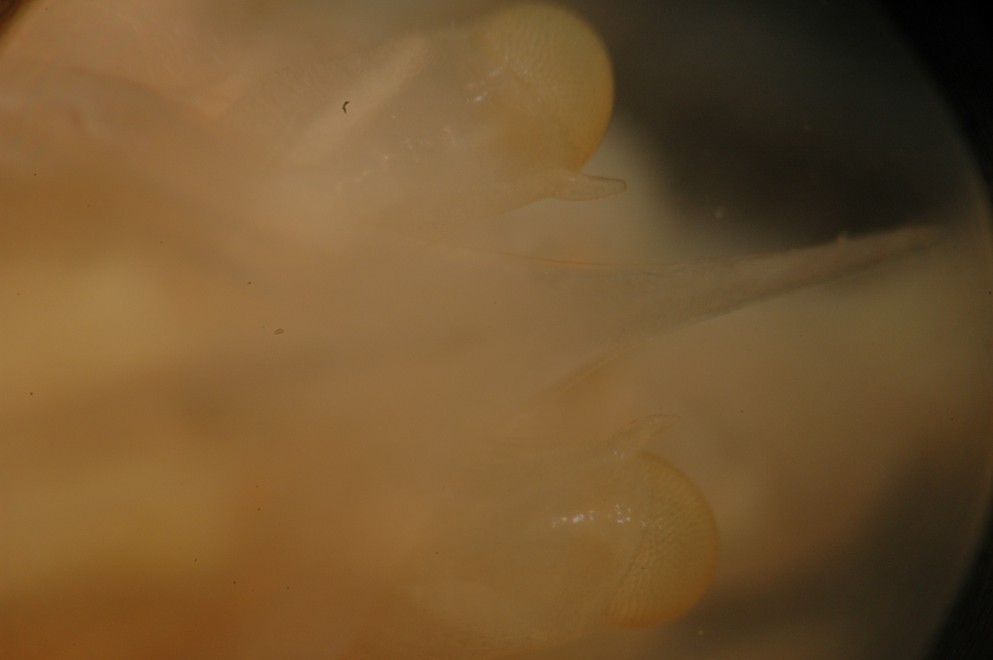
In this closeup dorsal view of the head, the median tubercles on the eyestalks near the corneas can be seen. From a presereved specimen.
-
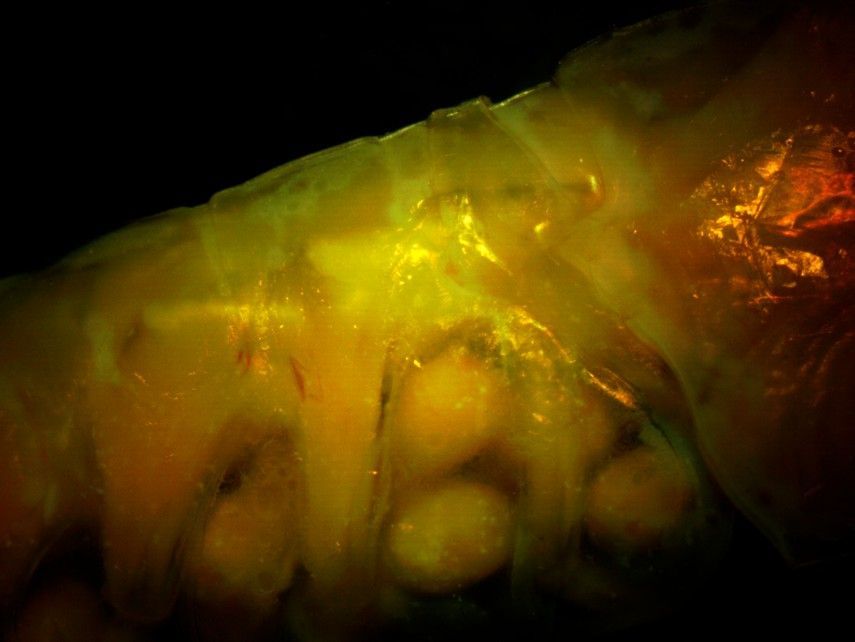
This right-side view of the anterior (first 3) abdominal segments (the posterior thorax is at the right side) shows that the second abdominal segment does not have a large posterior mid-dorsal spine. The large eggs can be seen attached to the pleopods.
-
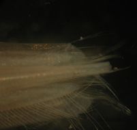
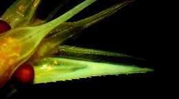
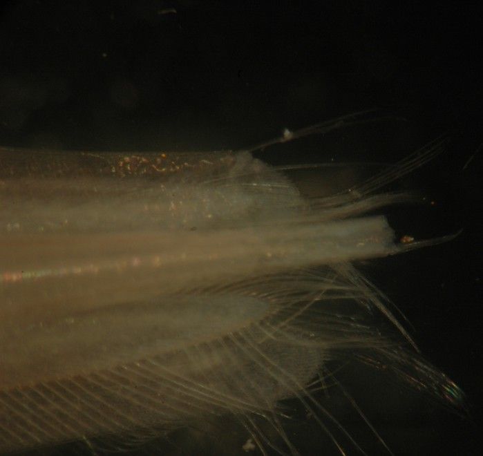
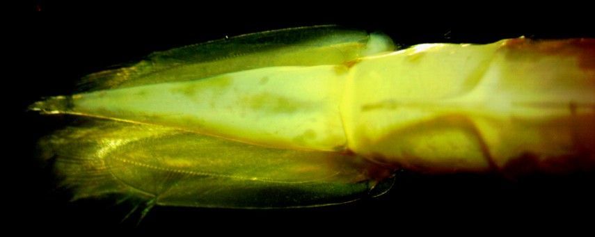
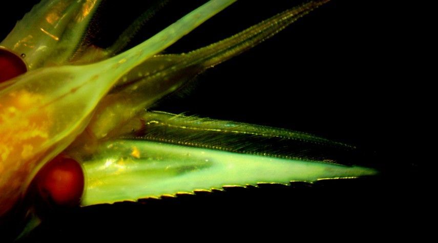
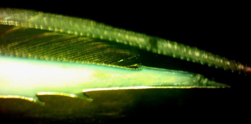
This is a closeup dorsal view of the telson and uropods. The uropods are shorter than the telson and fringed with long setae. The telson is truncate (not rounded) on the end, with two long spines at the corners (one of which is broken off on this individual). Photo of a preserved individual.
-
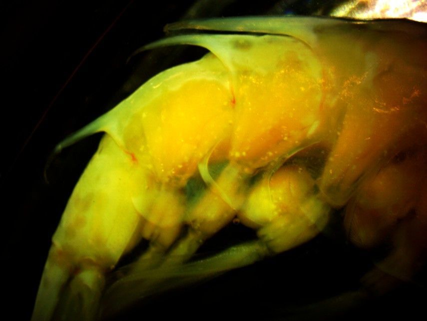
This right-side view of the posterior abdominal segments show the large posterior-pointing mid-dorsal spines on several of them. Abdominal segment 3 is at the top right, then segments 4,5,6, and the base of the telson is visible at the bottom left. Note that segment 6 is not dorsally carinate.
-
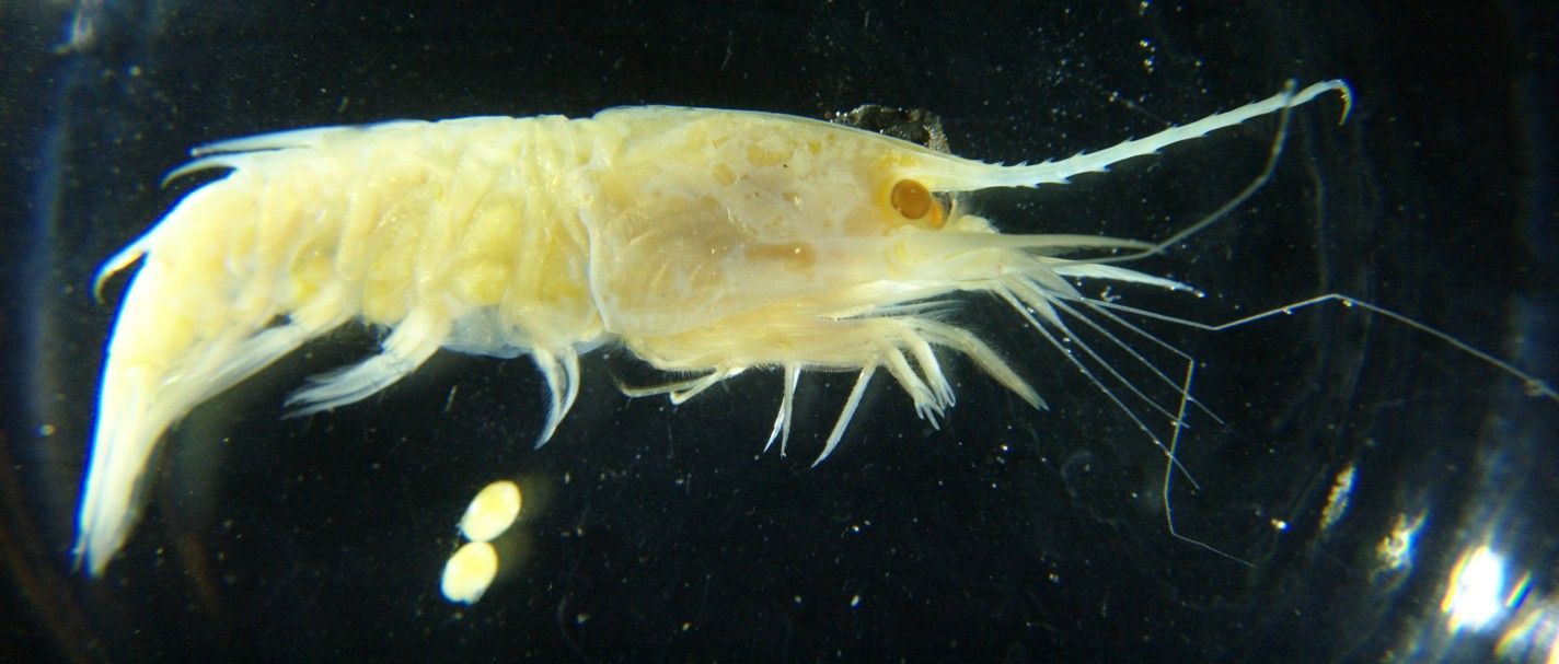
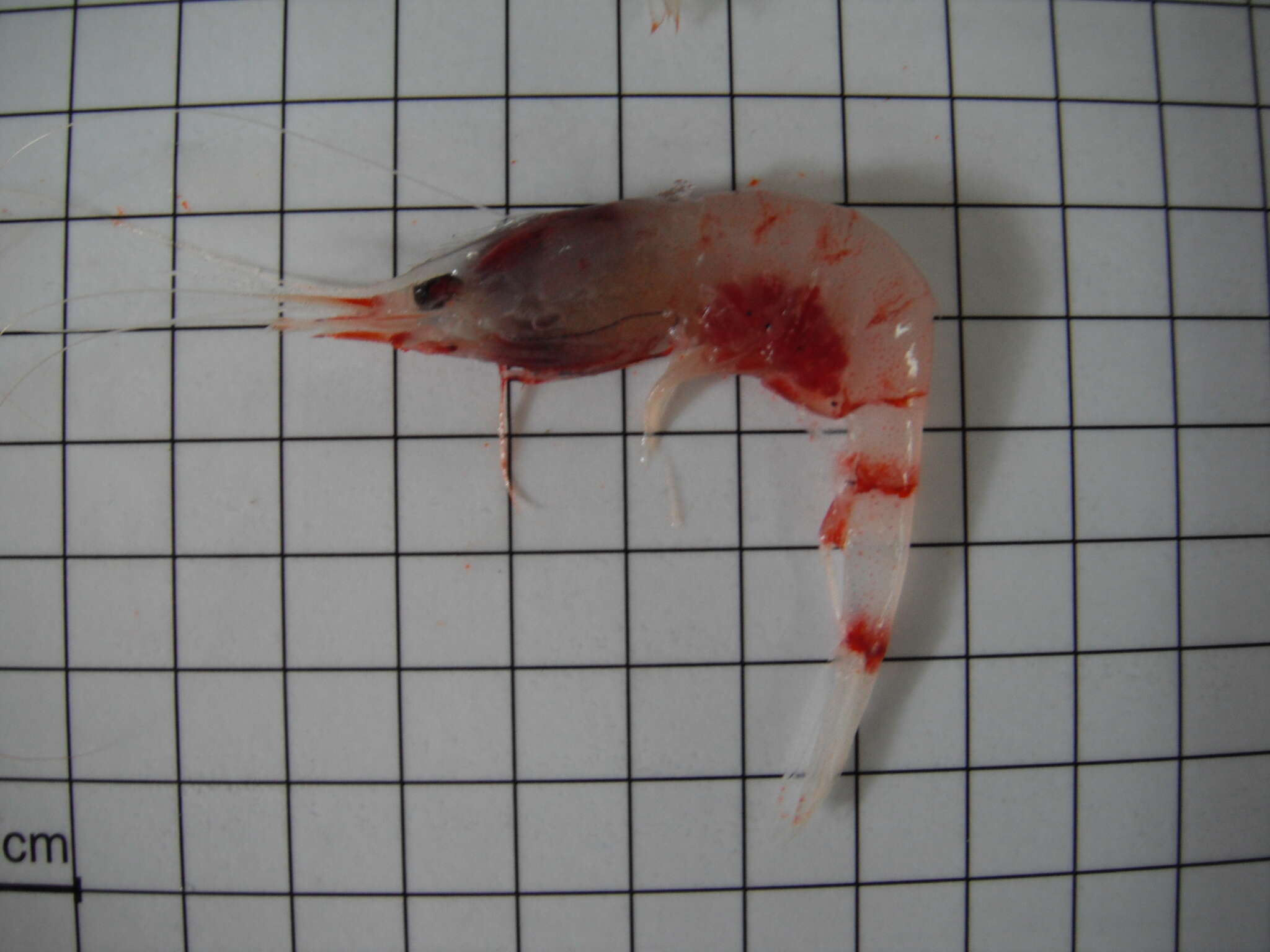
A female Hymenodora frontalis carrying eggs. Caught 1000-1500 m depth off Pt. Conception, CA. Note the yellowish eye, almost devoid of black pigment, and the large egg size. (Photo by: Dave Cowles, May 1995)
-
Dorsal view of the rear abdomen and telson. The rightmost segment is abdominal segment 5. Segment 6 is below the posterior spine from segment 5. To the left are the uropods and telson.
-
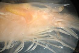
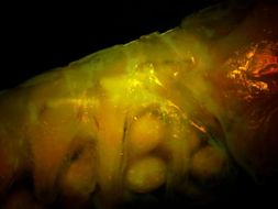
In O. spinosus there is no sharp tooth on the posterior margin of the ventralcarapace. In this view of the animal's right side, the ventral margin of the carapace plus the pereopods can be seen to the right and the first abdominal segment with pleopods and several eggs is visible to the left. The posterior margin of the ventralcarapace has an acutely rounded corner but no sharp tooth.
-
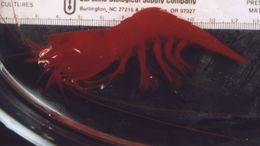
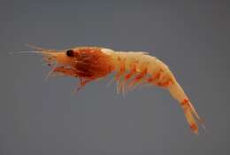
A female Oplophorus spinosus carrying eggs, carapace length 1.5 cm, captured in midwater off Hawaii in 1996. In real life this species would be partly pinkish-red and partly transparent. The specimen above has been preserved in formalin. Several eggs have dropped off the pleopods. (Photo by: Dave Cowles, 2012 )
-
-
-
-
-
-
-
SEFSC Pascagoula Laboratory; Collection of Brandi Noble, NOAA/NMFS/SEFSC.
Wikimedia Commons
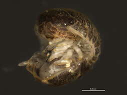
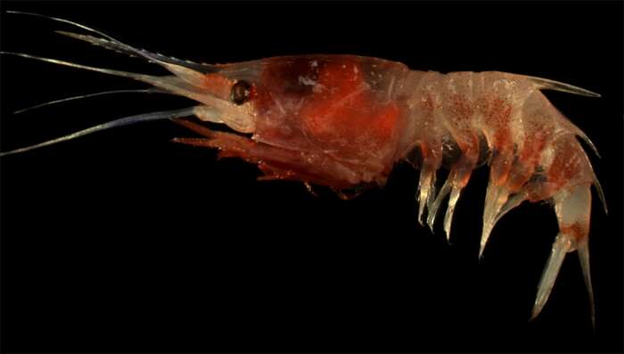
Summary.mw-parser-output table.commons-file-information-table,.mw-parser-output.fileinfotpl-type-information{border:1px solid #a2a9b1;background-color:#f8f9fa;padding:5px;font-size:95%;border-spacing:2px;box-sizing:border-box;margin:0;width:100%}.mw-parser-output table.commons-file-information-table>tbody>tr,.mw-parser-output.fileinfotpl-type-information>tbody>tr{vertical-align:top}.mw-parser-output table.commons-file-information-table>tbody>tr>td,.mw-parser-output table.commons-file-information-table>tbody>tr>th,.mw-parser-output.fileinfotpl-type-information>tbody>tr>td,.mw-parser-output.fileinfotpl-type-information>tbody>tr>th{padding:4px}.mw-parser-output.fileinfo-paramfield{background:#ccf;text-align:right;padding-right:0.4em;width:15%;font-weight:bold}.mw-parser-output.commons-file-information-table+table.commons-file-information-table,.mw-parser-output.commons-file-information-table+div.commons-file-information-table>table{border-top:0;padding-top:0;margin-top:-8px}@media only screen and (max-width:719px){.mw-parser-output table.commons-file-information-table,.mw-parser-output.commons-file-information-table.fileinfotpl-type-information{border-spacing:0;padding:0;word-break:break-word;width:100%!important}.mw-parser-output.commons-file-information-table>tbody,.mw-parser-output.fileinfotpl-type-information>tbody{display:block}.mw-parser-output.commons-file-information-table>tbody>tr>td,.mw-parser-output.commons-file-information-table>tbody>tr>th,.mw-parser-output.fileinfotpl-type-information>tbody>tr>td,.mw-parser-output.fileinfotpl-type-information>tbody>tr>th{padding:0.2em 0.4em;text-align:left;text-align:start}.mw-parser-output.commons-file-information-table>tbody>tr,.mw-parser-output.fileinfotpl-type-information>tbody>tr{display:flex;flex-direction:column}.mw-parser-output.commons-file-information-table+table.commons-file-information-table,.mw-parser-output.commons-file-information-table+div.commons-file-information-table>table{margin-top:-1px}.mw-parser-output.fileinfo-paramfield{box-sizing:border-box;flex:1 0 100%;width:100%}} Description: English: A deepsea shrimp ( Janicella spinicauda ). Gulf of Mexico. Date: 9 February 2007, 03:30. Source:
NOAA Photo Library:
fish4539. Author: SEFSC Pascagoula Laboratory; Collection of Brandi Noble, NOAA/NMFS/SEFSC.
-
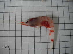
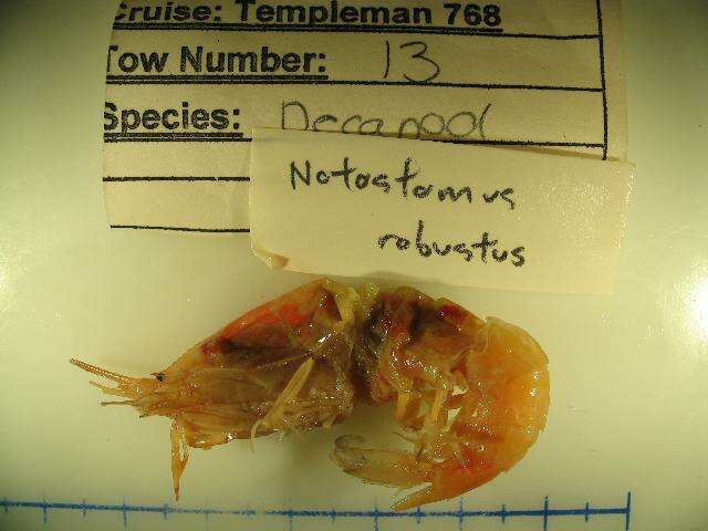
Lateral..
-
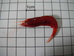
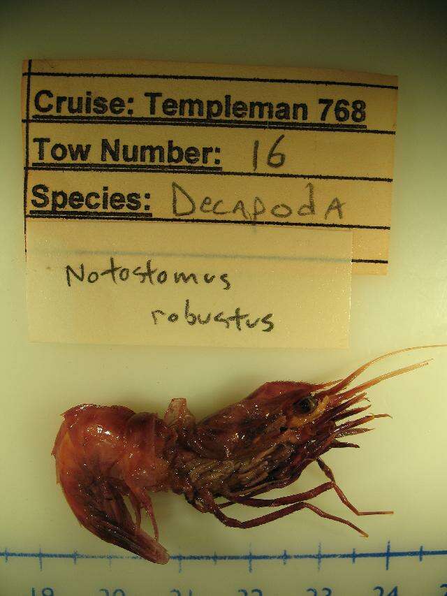
Lateral..
-
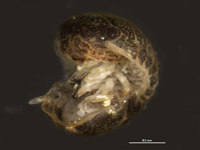
Lateral..
-