-
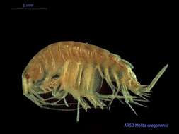
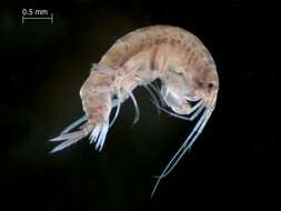
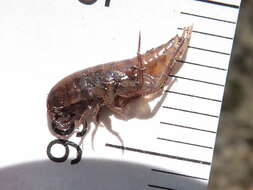
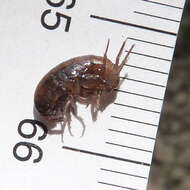
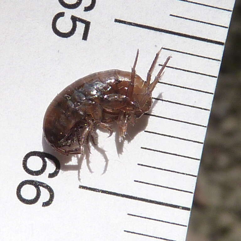
Collected from Puget Sound sediments and photographed by the Washington State Department of Ecologys Marine Sediment Monitoring Team. For more information about this teams work visit:
www.ecy.wa.gov/programs/eap/psamp/index.htm.
-
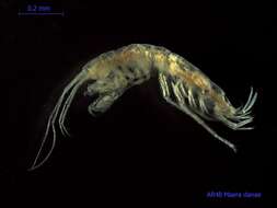
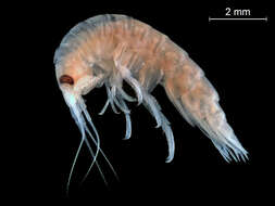
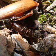
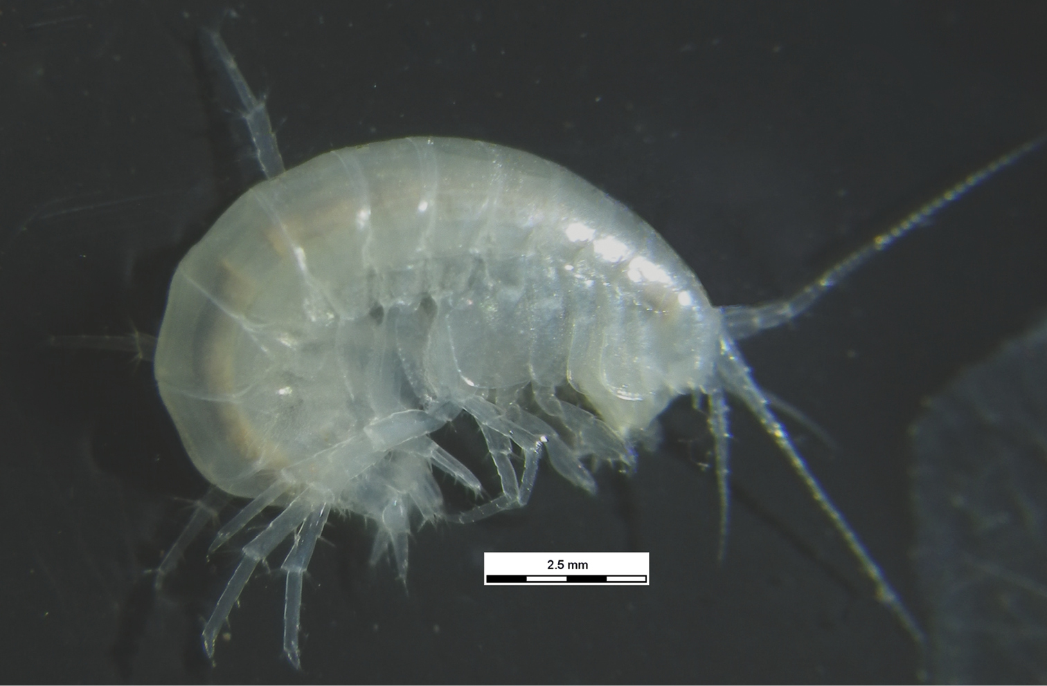
Collected from Puget Sound sediments and photographed by the Washington State Department of Ecologys Marine Sediment Monitoring Team. For more information about this teams work visit:
www.ecy.wa.gov/programs/eap/psamp/index.htm.
-
-
-
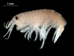
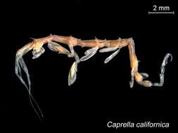
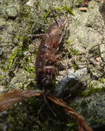
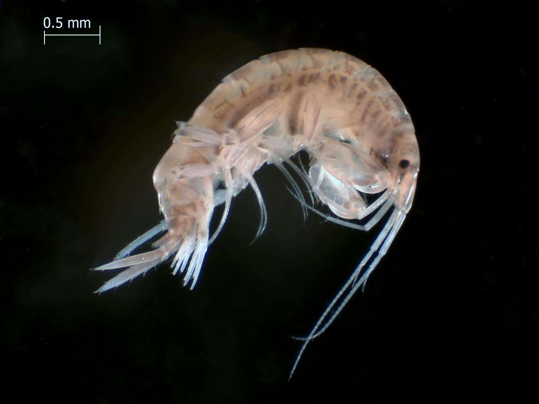
Collected from Puget Sound sediments and photographed by the Washington State Department of Ecologys Marine Sediment Monitoring Team. For more information about this teams work visit:
www.ecy.wa.gov/programs/eap/psamp/index.htm.
-
-
-
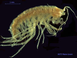
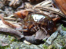
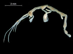
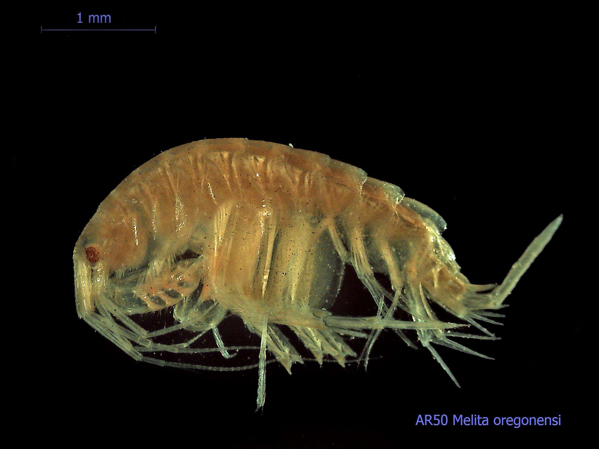
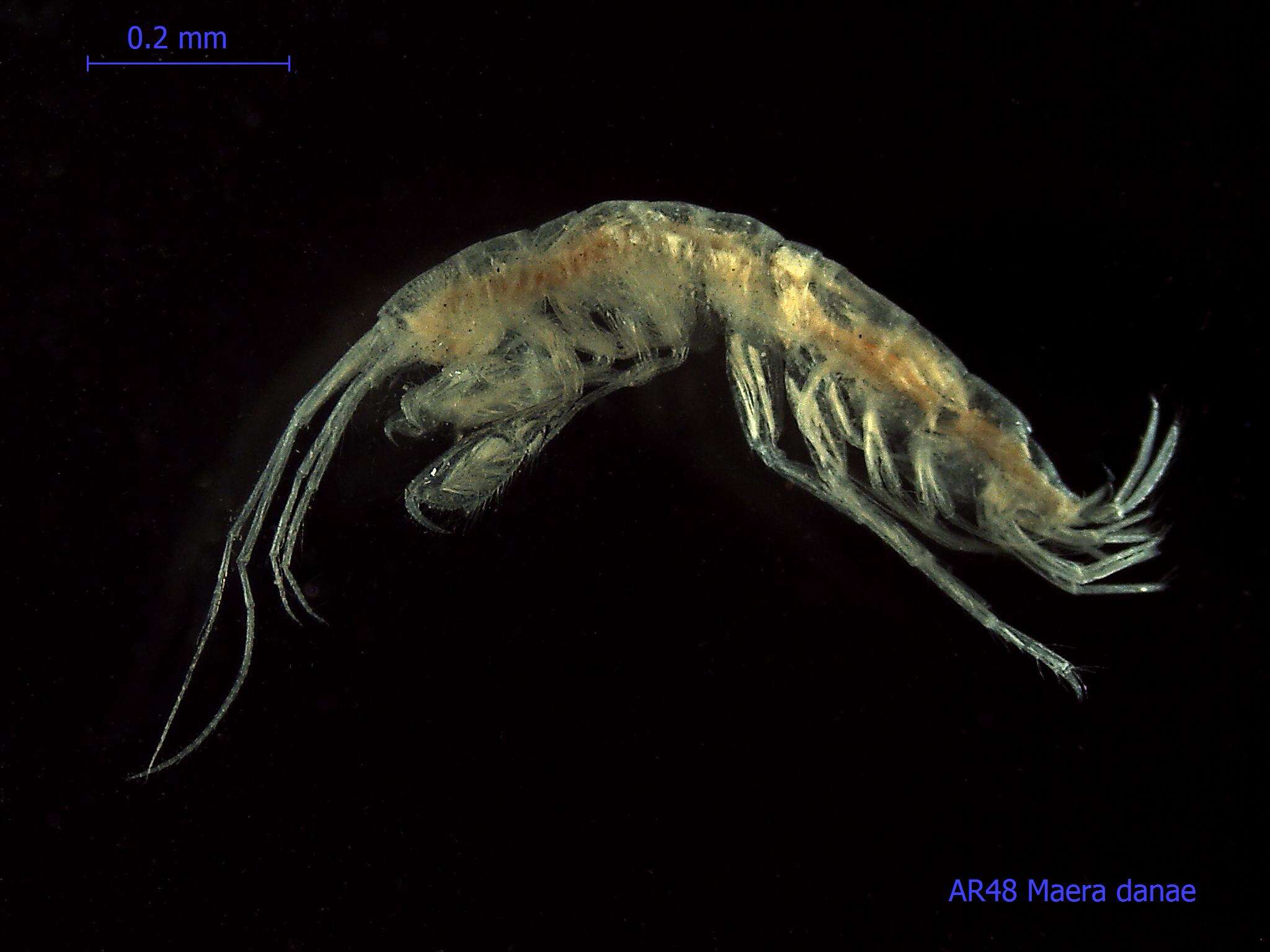
Eyes Under Puget SoundThis species image was collected from Puget Sound sediments and photographed by the Washington State Department of Ecologys Marine Sediment Monitoring Team. For more information about this teams work visit:
www.ecy.wa.gov/programs/eap/psamp/index.htm.Cant get enough benthos? Check out our Eyes Under Puget Sound Critter of the Month species profile blogs at
bit.ly/critterofthemonth
-
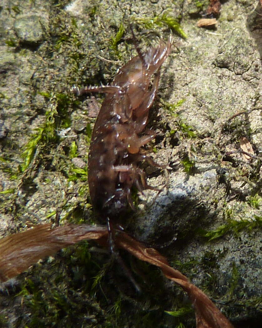
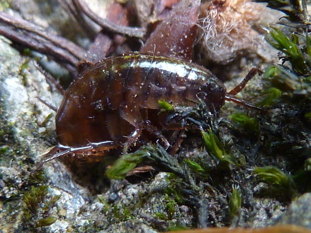
Bronwydd Arms, Wales, United Kingdom
-
-
Bronwydd Arms, Wales, United Kingdom
-
Bronwydd Arms, Wales, United Kingdom
-
Bronwydd Arms, Wales, United Kingdom
-
Bronwydd Arms, Wales, United Kingdom
-
Bronwydd Arms, Wales, United Kingdom
-
Bronwydd Arms, Wales, United Kingdom
-
Dmitry A. Sidorov, Andrey A. Gontcharov, Dmitry M. Palatov, Steven J. Taylor, Alexander A. Semenchenko
Subterranean Biology
Figure 2.Photograph of live specimen of Zenkevitchia yakovi sp. n. in the cave “Istočnik Tcebel’da”, from right side. Photography by A. Korotaev.
-
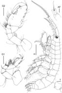
Figure 1.Elasmopus yucalpeten sp. n., holotype male, 6.6 mm, CYMX-1-EY; Yucalpeten harbor, Yucatan, Mexico. Scale bar for H represents 1 mm; scale bars for G1 and G2 represent 0.3 mm; scale bars for AF and T represent 0.1 mm.
-
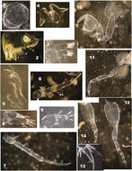
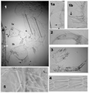
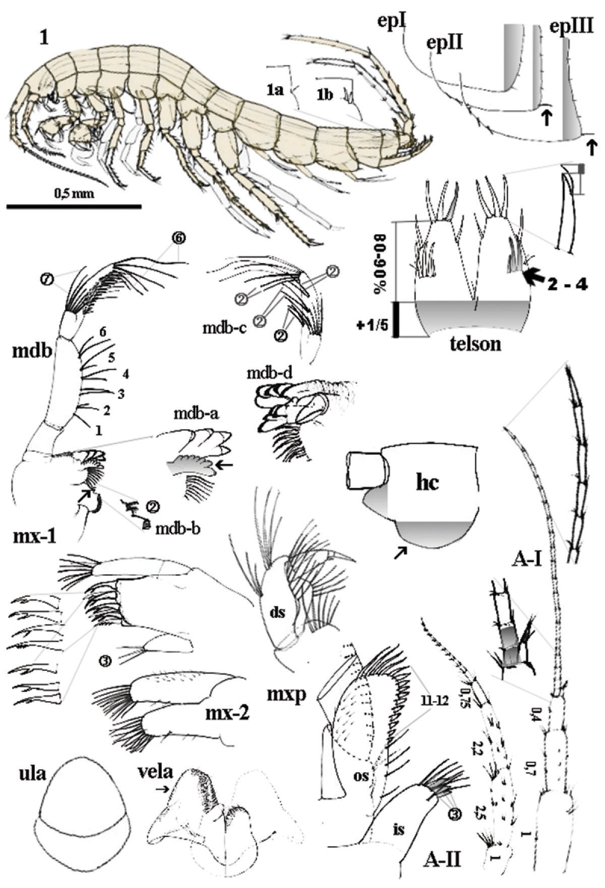
Figure 2.Niphargus plurispinosus sp. n.: 1 male, general view; 1a-1b dorso-later thorns; mdb - mandibula and details of mdb-a left incisor and lacina mobilis; mdb-b) two setae between bisserated thorns; mdb-c setae pattern on distal segment of mdb-palp; mdb-d, right incisor and lacina mobilis; mx-1 1st maxilla; mx-2 2nd maxilla; ula upper lip; vela ventral labium; mxp maxilliped: in inner segment os outer segment; ds distal segment of palp; epI-epIII epimeral plate I-III; A-I 1st antenna; A-II antenna; hc head capsula, left lateral view; telson, dorsal view. Not scaled, except of the general view of the male.
-
Figure 3.Niphargus plurispinosus sp. n.: 1 upper lip 2 right mandible 3 second maxilla 4 labium 5 half maxilliped (without inner portion) 6 3rd uropod (female) 7 3rd uropod (juvenile male) 8–9 telson (one lobe of male) 10 2nd gnathopod 11 4nd pereopod 12 7th pereopod 13 distal segment and 6th pereopod 14 6th pereopod: regeneration of segments behind basis (Photo: I. Hudec). Not drawn to scale.
-
Dmitry A. Sidorov, Andrey A. Gontcharov, Dmitry M. Palatov, Steven J. Taylor, Alexander A. Semenchenko
Subterranean Biology
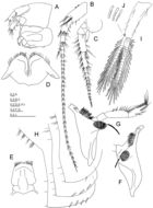
Figure 4.Zenkevitchia yakovi sp. n.: holotype, male (10.0 mm), X43382/Cr-1613-FEFU. A Head, lateral view B, C Antenna 1 and 2, lateral views D Labium, ventral view E Labrum, anterior view F, G Left and right mandibles, medial views H Epimeral plates 1–3, lateral views I pleopod 2, medial view J Coupling setae (retinacula), medial view.
-
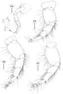
Figure 3.Elasmopus yucalpeten sp. n., holotype male, 6.6 mm, CYMX-1-EY; Yucalpeten harbor, Yucatan, Mexico. Scale bars represent 0.3 mm.
-
Figure 4.Niphargus plurispinosus sp. n. – “postreproductive” male: 1 gnathopods with detail of deformed gpI (1a) and normal developed gpII (1a) 2 gpII -setae on dactylus 3 telson and regenerated (?) upIII 4 segment setae on flagellum of AI 5 maxillae I: position of three setae on inner portion (Photo: I. Hudec). Not drawn to scale.
-
Dmitry A. Sidorov, Andrey A. Gontcharov, Dmitry M. Palatov, Steven J. Taylor, Alexander A. Semenchenko
Subterranean Biology
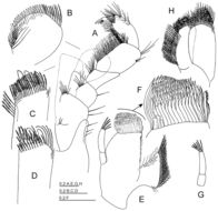
Figure 5.Zenkevitchia yakovi sp. n.: holotype, male (10.0 mm), X43382/Cr-1613-FEFU. A Maxilliped, ventral view B, C Enlarged outer and inner plates of maxilliped, ventral view D Enlarged inner plate of maxilliped, dorsal view E Left maxilla 1, dorsal view F Enlarged outer plate of maxilla 1 G Palp of right maxilla 1, dorsal view H Maxilla 2, dorsal view.