-
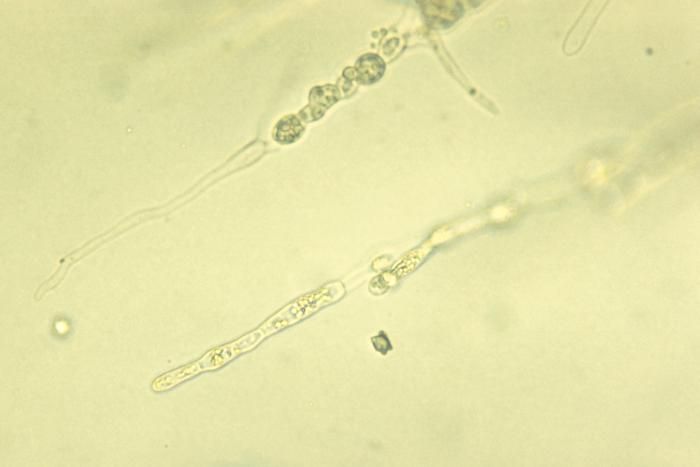
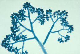
This photomicrograph shows a clusters of asexual spore-containing conidia of a Botrytis sp. fungus.Created: 1955
-
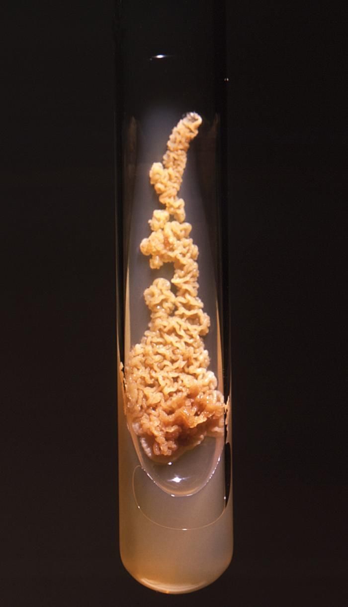
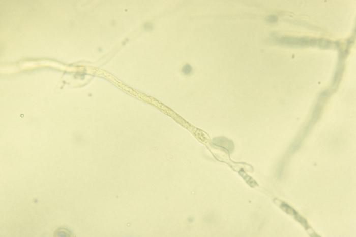
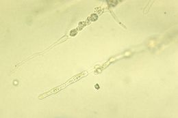
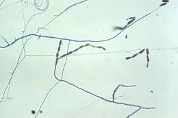
This 1971 image depicted a Sabourauds dextrose agar slant culture, which had cultivated a colony of Prototheca filamenta algal organisms. This alga lacks the presence of plastids, and is therefore, achlorophyllous.Created: 1971
-
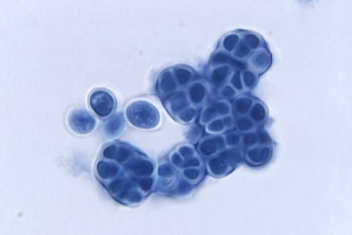
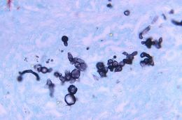
Magnified 850X, this Gamori-stained photomicrograph of a canine liver tissue specimen, revealed the presence of budding Blastomyces dermatitidis fungal cells of various sizes. Note the accompanying filaments, or mycelium.What is blastomycosis?Blastomycosis is disease caused by a fungus, Blastomyces dermatitidis, which is found in parts of the south-central, south-eastern and mid-western United States. Microfoci are also found in Central and South America and parts of Africa. The fungus can be found in moist soil enriched with decomposing organic debris.Created: 1971
-
This photomicrograph shows a clusters of asexual spore-containing conidia of a Botrytis sp. fungus.Created: 1955
-
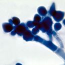
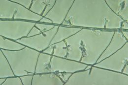
This photomicrograph depicted numbers of the colorless alga, Prototheca filamenta. This alga lacks the presence of plastids, and is therefore, achlorophyllous. Under microscopic analysis, Prototheca spp. resemble a fungal organism, and can therefore, be mistaken when attempting to identify these algae.Similar to the members of the genus Chlorella, Prototheca spp. are heterotrophic, , which means these organisms require carbon in order to thrive, and obtains this nutrient through its consumption of organic substrates. This algal culture was prepared using a lactophenol cotton blue mount fixation technique.Created: 1972
-
Magnified 500X, this Gamori-stained photomicrograph of a canine liver tissue specimen, revealed the presence of budding Blastomyces dermatitidis fungal cells of various sizes. Note the accompanying filaments, or mycelium.What is blastomycosis?Blastomycosis is disease caused by a fungus, Blastomyces dermatitidis, which is found in parts of the south-central, south-eastern and mid-western United States. Microfoci are also found in Central and South America and parts of Africa. The fungus can be found in moist soil enriched with decomposing organic debris.Created: 1971
-
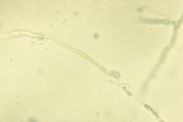
Note the resemblance between the algae Prototheca filamenta to a fungal organism due to its mycelia-like filaments.Created: 1973
-
Magnified 850X, this Gamori-stained photomicrograph of a canine liver tissue specimen, revealed the presence of budding Blastomyces dermatitidis fungal cells of various sizes. Note the accompanying filaments, or mycelium.What is blastomycosis?Blastomycosis is disease caused by a fungus, Blastomyces dermatitidis, which is found in parts of the south-central, south-eastern and mid-western United States. Microfoci are also found in Central and South America and parts of Africa. The fungus can be found in moist soil enriched with decomposing organic debris.Created: 1971
-
Note the resemblance between the algae Prototheca filamenta to a fungal organism due to its mycelia-like filaments.Created: 1973
-
Magnified 500X, this Gamori-stained photomicrograph of a canine liver tissue specimen, revealed the presence of budding Blastomyces dermatitidis fungal cells of various sizes. Note the accompanying filaments, or mycelium.What is blastomycosis?Blastomycosis is disease caused by a fungus, Blastomyces dermatitidis, which is found in parts of the south-central, south-eastern and mid-western United States. Microfoci are also found in Central and South America and parts of Africa. The fungus can be found in moist soil enriched with decomposing organic debris.Created: 1971
-
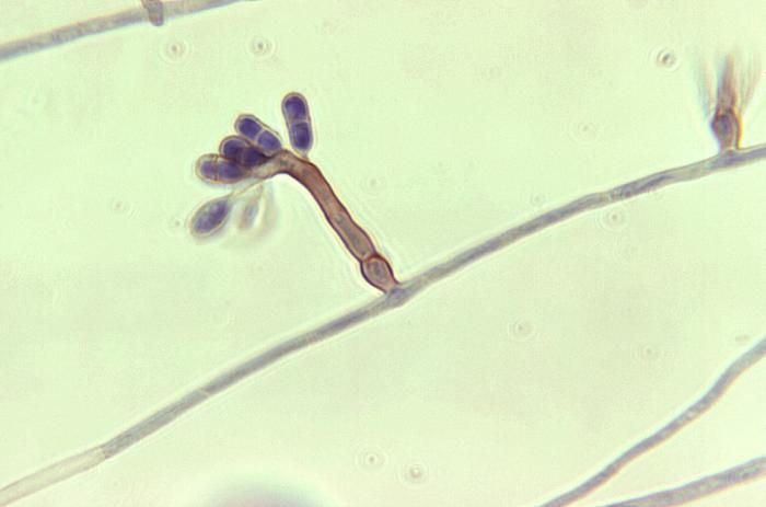
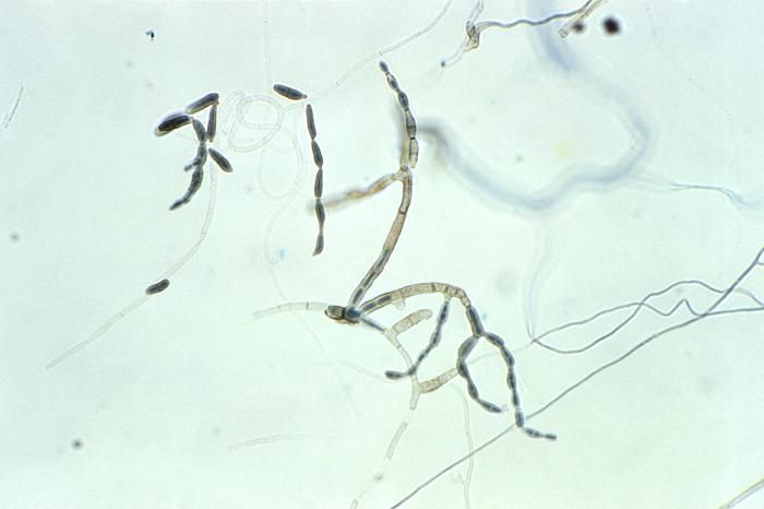
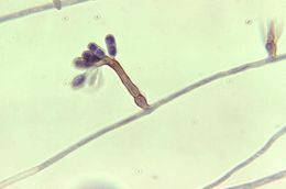
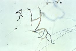
Magnified 1125X, this photomicrograph reveals some of the ultrastructural morphology of a cluster of Ochroconissp. conidia, formerly known as Dactylaria.Created: 1973
-
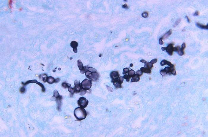
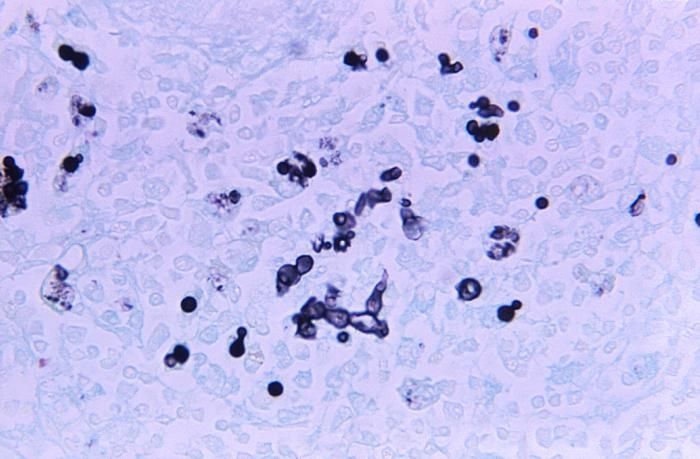
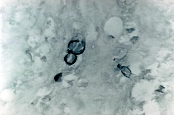
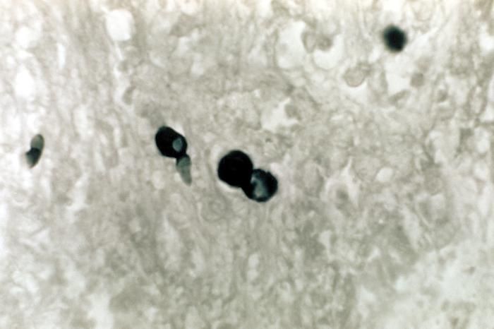
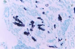
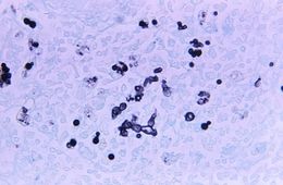
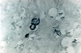
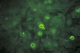
Note the histopathologic changes seen in blastomycosis due to Blastomyces dermatitidis using methenamine silver stain.Created: 1972
-
This photomicrograph reveals the conidia of a member of the fungal genus Ochroconis, formerly known as Dactylaria.Created: 1973
-
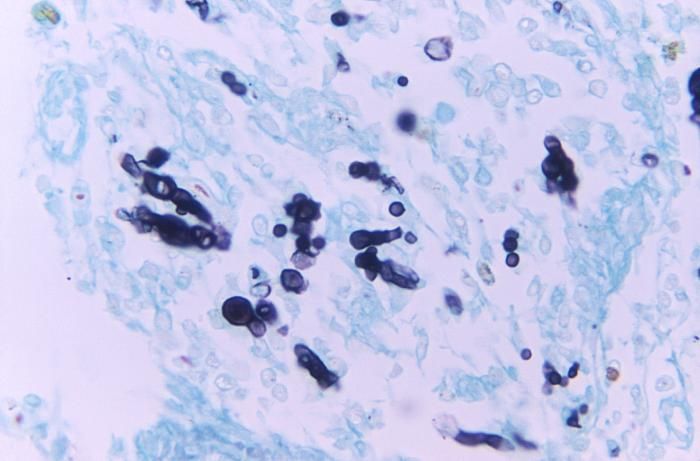
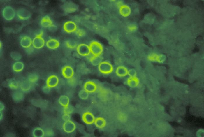
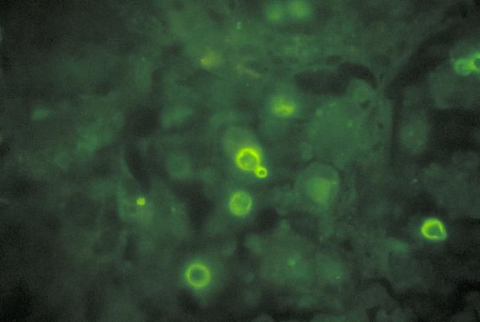
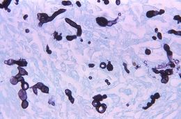
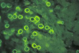
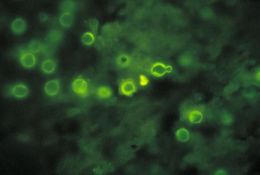
Direct FA stain revealing the histopathology of lung tissue blastomycosis due to the organism Blastomyces dermatitidis.Created: 1977
-
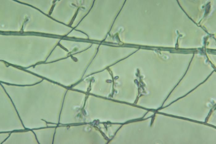
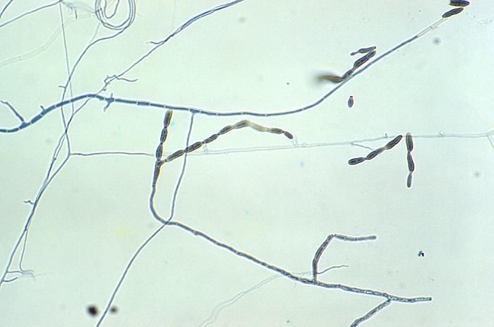
Magnified 400X, this slide culture photomicrograph highlighted some of the ultrastructural morphology exhibited by numbers of Bispora betulina, also known as Dicoccum betulinum, fungal organisms.Created: 1974
-
Direct FA stain revealing the histopathology of lung tissue blastomycosis due to the organism Blastomyces dermatitidis.Created: 1977
-
Note the histopathologic changes seen in blastomycosis due to Blastomyces dermatitidis using methenamine silver stain.Created: 1972