-
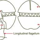
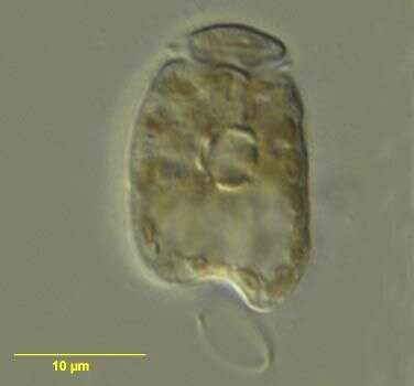
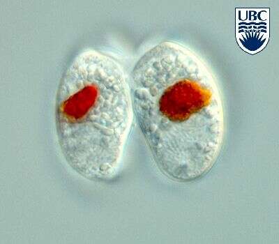
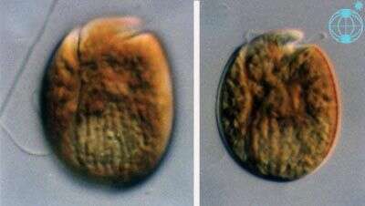
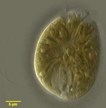
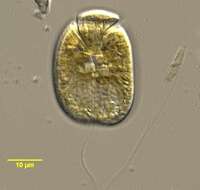
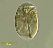
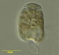
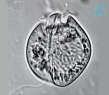
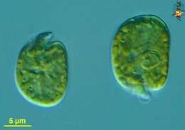
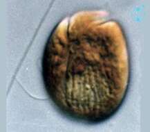
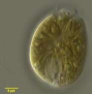
Amphidinium (am-fee-din-ee-um) herdmanii Kofoid & Swezy 1921. The image shows a cell in ventral view (side-reversed ). The epicone is small and turned to the left. The plastids are radiating from the centre.
-

Cells rounded oblong to oval from the ventral side, dorso-ventrally flattened. Length 20 - 31 microns, width 15 - 25 microns, breadth (lateral) approximately 10 microns, length to width ratio 1.2 - 1.5. Epicone large triangle from the ventral side, 8 - 13 microns wide at its widest, slightly deflected to the left. From the dorsal side epicone symmetrical, protruding over the hypocone, apically flat or with the sides sloping down away from the apex. Short (3 - 4 microns) groove begins just above the proximal end of the cingulum, leading towards the apex. Cingulum beginning approximately 0.3 of the cell length from the apex, ascending initially, continuing in a straight path across the dorsal side then descending on the ventral side, distal end slightly higher than proximal. Large (2 - 3 microns) pusule present, just to the right of the start of the sulcus, smaller pusule, difficult to observe, just below the proximal end of the cingulum. Longitudinal flagellum originating in pocket below the proximal end of the cingulum. Sulcus beginning below the proximal end of the cingulum, initially narrow then widening towards its posterior end, left side much more distinct than right. Hypocone slightly asymmetrical, with the left side longer than the right, in high focus appearing indented at the antapex by the sulcus. Nucleus in the posterior part of the hypocone, crescent shaped, 11 - 17 x 3 - 7 microns. Chloroplast single, radiating from the centre. Round pyrenoid-like structure, 5 - 6 microns diameter, sometimes apparent in the centre. Small lipid globules and reddish bodies, possibly food particles, occasionally present.
-
Amphidinium herdmanii observed in marine muds and sandy sediments in the vicinity of Broome, Western Australia in September 2003. This image was taken using differential interference contrast optics. This work was supported by the Australian Biological Resources Study.
-
Amphidinium herdmanii observed in marine muds and sandy sediments in the vicinity of Broome, Western Australia in September 2003. This image was taken using differential interference contrast optics. This work was supported by the Australian Biological Resources Study.
-
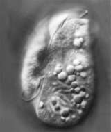
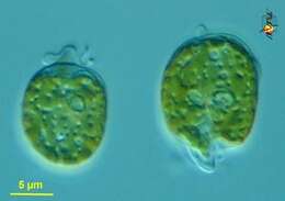
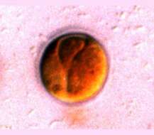
Amphidinium herdmanii, showing its pyrenoid, observed in marine muds and sandy sediments in the vicinity of Broome, Western Australia in September 2003. This image was taken using differential interference contrast optics. This work was supported by the Australian Biological Resources Study.
-
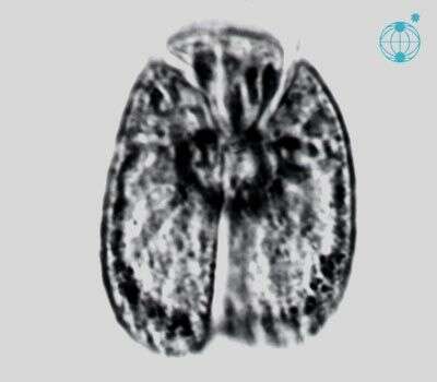
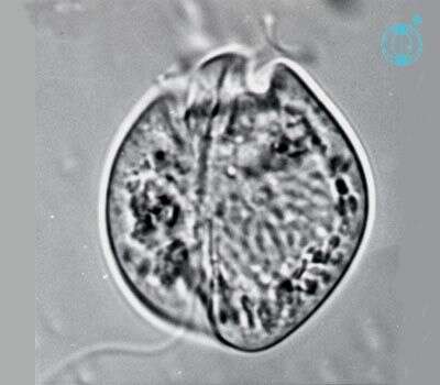
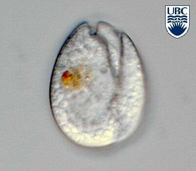
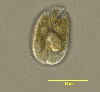
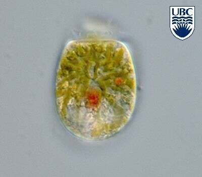
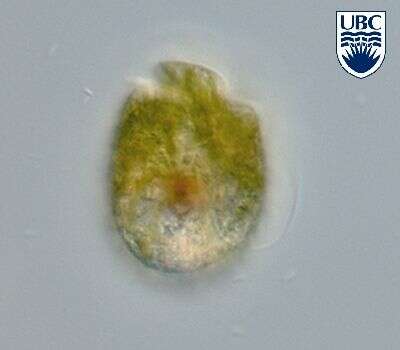
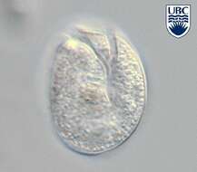
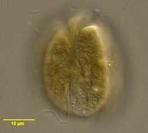
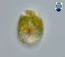
Amphidinium (am-fee-din-ee-um) incoloratum Campbell1973. The image shows a cell in ventral view. The epicone is small and turned to the left. The cell is colourless (contains no plastids). The large nucleus is in the middle of the left cell side.
-
Cells broadly oval to egg-shaped, with a relatively straight left side and a convex right side, dorso-ventrally flattened. Length 24 - 38 microns, width 17 - 24 microns, length to width ratio 1.3 - 1.6. Epicone 5 - 10 microns at its widest, deflected to the left. Cingulum becoming relatively deep and wide (approximately 2 microns), distal end approximately 4 microns lower than proximal. Transverse flagellum originating in a pocket just below the proximal end of the cingulum. Narrow ventral ridge beginning just to the right of the origin of the cingulum, contininuing down the cell in a straight path, past the distal end of the cingulum to the origin of the sulcus. Sulcus beginning 0.6 - 0.7 of the cell length from the apex, to the left of the mid-ventral line, initially narrow, the left side broadening posteriorly, the right side continuing in a straight line to the posterior end. Longitudinal flagellum arising in a pocket just before the anterior end of the sulcus, approximately 60 microns. Two pusules present, a large (approximately 2 microns diameter) obvious one to the right of the anterior end of the sulcus, and a smaller, less distinct one just below the origin of the cingulum. Apical groove not present. Nucleus in the posterior part of the hypocone, round, 10 -14 microns diameter. Chloroplasts not present. Cytoplasm filled with large colourless lipid globules and occasionally food particles. Non-motile cells round, approximately 45 microns diameter, surrounded by a hyaline layer, showing no detail of cingulum or sulcus.
-
Amphidinium incoloratum observed in marine muds and sandy sediments in the vicinity of Broome, Western Australia in September 2003. This image was taken using differential interference contrast optics. This work was supported by the Australian Biological Resources Study.
-
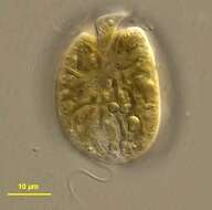
Amphidinium incoloratum Campbell 1973.
-
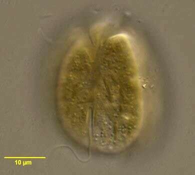
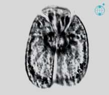
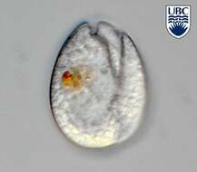
Amphidinium incoloratum Campbell 1973. Just divided cell, daughter cells still connected to each other.
-
Amphidinium incoloratum Campbell 1973.
-
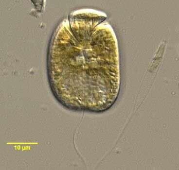
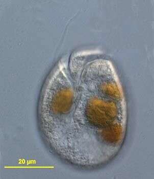
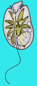
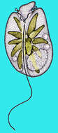
Amphidinium (am-fee-din-ee-um) operculatum, an autotrophic dinoflagellate, girdle located very close to anterior pole of cell such that the epicone is a small protrusion. The plastid, pyrenoid and flagella are evident. Differential interference microscopy.
data on this strain.
-
Amphidinium (am-fee-din-ee-um) operculatum, an autotrophic dinoflagellate, girdle located very close to anterior pole of cell such that the epicone is a small protrusion. The plastid, pyrenoid and flagella are evident. Differential interference microscopy.
data on this strain.
-
-
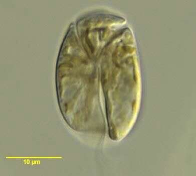
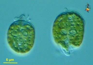
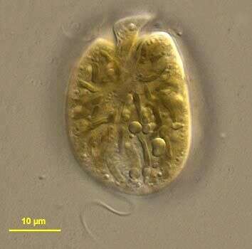
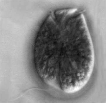
Amphidinium (am-fee-din-ee-um) operculatum Claparede & Lachmann 1858. The images show cells in ventral view. The epicone is small and turned to the left. Many yellow-brown plastids are present. The lighter area at the posterior of the cell is the nucleus.
-
Amphidinium (am-fee-din-ee-um) operculatum Claparede & Lachmann 1858. The image shows a cell in ventral view. The epicone is small and turned to the left. Many yellow-brown plastids are present. The lighter area at the posterior of the cell is the nucleus.
-
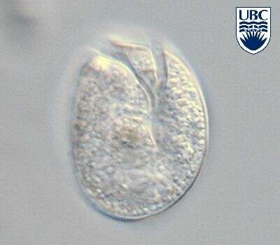
Amphidinium (am-fee-din-ee-um) operculatum Claparede & Lachmann 1858. The image shows a non-motile stage (temporary cyst) in ventral view. The cell is surrounded by a hyaline layer. Yellow brown plastids are present.
-
Cells ovoid to egg-shaped, length 29 - 48 microns, width 21 - 28 microns, length to width ratio 1.4 - 1.6. Dorso-ventrally flattened. Epicone 7 - 10 microns wide, anteriorly flat, with the right anterior corner almost a 90 degree angle and the left corner extended into a 30 degree- 45 degree angle. Cingulum relatively deep, beginning 0.2 - 0.3 of the cell length from the apex, initially rising abruptly, continuing almost horizontally around the cell, proximal end slightly higher than distal. Hypocone rounded, slightly asymmetrical, right side more curved than left. Sulcus relatively deep, beginning just to the right of the mid-ventral line, 0.6 - 0.7 of the cell length from the apex, initially narrow, then widening at the posterior end. Apical groove not present. A narrow ventral ridge runs between the two points of flagellar insertion. Two pusules present: one obvious, approximately 2 microns diameter, to the right of the origin of the sulcus, the second less obvious, also 2 microns diameter, just below and to the left of the proximal end of the cingulum. Chloroplasts in long thin strands, yellow-brown. Nucleus in the posterior part of the hypocone, crescent shaped or oval 8 - 10 x 12 - 18 microns, containing very fine chromosomes. Division is by binary fission in the motile cell. A red-orange body, either a food particle or eyespot, occasionally present just anterior to the nucleus. Colourless globules present in some cells.
-
Amphidinium operculatum observed in marine muds and sandy sediments in the vicinity of Broome, Western Australia in September 2003. This image was taken using differential interference contrast optics. This work was supported by the Australian Biological Resources Study.
-
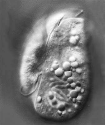
Amphidinium operculatum, showing its posterior longitudinal flagellum, observed in marine muds and sandy sediments in the vicinity of Broome, Western Australia in September 2003. This image was taken using differential interference contrast optics. This work was supported by the Australian Biological Resources Study.
-
Amphidinium operculatum, showing its elongated plastids, observed in marine muds and sandy sediments in the vicinity of Broome, Western Australia in September 2003. This image was taken using differential interference contrast optics. This work was supported by the Australian Biological Resources Study.
-
Amphidinium operculatum, observed in marine muds and sandy sediments in the vicinity of Broome, Western Australia in September 2003. This image was taken using differential interference contrast optics. This work was supported by the Australian Biological Resources Study.
-
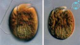
This is Amphidinium cf. operculatum in that it looks like but is not fully identical with the usual concept of this species.
-
This is Amphidinium cf. operculatum in that it looks like but is not fully identical with the usual concept of this species.