-
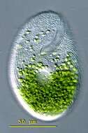
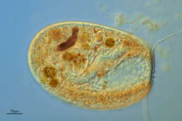
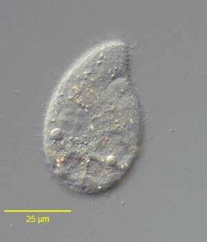
Bryometopus (bry-o-me-toe-puss) is a uniformly ciliated and the cell is rounded at both poles and is slightly reniform. The conspicuous peristome lies obliquely across the ventral surface. The single contractile vacuole is located approximately in the middle of the cell on the ventral side. The macronucleus may be oval or elongate with several micronuclei. Bryometopus can be confused with Balantidioides which does not have an undulating membrane and with Condylostoma which has a wide triangular peristome and a highly conspicuous undulating membrane. This cell with a buccal cavitiy up to the first third of the cell. The posterior half of the cell is filled with endosymbitic algae. Measuring 120 microns. This specimen was collected in freshwater ponds near Konstanz, Germany. Differencial interference contrast.
-

Infraciliature (posterior view, dorsal at top) of the colpodid ciliate, Maryna ovata GELEI, 1950. Cell size is highly variable (length 25-110 mm). The cells are mushroom or champagne cork-shaped. The conical anterior end is termed the calyx and the truncate cylindrical posterior part the uvula. There is a definite horseshoe-shaped sulcus oriented perpendicular to the long axis between the calyx and uvula. The infraciliature is complex, twisting around the calyx and running posteriorly down onto the uvula (as seen here). There is a semicircular array of longer terminal cilia along the posterior margin of the uvula (their kinetids are seen here). Some descriptions (e.g. Curds, C.R. British and Other Freshwater Ciliated Protozoa. Part I, p.182. Cambridge, Bath 1982.) erroneously describe the uvula as being anterior. This is probably due to the organism's habit of residing in its dwelling tube or lorica anterior end innermost leaving the uvula protruding. Interestingly, cells turn around in the dwelling tube in order to exit front end first. The cytostome is located in the sulcus between calyx and uvula. It is flanked on it right and left by dense polykinetids (seen to the viewer's left at 8 o'clock position here). The location of the large spherical macronucleus and single adjacent micronucleus is variable. The contractile vacuole is located in the posterior uvula. Many refractile yellow crystals are found in the cytoplasm. These impart a blackish color to the cells in vivo under low magnification. The tubular organic test is attached to the substrate and may be nearly 1000 μ long. The cell flees the test at the slightest disturbance so most cells are found swimming freely when examined under a coverslip. Maryna ovata feeds on bacteria and algae. This specimen stained with silver carbonate (see Foissner, W.Europ. J. Protistol.27,313-330;1991). Collected from a eutrophic pond near Boise, Idaho August 2004. Brightfield optics.
-
Right lateral view of the colpodid ciliate, Exocolpoda augustini (Foissner, 1987) Foissner, Agatha and Berger, 2002. Foissner erected the family Exocolpodidae based on the life cycle of its members, namely, cell division in free-swimming individuals instead of reproduction in division cysts as seen in the Colpodidae. He felt this life cycle characteristic,the unique boomerang-shaped left oral polykinetid and the unique thick-walled resting cyst of this species warranted its transfer to the new genus, Exocolpoda. The anterior of the cell is cone-shaped and the posterior globular.The small cytostome is in the anterior 1/4 of the cell.There are 25-35 somatic kineties composed of doubly ciliated dikinetids.The right somatic kineties spiral slightly on the long axis to end on the short preoral suture. The left kineties curve more strongly to perpendicularly abut the suture.There are two oral poykinetids. The lekt polykinetid has a unique angulated shape like a boomerang.The macronucleus is spherical.The nucleolus is ribbon-like.There is a single posterior contractile vacuole with a solitary excretory pore.Collected near Boise, Idaho (43°38'21.10"N 116°11'10.78"W elev. 2908 ft.) from an ice-covered temporary puddle containing leaf litter and dead grass.November, 2005.DIC.
-
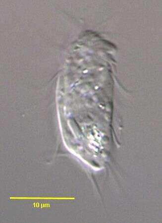
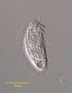
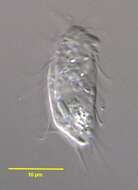
Pseudoplatyophrya nana (KAHL,1926) FOISSNER,1980.DIC.
-
Villoslada de Cameros, La Rioja, Spain
-
Mellanes, Castille and Leon, Spain
-
Los Cotos, Madrid, Spain
-
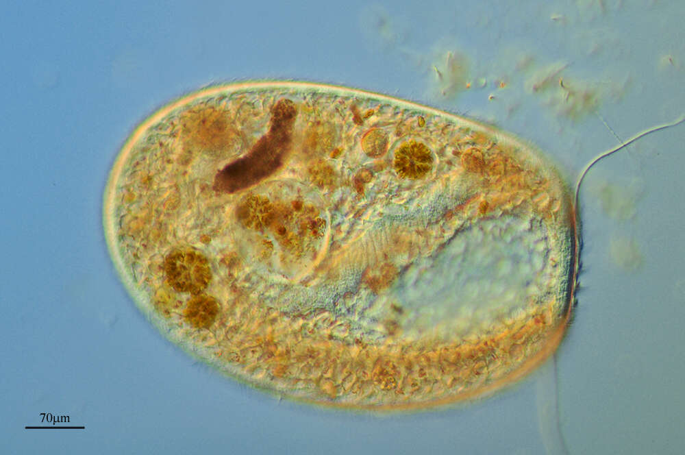
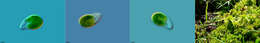
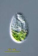
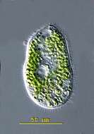
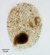
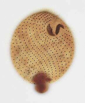
Thylakidium (thigh-lack-kid-ee-um) has an ovoid body that is uniformly ciliated, anteriorly truncated and posteriorly rounded. The cytoplasm is usually full of green endosymbiotic algae. The deep peristomial cavity opens apically and is lined out with membranelles which wind clockwise around the apex of the cell. The cytopharynx is bent towards the animal's left. The macronucleus is rounded and there is a single laterally placed contractile vacuole. Thylakidium can be confused with Bursaria and Bursaridium. This specimen was collected in freshwater ponds near Konstanz, Germany. This cell in ventral view with a truncated anterior end. The peristomial cavity extends up to the middle of the cell. Differential interference contrast.
-
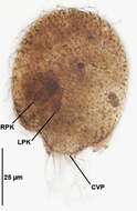
Infraciliature (left ventrolateral view) of the colpodid ciliate, Maryna ovata GELEI, 1950 (left ventrolateral view. LPK=left oral polykinetid.RPK=right oral polykinetid.The contractile vacuole pore (CVP) is located at the end of the uvula.This specimen stained with Protargol (Wilbert modification) (see Foissner, W.Europ. J. Protistol.27,313-330;1991). Collected from a eutrophic pond near Boise, Idaho August 2008. Brightfield.
-

Right lateral view of the colpodid ciliate Exocolpoda augustini (Foissner, 1987) Foissner, Agatha and Berger, 2002.Foissner erected the family Exocolpodidae based on the life cycle of its members, namely, cell division in free-swimming individuals instead of reproduction in division cysts as seen in the Colpodidae. He felt this life cycle characteristic,the unique boomerang-shaped left oral polykinetid and the unique thick-walled resting cyst of this species warranted its transfer to the new genus, exocolpoda. The anterior of the cell is cone-shaped and the posterior globular.The small cytostome is in the anterior 1/4 of the cell.There are 25-35 somatic kineties composed of doubly ciliated dikinetids (the paired cilia are seen well to viewer's right here).The right somatic kineties spiral slightly on the long axis to end on the short preoral suture. The left kineties curve more strongly to perpendicularly abut the suture.There are two oral poykinetids. The left oral polykinetid has a unique angulated shape like a boomerang.The macronucleus is spherical.The nucleolus is ribbon-like.In this specimen the macronucleus has extruded posteriorly during fixation.There is a single posterior contractile vacuole with a solitary excretory pore.Collected near Boise, Idaho (43°38'21.10"N 116°11'10.78"W elev. 2908 ft.) from an ice-covered temporary puddle containing leaf litter and dead grass.November, 2005.Stained by the silver carbonate technique (see Foissner, W. Europ. J. Protistol., 27:313-330;1991).DIC.
-
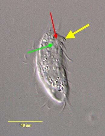
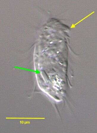
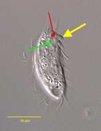
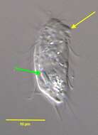
Pseudoplatyophrya nana (KAHL,1926) FOISSNER,1980.The large yellow arrow indicates the feeding tube specialized for the puncture of fungal cells upon which P. nana feeds. This tube projects from the ventral surface of the cell.The red arrow indicates the small adoral organelle composed of about 7 ciliated kinetids just posterior to the base of the feeding tube. The green arrow indicates the curved paraoral membrane to the right of the feeding tube. From a non-flooded Petri dish culture of topsoil collected at a public park in Boise, Idaho.November 2006.DIC.
-
Mahide, Castille and Leon, Spain
-
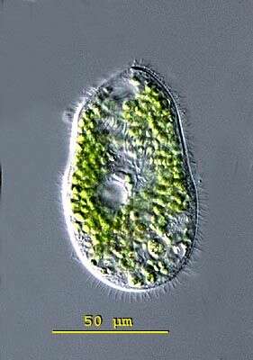
Thylakidium (thigh-lack-kid-ee-um) has an ovoid body that is uniformly ciliated, anteriorly truncated and posteriorly rounded. The cytoplasm is usually full of green endosymbiotic algae. The deep peristomial cavity opens apically and is lined out with membranelles which wind clockwise around the apex of the cell. The cytopharynx is bent towards the animal's left. The macronucleus is rounded and there is a single laterally placed contractile vacuole. Thylakidium can be confused with Bursaria and Bursaridium. This specimen was collected in freshwater ponds near Konstanz, Germany. The contractile vacuole of Thylakidium pituitosum is located ventrally in the middle of the cell. The cell is filled up with green endosymbiotic algae. Differential interference contrast.
-
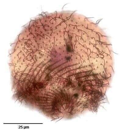
Infraciliature (dorsolateral view) of the colpodid ciliate, Maryna ovata GELEI, 1950. Cell size is highly variable (length 25-110 mm). The cells are mushroom or champagne cork-shaped. The conical anterior end is termed the calyx and the truncate cylindrical posterior part the uvula. There is a definite horseshoe-shaped sulcus oriented perpendicular to the long axis between the calyx and uvula. The infraciliature is complex, twisting around the calyx and running posteriorly down onto the uvula (as seen here). Doubly ciliated dikinetids are seen here over the anterior calyx. There is a semicircular array of longer terminal cilia along the posterior margin of the uvula. Some descriptions (e.g. Curds, C.R. British and Other Freshwater Ciliated Protozoa. Part I, p.182. Cambridge, Bath 1982.) erroneously describe the uvula as being anterior. This is probably due to the organism's habit of residing in its dwelling tube or lorica anterior end innermost leaving the uvula protruding. Interestingly, cells turn around in the dwelling tube in order to exit front end first. The cytostome is located in the sulcus between calyx and uvula. It is flanked on it right and left by dense polykinetids (not seen in this view). The location of the large spherical macronucleus and single adjacent micronucleus is variable. The contractile vacuole is located in the posterior uvula. Many refractile yellow crystals are found in the cytoplasm. These impart a blackish color to the cells in vivo under low magnification. The tubular organic test is attached to the substrate and may be nearly 1000 μ long. The cell flees the test at the slightest disturbance so most cells are found swimming freely when examined under a coverslip. Maryna ovata feeds on bacteria and algae. This specimen stained with silver carbonate (see Foissner, W.Europ. J. Protistol.27,313-330;1991). Collected from a eutrophic pond near Boise, Idaho August 2004. Brightfield optics.
-

Right lateral view of the colpodid ciliate, Exocolpoda augustini (Foissner, 1987) Foissner, Agatha and Berger, 2002. Foissner erected the family Exocolpodidae based on the life cycle of its members, namely, cell division in free-swimming individuals instead of reproduction in division cysts as seen in the Colpodidae. He felt this life cycle characteristic,the unique boomerang-shaped left oral polykinetid and the unique thick-walled resting cyst of this species warranted its transfer to the new genus, Exocolpoda. The anterior of the cell is cone-shaped and the posterior globular.The small cytostome is in the anterior 1/4 of the cell.There are 25-35 somatic kineties composed of doubly ciliated dikinetids.The right somatic kineties spiral slightly on the long axis to end on the short preoral suture. The left kineties curve more strongly to perpendicularly abut the suture.There are two oral poykinetids. The lekt polykinetid has a unique angulated shape like a boomerang.The macronucleus is spherical.The nucleolus is ribbon-like.There is a single posterior contractile vacuole with a solitary excretory pore.Collected near Boise, Idaho (43°38'21.10"N 116°11'10.78"W elev. 2908 ft.) from an ice-covered temporary puddle containing leaf litter and dead grass.November, 2005.Phase contrast.
-
Pseudoplatyophrya nana (KAHL,1926) FOISSNER,1980.DIC.
-

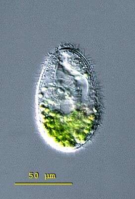
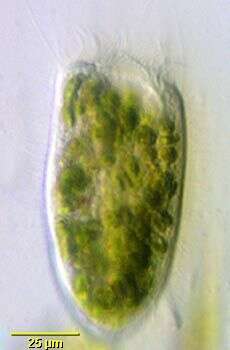
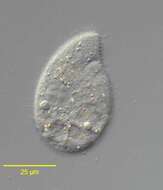
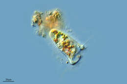
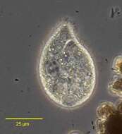
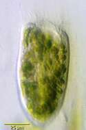
Paracondylostoma setigerum, a colpodid ciliate originally recovered from an alpine pond in central Austria and described by Foissner and Kreutz (Foissner, W., Kreutz, M.: Systematic position and phylogentic relationship of the genus Bursaridium, Paracondylostoma, Thylakidium, Bryometopus, and Bursaria (Ciliphora: Colpoda). Acta Protozoologica 37, 227 - 240 (1998). Overall shape is fusiform with truncate anterior. Oral aperture is anterior, funnel-shaped and extends about 1/3 the length of the cell. A line of membranelles extends completely along the margin of the aperture on the left. Somatic ciliature is uniform in longitudinal kineties. Prominent long bristle-like cilia radiate from the anterior end. The cells occupy a mucus sheath which is transparent and very difficult to see unless bacteria and debris adhere. Cells often flee the sheath when placed on the slide. This individual contains large numbers of zoochlorellae. Centrally located macronucleus is spherical. From freshwater pond near Boise, Idaho. Brightfield illumination.
-

Portrait (right lateral view) of the colpodid ciliate, Maryna ovata GELEI, 1950 that has fled its organic test or dwelling tube. This cell is slightly squashed beneath the coverslip. Cell size is highly variable (length 25-110 mm). The cells are mushroom or champagne cork-shaped. The large conical anterior end is termed the calyx and the smaller truncate cylindrical posterior part the uvula. There is a definite horseshoe-shaped sulcus oriented perpendicular to the long axis between the calyx and uvula. The infraciliature is complex, twisting around the calyx and running posteriorly down onto the uvula. There is a semicircular array of longer terminal cilia along the posterior margin of the uvula (seen here). Some descriptions (e.g. Curds, C.R. British and Other Freshwater Ciliated Protozoa. Part I, p.182. Cambridge, Bath 1982.) erroneously describe the uvula as being anterior. This is probably due to the organism's habit of residing in its dwelling tube or lorica anterior end innermost leaving the uvula protruding. Interestingly, cells turn around in the dwelling tube in order to exit front end first. The cytostome is located in the sulcus between calyx and uvula (seen here to viewer's right). It is flanked on it right and left by dense polykinetids. The location of the large spherical macronucleus and single adjacent micronucleus is variable. The contractile vacuole is located in the posterior uvula (just visible here). Many refractile yellow crystals are found in the cytoplasm (seen here in the posterior calyx). These impart a blackish color to the cells under low brightfield magnification. The tubular organic test is attached to the substrate and may be nearly 1000 μ long. The cell flees the test at the slightest disturbance so most cells are found swimming freely when examined under a coverslip as seen here. Maryna ovata feeds on algae and bacteria. Collected from a eutrophic pond near Boise, Idaho August 2004. DIC optics.
-

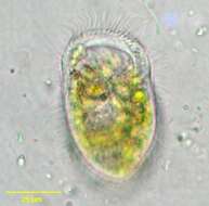
Ventral view of the colpodid ciliate, Exocolpoda augustini (Foissner, 1987) Foissner, Agatha and Berger, 2002.Foissner erected the family Exocolpodidae based on the life cycle of its members, namely, cell division in free-swimming individuals instead of reproduction in division cysts as seen in the Colpodidae. He felt this life cycle characteristic,the unique boomerang-shaped left oral polykinetid and the unique thick-walled resting cyst of this species warranted its transfer to the new genus, Exocolpoda. The anterior of the cell is cone-shaped and the posterior globular.The small cytostome is in the anterior 1/4 of the cell.There are 25-35 somatic kineties composed of doubly ciliated dikinetids (the paired cilia are seen well to viewer's right here).The right somatic kineties spiral slightly on the long axis to end on the short preoral suture. The left kineties curve more strongly to perpendicularly abut the suture.There are two oral poykinetids. The left oral polykinetid has a unique angulated shape like a boomerang.The macronucleus is spherical.The nucleolus is ribbon-like.In this specimen the macronucleus has extruded posteriorly during fixation.There is a single posterior contractile vacuole with a solitary excretory pore.Collected near Boise, Idaho (43°38'21.10"N 116°11'10.78"W elev. 2908 ft.) from an ice-covered temporary puddle containing leaf litter and dead grass.November, 2005.Stained by the silver carbonate technique (see Foissner, W. Europ. J. Protistol., 27:313-330;1991).Brightfield.
-
Pseudoplatyophrya nana (KAHL,1926) FOISSNER,1980.The yellow arrow indicates the feeding tube by which P. nana punctures and feeds on fungi and yeast cells. The green arrow indicates the unusually large oblong micronucleus.DIC.
-
Paracondylostoma setigerum. This image demonstrates the membranelles around the funnel-shaped oral aperture and the long anterior bristle cilia (indistinct here but seen on the organism's right). This individual contains many zoochlorellae. From freshwater pond near Boise, Idaho. Oblique illumination.
-
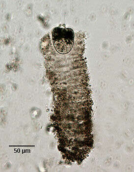
Portrait of the colpodid ciliate, Maryna ovata GELEI, 1950. Cell size is highly variable (length 25-110 mm). The cells are mushroom or champagne cork-shaped. The conical anterior end is termed the calyx and the truncate cylindrical posterior part the uvula. There is a definite horseshoe-shaped sulcus oriented perpendicular to the long axis between the calyx and uvula. The infraciliature is complex, twisting around the calyx and running posteriorly down onto the uvula. There is a semicircular array of longer terminal cilia along the posterior margin of the uvula. Some descriptions (e.g. Curds, C.R. British and Other Freshwater Ciliated Protozoa. Part I, p.182. Cambridge, Bath 1982.) erroneously describe the uvula as being anterior. This is probably due to the organismâs habit of residing in its dwelling tube or lorica anterior end innermost leaving the calyx protruding (seen here). Interestingly, cells turn around in the dwelling tube in order to exit front end first. The cytostome is located in the sulcus between calyx and uvula. It is flanked on it right and left by dense polykinetids. The location of the large spherical macronucleus and single adjacent micronucleus is variable. The contractile vacuole is located in the posterior uvula (just visible here). Many refractile yellow crystals are found in the cytoplasm. These impart a blackish color to the cells under low magnification (seen here). The tubular organic test is attached to the substrate and may be nearly 1000 mm long. The cell flees the test at the slightest disturbance so most cells are found swimming freely when examined under a coverslip. This specimen was photographed without a coverslip resulting in some degradation of the image. Maryna ovata feeds on bacteria and algae. Collected from a eutrophic pond near Boise, Idaho August 2004. Brightfield optics.
-
Ventral view of the colpodid ciliate, Exocolpoda augustini (Foissner, 1987) Foissner, Agatha and Berger, 2002.Foissner erected the family Exocolpodidae based on the life cycle members, namely, cell division in free-swimming individuals instead of reproduction in division cysts as seen in the Colpodidae. He felt this life cycle characteristic,the unique boomerang-shaped left oral polykinetid and the unique thick-walled resting cyst of this species warrented its transfer to the new genus, Exocolpoda. The anterior of the cell is cone-shaped and the posterior globular.The small cytostome is in the anterior 1/4 of the cell.There are 25-35 somatic kineties composed of doubly ciliated dikinetids (the paired cilia are seen well to viewer's right here).The right somatic kineties spiral slightly on the long axis to end on the short preoral suture. The left kineties curve more strongly to perpendicularly abut the suture.There are two oral poykinetids. The left oral polykinetid has a unique angulated shape like a boomerang.The macronucleus is spherical.The nucleolus is ribbon-like.In this specimen the macronucleus has extruded posteriorly during fixation.There is a single posterior contractile vacuole with a solitary excretory pore.Collected near Boise, Idaho (43°38'21.10"N 116°11'10.78"W elev. 2908 ft.) from an ice-covered temporary puddle containing leaf litter and dead grass.November, 2005.Stained by the silver carbonate technique (see Foissner, W. Europ. J. Protistol., 27:313-330;1991).Brightfield.
-
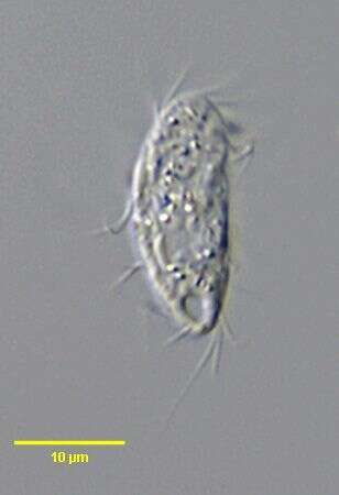
Pseudoplatyophrya nana (KAHL,1926) FOISSNER,1980.DIC.