-
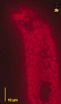
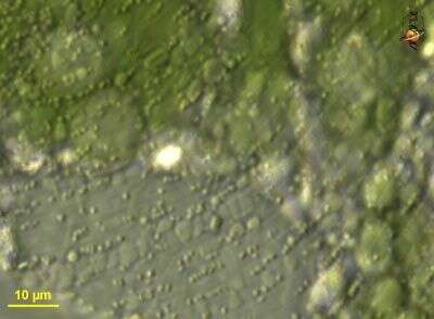
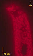
Euglena oxyuris (you-glean-a ox-ee-your-is), is a moderate to large euglena, with a stiff pellicle that has well developed ridges. Flagella can be short. Although it can squirm it is not very actively metabolic. With large numbers of small plastids. This cell was illuminated using high energy ultraviolet light and the red autofluorescence then photographed. The fluorescence comes from the chlorophylls in the chloroplast and so we can use this to get a sense of the number and size of the plastids. Fluorescence.
-
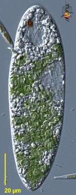
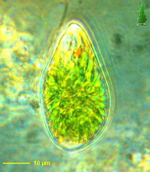
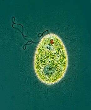
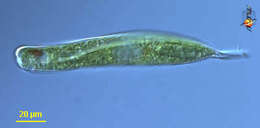
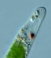
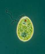
Euglena (you-glean-a) is the iconic genus of euglenoid flagellates. The body is typically spindle-shaped, although two flagella arise in a pocket within the cell only one emerges (and sometimes none). The body can squirm, and the cell has one to many chloroplasts. At the anterior of the body, a thin channel (flagellar canal) leads to the flagellar pocket, and alongside this is a contractile vacuole. A red eyespot or stigma is associated with the bottom of the flagellar canal. Phase contrast.
-
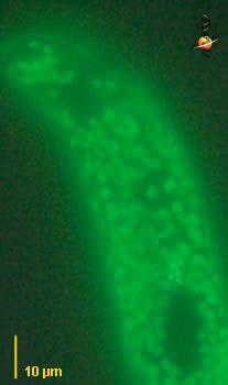
Euglena oxyuris (you-glean-a ox-ee-your-is), is a moderate to large euglena, with a stiff pellicle that has well developed ridges. Flagella can be short. Although it can squirm it is not very actively metabolic. With large numbers of small plastids. This cell was illuminated using high energy ultraviolet light and the green autofluorescence then photographed. Fluorescence.
-
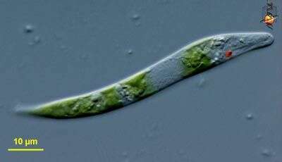
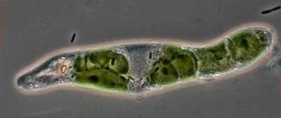
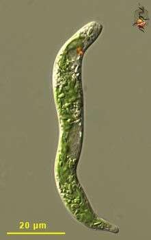
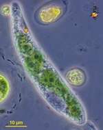
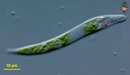
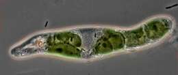
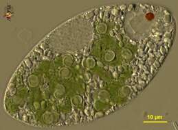
Euglena (you-glee-na) is the iconic representative of the euglenids, a group of flagellates common in freshwaters and marine sediments. Some euglenids have bright green chloroplasts, such as this one, and there is also a small red eyespot located close to the anterior (to the right, here) of the cell. This species, probably E. mutabilis, is worm-like, squirms and has no emergent flagella. Differential interference contrast. Material from Nymph Creek and Nymph Lake, thermal sites within Yellowstone National Park, photograph by Kathy Sheehan and David Patterson.
-
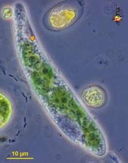
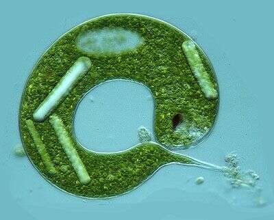
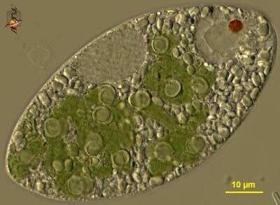
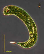
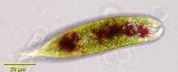
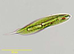
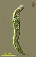
Euglena oxyuris (you-glean-a ox-ee-your-is), is a moderate to large euglena, with a stiff pellicle that has well developed ridges, and often - as we can see here - with a fold in the cell. Flagella can be short. Although it can squirm it is not very actively metabolic. With large numbers of small plastids. Large red eyespot near the anterior (right). Differential interference contrast.
-
-
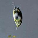
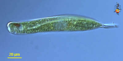
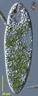
Euglena oxyuris, one of the larger (but not largest) euglenids, broad front end, and with a posterior spike. No emergent flagellum, moves by gliding across the substrate. With obvious red eye spot and large paramylon granules. Differential interference contrast.
-
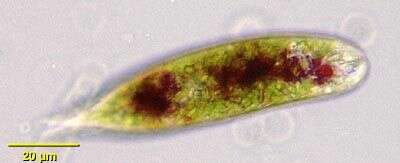
Euglena sanguinea - a brightfield portrait of this slow swimming species pigmented with hematochrome granules. Granules aggregate as seen here in low light conditions and disperse with increases in either water temperature or light intensity. Flagellum typically body length but not seen here. Small spindle shaped chloroplasts often spirally aligned with pellicular striations. Also referred to as E. rubra. Collected from freshwater pond near Boise, Idaho.
-
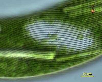
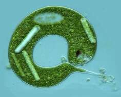
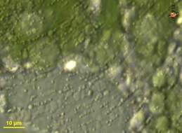
Euglena oxyuris, one of the larger (but not largest) euglenids, broad front end, and with a posterior spike. No emergent flagellum, moves by gliding across the substrate. With obvious red eye spot and large paramylon granules. This image is of the pellicle, showing the folded nature that is distinctive for euglenids. Differential interference contrast.
-
Detail of the anterior end of a Euglena cell, collected at Beaver Lake, showing the flagellar pocket, a very short flagellum with a swollen basal region (the flagellum is not long enough even to project from the front of the cell). The eyespot is closely associated with the flagellum.
-
One of the larger species in the genus. The cortical region of the cell is thick and thrown into fine folds. There are several large rod-like paramylon granules, ovoid nucleus, and dark red spot near the front of the cell. There is no emergent flagellum. Nomarski optics.
-
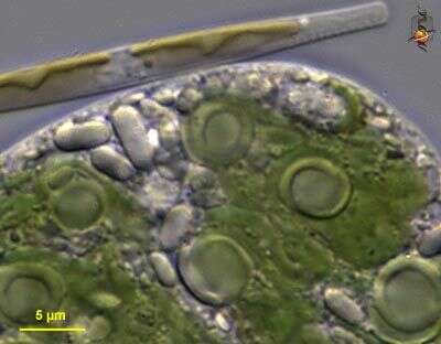
This image of Euglena, collected from Beaver Lake, emphasizes the disk- shaped chloroplasts. The front of the cell is to the left. The light area is called the reservoir. Adjacent to this region is the red eyespot that helps to control the direction in which the cells move. The granular region in the center of the cell is the nucleus.
-
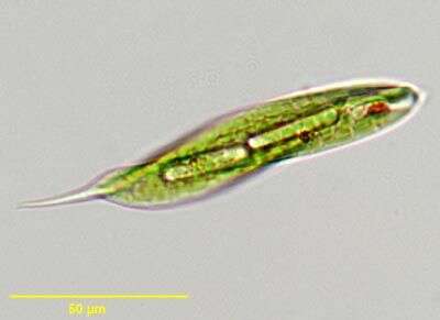
Euglena tripteris, elongate swimming euglenid, distinguished by triangular body profile in cross-section. Image shows elongate paramylon granules, and anterior eyespot. From freshwater pond near Boise, Idaho.
-
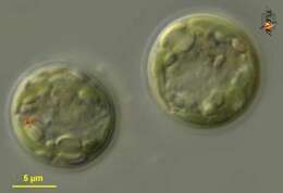
Euglena. Cyst observed in freshwater habitats in the vicinity of Broome, Western Australia in September 2003. This image was taken using differential interference contrast optics. This work was supported by the Australian Biological Resources Study.
-
Euglena velata. Cell observed in freshwater habitats in the vicinity of Broome, Western Australia in September 2003. Animations by Rosemary Arbur of flagellar beat patterns are available
here.This image was taken using differential interference contrast optics. This work was supported by the Australian Biological Resources Study.
-
Euglena. Cyst observed in freshwater habitats in the vicinity of Broome, Western Australia in September 2003. This image was taken using differential interference contrast optics. This work was supported by the Australian Biological Resources Study.
-
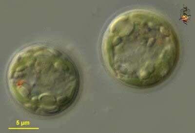
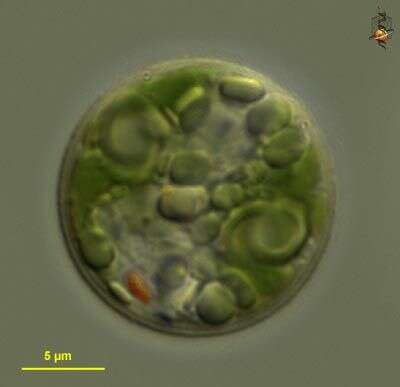
Euglena velata. Plastids with pyrenoids. Cell observed in freshwater habitats in the vicinity of Broome, Western Australia in September 2003. This image was taken using differential interference contrast optics. This work was supported by the Australian Biological Resources Study.
-
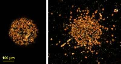
Euglena. The picture to the left shows euglenids and some diatoms concentrated within a narrow beam of light using photokinetic reactions to changing light intensities. The image to the right shows the cells beginning to move outwards after the beam of light has been 'opened up'. This work was supported by the Australian Biological Resources Study.
-
Euglena velata. Cell observed in freshwater habitats in the vicinity of Broome, Western Australia in September 2003. This image was taken using differential interference contrast optics. This work was supported by the Australian Biological Resources Study.
-
Single cell without an emergent flagellum. The eyespot is the red structure near the front of the cell, and there is a contractile vacuole near it. the clear, slightly speckled, region near the center of the cell is the nucleus.
-
Euglena velata. Cell observed in freshwater habitats in the vicinity of Broome, Western Australia in September 2003. This image was taken using differential interference contrast optics. This work was supported by the Australian Biological Resources Study.
-
Collected from Cumloden Swamp on July 8, 2002.
-
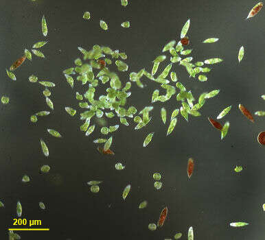
This image shows two species of Euglena, Euglena velata (the green cells) and Euglena sanguinea (the red cells). Low magnification image.
-
This phase contrast micrograph of an un-named species of euglena shows the way that euglenid flagella beat. The flagellum is thrown into loops and these are pushed along the flagellum from base to tip. Several loops can be seen here. Cell with grass-green plastids and red eyespot.