-
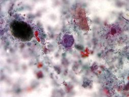
Magnified 675X, this photomicrograph revealed the presence of a parasitic Entamoeba histolytica trophozoite, which contained vacuolated cytoplasm, within which were two red blood cells (RBCs), and a pyknotic body. Entamoeba histolytica/Entamoeba dispar trophozoites have a single nucleus, which have a centrally placed karyosome and uniformly distributed peripheral chromatin. This typical appearance of the nucleus is not always observed as some trophozoites can have nuclei with an eccentric karyosome and unevenly distributed peripheral chromatin. The cytoplasm has a granular or "ground-glass" appearance. E. histolytica/E. dispar trophozoites usually measure 15µm - 20µm (range 10µm - 60µm), tending to be more elongated in diarrheal stool.Created: 1971
-
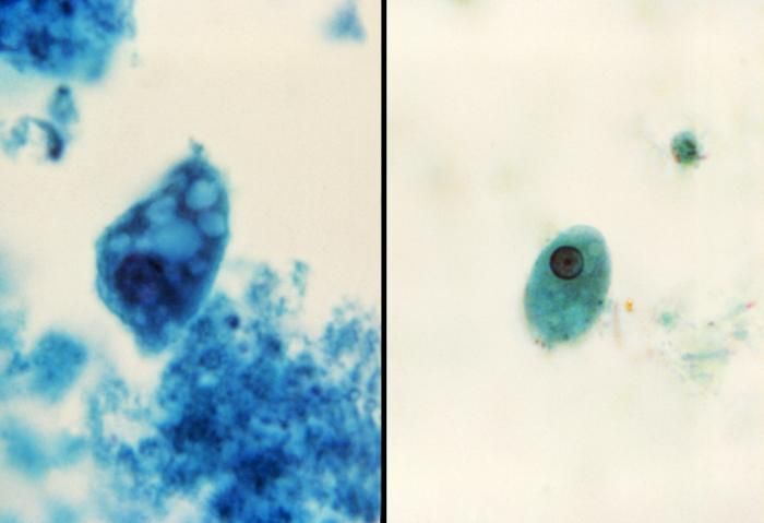
Using a trichrome stain, this photomicrograph depicted a cyst of the single-celled parasite, Entamoeba histolytica. Stained a blue color, the cyst, see here in the center of the micrograph, is one of the life cycle phases through which a protozoan organism passes as it matures. In this phase, due to the protective cyst wall, the organism is extremely resilient to the elements and is able to survive from days to weeks in the external environment. The cyst represents the highly infective phase of the life cycle. Note the presence of an elongated, blunt ended chromatoid body within the cyst A, and a well-defined nucleus B.Created:
-
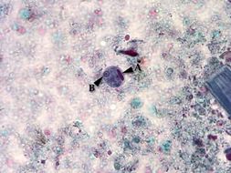
Using a trichrome stain, this photomicrograph depicted a trophozoite of the single-celled parasite, Entamoeba histolytica. Stained purple, the trophozoite, see here in the center of the micrograph, is one of the life cycle phases through which a protozoan organism passes as it matures, and is the active-feeding phase of its growth. The other particulates surrounding the trophozoite represent debris from the slide specimen.Created:
-
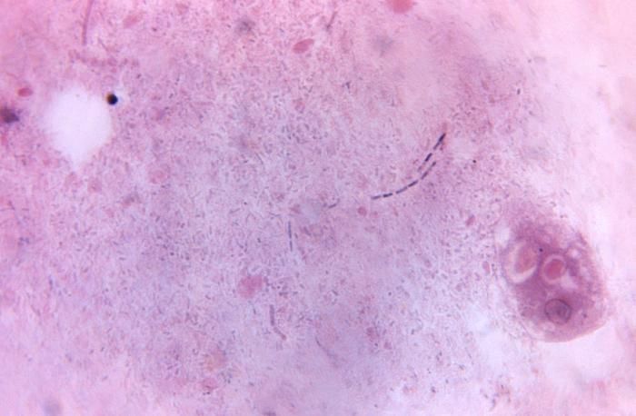
This photomicrograph revealed Entamoeba histolytica cysts that when mature, will display four identifiable nuclei.Created: 1980
-
This split-screen image revealed two parasitic Entamoeba histolytica protozoan trophozoites.Created: 1980